

Dr.Amrita Sawhney, Dr.Ashok Kumar Grover,Dr.Shaloo Bageja
Optic Nerve Sheath Fenestration : creating an effective window!!
Purpose :To evaluate the clinical outcomes of optic nerve sheath fenestration in patients presenting with papilledema with progressive loss of vision .
Optic nerve sheath fenestration(ONSF) is a surgical procedure used to reduce hydrostatic pressure within subarachnoid space of the optic nerve to relieve optic nerve head (ONH) edema.1
Hayreh validated the use of ONSF in cases of ONH edema secondary to elevated intracranial pressure (ICP), also known as papilledema.. 2,3,4This occurs due to disturbance in axoplasmic transport system leading to axonal swelling and nerve fibre dysfunction, ultimately resulting in loss of peripheral as well as central vision . 5,6
Studies have shown chemical and radiographic evidence of compartmentation of the subarachnoid space (SAS) of the optic nerve, suggesting that the optic neuropathy due to increased ICP may occur as a result of CSF collection at the ONH.7
Computed tomography-cisternography performed in patients with papilledema has shown reduced contrast-loaded CSF in the SAS of the optic nerve compared with the basal cistern, suggesting sequestration of CSF in the SAS of the optic nerve.Patients with papilledema have a higher concentration of lipocalin-like prostaglandin D synthase (L-PGDS) in the SAS of the optic nerve compared with the lumbar CSF, which further supports the CSF sequestration theory.8
The main indication of ONSF is papilledema with progressive visual loss with minimal headache, despite of maximal medical therapy or in patients who are unable tolerate the medical therapy.
Causes of papilledema with progressive visual field loss are:
- Idiopathic Intracranial hypertension ( most common)
- Venous sinus thrombosis
- Unresectable CNS tumour
- Arteriovenous malformation of vein of gallen
The most accepted theory of ONSF is that it works by creating a CSF filter from the SAS of the optic nerve into the surrounding orbital tissue, thereby reducing the CSF volume and pressure surrounding the ONH. This theory has been supported by MRI findings in the early postoperative period which demonstrated a fluid collection or a cyst like structure adjacent to the fenestration site, implying CSF filtration.9,10
Material and Method :It was a prospective interventional case series of 6 eyes of 5 patients presenting with progressive loss of vision caused by papilledema due to Idiopathic intracranial hypertension ( 4/5 patients or 4/6 eyes) and cerebral venous thrombosis ( 1/5 patients or 2/6 eyes). Visual acuity, visual fields (if possible), fundus photography ( Figure 1a,b) , orbital and head imaging ( Figure 2a,b) were recorded.
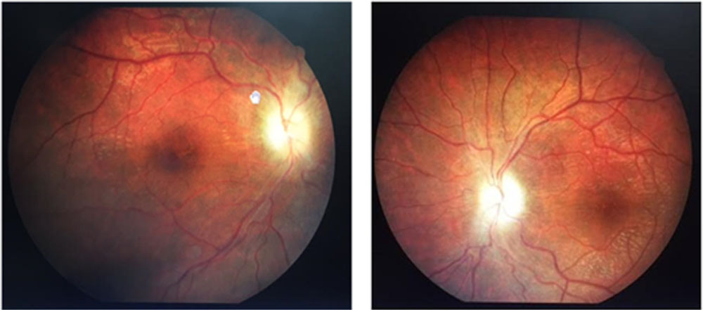
Fig1a Fig 1b
Figure 1 a,b- Fundus photographs of a 19 year old male ,with cerebral venous sinus thrombosis, showing papilledema with disc pallor LE>RE.Visual acuity RE- FC 2mt with projection of rays (PR) accurate in all 4 quadrants LE- Perception of light present , PR accurate only in temporal quadrant
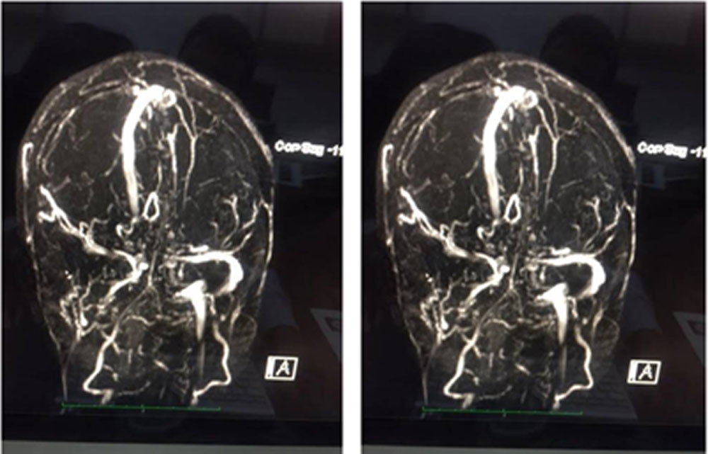
Figure 2a,b – Magnetic resonance venography of the same patient showing loss of normal flow related enhancement in basal veins, straight sinus, right transverse and sigmoid sinus suggestive of venous sinus thrombosis.
All the patients underwent transconjunctival medial orbitotomy approach to optic nerve sheath fenestration under general anaesthesia.
Preoperative medical management was given in all the patients – Intravenous Methlyprednisolone 1gm once a day for 3 days, diuretic, intravenous mannitol and anticoagulant (LMWH) injection ( in case of Cerebral venous thrombosis).
Medial transconjunctival approach was described by Galbraith and Sullivan in 1973.
Technique of medial transconjunctival approach used is asfollows:
- A medial limbal conjunctival peritomy was performed and the conjunctival incision was extended superiorly and inferiorly.( Figure 3)
- The medial rectus muscle was isolated and the tendon was secured with a double armed 6–0 polyglactin (Figure 4)
- The muscle was detached from the globe using scissors, leaving a small remnant of muscle tendon attached to the globe. ( Figure 5)
- 4–0 non-absorbable traction suture was placed through the muscle tendon, and the globe was retracted laterally. ( Figure 6)
- Long posterior ciliary arteries were then identified between the superior and inferior poles of the insertion of the medial rectus muscle. ( Figure 7)
- Retrobulbar optic nerve was approached through the posterior reflection of tenon’s capsule and retrobulbar orbital fat. (Figure 8)
- A sharp blade on a long handle was used to incise the optic nerve sheath approximately 2 mm posterior to the globe with careful attention to avoid any blood vessels on the surface of the nerve.( Figure 9)
- Incision was extended posteriorly upto 3-5mm. A cyclodialysis spatula was inserted into the subarachnoid space and moved in the anterior– posterior direction to lyse any arachnoidal trabeculations and adhesions.
- The traction suture was removed, and the medial rectus was reattached to the globe. ( Figure 10)
- Conjunctiva was closed with 8–0 polyglactin sutures.
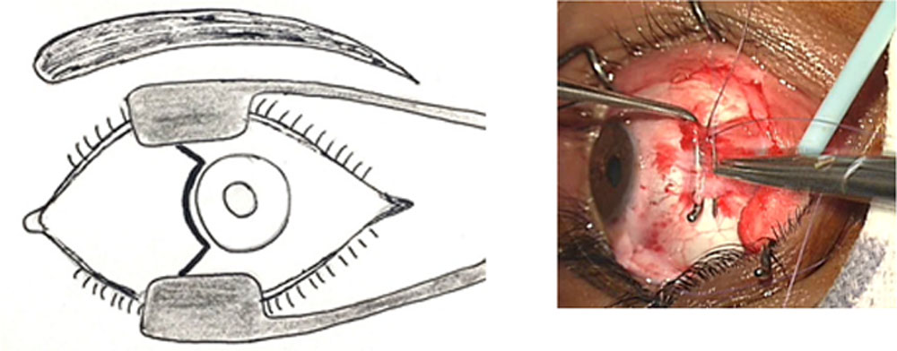
Figure 3 Figure 4
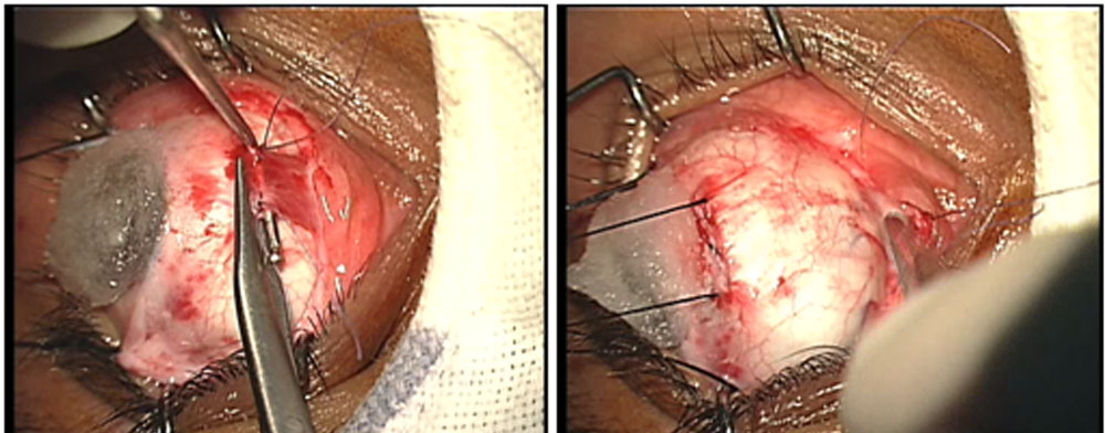
Figure 5 Figure 6
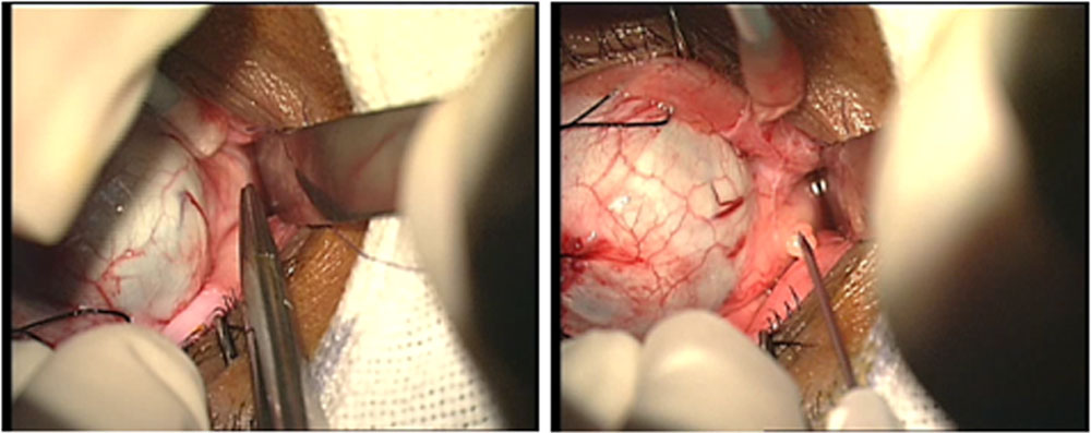
Figure 7 Figure 8
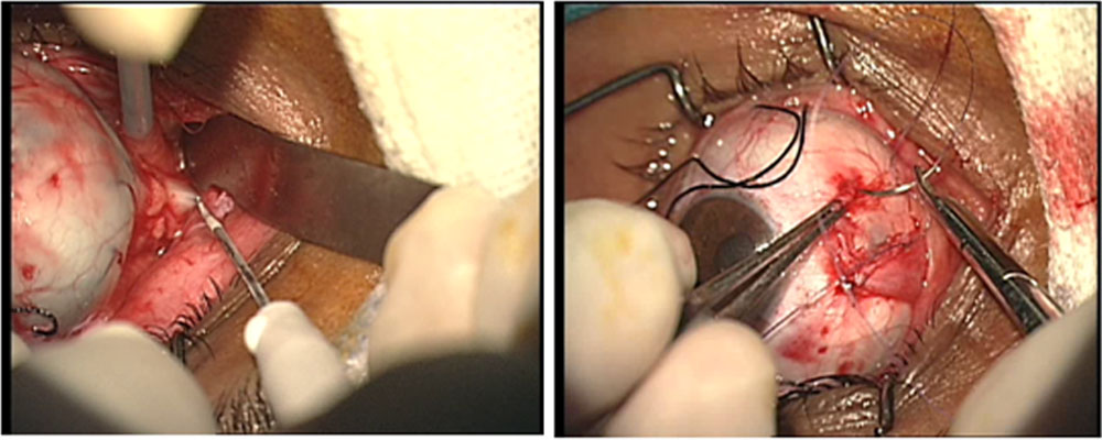
Results:
The average age at presentation in our study was30 years(18-50 years) and there were 3 males and 2 females in the cohort.
The procedure provides an excellent access and exposure to the optic nerve .
ONSF is a relatively safe and effective surgical procedure in patients with visual loss due to papilledema.
| Diagnosis | Preop visual acuity | Postop visual acuity
Day 1 1 month |
|
| Patient 1 | Cerebral venous thrombosis | RE : Finger counting 2 mt
LE: Perception of light +/- |
RE: FC 2 mt RE: 6/60
LE : PL + LE: HM +, PR accurate |
| Patient 2 | IIH | LE: PL positive , PR accurate | LE: FCCF LE: FC 1 mt |
| Patient 3 | IIH | LE : FC 2mt | LE: FC 2mt LE : FC 2mt |
| Patient 4 | IIH | RE : FC 1mt | RE: FC 2mt RE: 6/36 |
| Patient 5 | IIH | LE : HM , PR accurate | LE: FCCF LE: 6/60 |
Discussion
In our study 100 percent patients showed significant improvement in visual parameters after surgical procedure with no adverse events.
In a meta-analysis based on retrospective data of ONSF performed on 423 eyes in patients with IIH, visual acuity improved in 50 % of eyes and visual field improved in 72 % of eyeswith worsening of the visual field or visual acuity in only 11 % of cases.11
Alsuhaibani et al conducted a study on 62 IIH patients with bilateral papilledema who underwent unilateral ONSF were found to have a decrease in the median grade of papilledema in both the operated and the nonoperated eye. The median grade of papilledema in the operated eye decreased from grade 3 preoperatively to grade 0.5 by 12 months. The median grade of papilledema in the nonoperated eye decreased from grade 2 before surgery to a grade 1 12 months postoperatively.12
In 2004, Friedman et al , showed that the complication rate of ONSF was between 4.8% – 45%.( mean 12.9%) . In the same study failure rate was 13% ( progressive visual loss despite the surgery).13
ONSF has also been reported to improve the visual outcome of optic neuropathies due to other etiologies. Mohamed IG et al, conducted a case series of three patients with progressive visual loss due to radiation-induced optic neuropathy, ONSF improved visual function in all the patients.14
Guy et al. demonstrated reversal of progressive visual loss after ONSF in two patients with traumatic optic neuropathy .One of the patients had an optic nerve hematoma and the other developed delayed visual loss from an arachnoid cyst of the optic nerve sheath.15
References
- DeweckerL, On incision of the optic nerve in cases of neuroretinitis, Int Ophthalmol Cong Rep, 1872;4:11–14.
- Hayreh SS, Optic disc edema in raised intracranial pressure. v. pathogenesis, Arch Ophthalmol, 1977;95(9):1553–65.
- Hayreh SS, pathogenesis of oedema of the optic disc, Doc Ophthalmol, 1968;24(2):289–411.
- HayrehMS, Hayreh SS, Optic disc edema in raised intracranial pressure. i. evolution and resolution, Arch Ophthalmol, 1977;95(7):1237–44.
- Tso MO, Hayreh SS, Optic disc edema in raised intracranial pressure. iv. axoplasmic transport in experimental papilledema, Arch Ophthalmol, 1977;95(8):1458–62.
- Schirmer CM, Hedges TR, 3rd, mechanisms of visual loss in papilledema, Neurosurg Focus, 2007;23(5):E5.
- Killer HE, Jaggi GP, Flammer J, et al., cerebrospinal fluid dynamics between the intracranial and the subarachnoid space of the optic nerve. is it always bidirectional?, Brain, 2007;130 (pt 2):514–20.
- Killer HE, Jaggi GP, Miller NR, et al., cerebrospinal fluid dynamics between the basal cisterns and the subarachnoid space of the optic nerve in patients with papilloedema, Br J Ophthalmol, 2011;95(6):822–7.
- Yazici Z, Yazici B, TuncelE, Findings of magnetic resonance imaging after optic nerve sheath decompression in patients with idiopathic intracranial hypertension, Am J Ophthalmol, 2007;144(3):429–35.
- Hamed LM, Tse DT, Glaser JS, et al., Neuroimaging of the optic nerve after fenestration for management of pseudotumor cerebri, Arch Ophthalmol, 1992;110(5):636–9.
- Feldon SE, Visual outcomes comparing surgical techniques for management of severe idiopathic intracranial hypertension, Neurosurg Focus, 2007;23(5):E6.
- lsuhaibani ah, carter KD, NeradJa, lee aG, effect of optic nerve sheath fenestration on papilledema of the operated and the contralateral nonoperated eyes in idiopathic intracranial hypertension, Ophthalmology, 2011;118(2):412–14.
- Friedman DI, Jacobson DM, Idiopathic Intracranial hypertension, J Neuroophthalmol, 2004;24(2):138–45.
- Mohamed IG, RoaW, Fulton D, et al., Optic nerve sheath fenestration for a reversible optic neuropathy in radiation oncology, Am J Clin Oncol, 2000;23(4):401–5.
- Guy J, Sherwood M, Day AL, Surgical treatment of progressive visual loss in traumatic optic neuropathy. report of two cases, JNeurosurg, 1989;70(5):799–801
Leave a Comment