

Dr. Nilutpal Borah, Dr. Nilutpal Borah, Dr. Rup Kumar Phukan
Aim:
To identify the causes and visual prognosis of acute vision loss following caesarian or normal vaginal delivery.
Materials and Methods:
12 eyes(10 female patients) were studied(Jan, 2010- Dec,2017).Inclusion: sudden unilateral/ bilateral vision loss within 6 wks of delivery. Pre, post treatment BCVA, IOT, slit lamp, fundus examinations, FFA , OCT, perimetry were done. VECP, Biochemical investigations, B.P., neurological evaluation with CT/MRI were done(as necessary).
Results:
Total 12 eyes(Unilateral 8,bilateral 2 patients) were examined. Mean age-28.2 years. Mean follow up periodwas 7 months. 5 patients were primy& 3 were multi gravida. Presenting BCVA, 6/12- FCCF (4 eyes). Final BCVA 6/6 – FCCF(4 eyes). Fundus examination revealed- soft exudates, retinal hem, ME and disc edema, attenuated arteries and dilated veins(n=12). Causes were- pregnancy induced hypertension and preeclampsia in -4, anaemia in -1, anterior ischaemic optic neuropathy in -1 and unexplained(possiblePPD 1 patient) in – 4. Spontaneous resolution(after systemic treatment)in- 6 patients,after oral steroid-1patient, after IVMPin 1 patient(both eyes). 4 eyes(in 2 patients) had developed optic atrophy , retinal & RPE atrophy and hyperpigmentation in the macula.
Conclusion:
Although rare, acute unilateral or bilateral loss of vision may occur in postpartum period(n=10). Most patient recover spontaneously or after correction of systemic causes(n=8) but in a few irreversible moderate to profound vision loss may occur(n=2). Pregnancy induced hypertension, preeclampsia, anaemia, unexplained(possible PPD) were primary cause. Bilateral involvement and initial profound loss of vision were indicators of poor prognosis. Early detection is important for good recovery.
In postpartum periodacute loss of vision may occur due to various causes. Some important causes of vision loss are pregnancy induced hypertension,preeclampsia, anaemia, ischaemic optic neuropathy(anterior or posterior), central serous choroidopathy, posterior reversible encephalopathy syndrome(PRES), pituitary apoplexy1, postpartum depression(PPD),and sometimesunexplained.
We report a series of consecutive10 female patients who reported at our institute with the complaint of acute vision loss following caesarean or normal vaginal delivery.Patients were both primygravida ormultigravida.Aimof the study was to identify the causes and visual prognosis of postpartum acute loss of vision.
Although there are singlecase reports on postpartum loss of vision, a pubmed search for related articles on similar study did not yield positive results. We believe this is a first of its kind report about the causes and visual prognosis of postpartum acute loss of vision.
Materials and Methods:
Between January 2010 to December 2017, 12 eyes of 10 female patients who had reported or referredto our institute with dimness of vision following delivery were retrospectively studied. Patients who had complained of sudden unilateral or bilateral loss of vision within 6 weeks of vaginal or caesarean delivery were only included in the study. The patients underwent pre and post treatment best corrected visual acuity(Snellen’s) assessment,intraocular pressure measurement(applanation tonometry),slit lamp andfundus examination with +90 dioptre and +20 dioptre lens, digital fundus fluorescein angiography(Visucam 524, Carl Zeiss meditec),optical coherence tomography(Cirrus HD OCT 500 ,Carl Zeiss Meditec) and perimetry(HFA 720i, Carl Zeiss Meditec).Electrocardiography, visual evoked cortical potential (VECP/VEP), neurological evaluation, CT/MRI were done(when possible). Blood pressure andpulse were recorded. Biochemical investigations included routine blood investigations(TC,DLC,ESR,Hb), FBS and PPBS,urine examinations and serum creatinine. Follow up of patients were done every month till 9 months.
Results:
12 eyes of 10 female patients were studiedfrom January 2010 to December 2017. 8 patients had sudden unilateral and 2 patients had sudden bilateral loss of vision within 6 weeks after delivery. Mean age of the patients was 28.2 years(25 -31 years). Mean follow up period was 7 months(1 –9 months). 7 patients were primygravida and 3 were multi gravida. Best corrected visual acuity(BCVA) at presentation; from 6/12 toFCCF(4 eyes). Final BCVA from;6/6 to FCCF(4 eyes). Fundus examination revealed tortuous arteries,dilated veins, multiple soft exudates & retinal haemorrhage(deep and superficial), macular edemaand disc edema(very mild to severe) in 12 eyes. Fundus changes involved mostly the posterior fundus and there was great variability in presentation in different patients/eyes (i.e. very mild to severe).
Causes of loss of vision were, pregnancy induced hypertension and preeclampsia-4 patients, severe anaemia-1 patient, anterior ischaemic optic neuropathy-1patientand unexplainedin 4 patients(possible PPD-1 patient). Complete resolution of retinal haemorrhage, exudatesand discedemawith vision improvement hadoccurred in 6 patients spontaneously(within 3 months after treatment of systemic condition only).Vision had improved completely after treatment withoral corticosteroid(1mg/kg/day in tapering doses) in 1patient andpartially after IVMP in 1 patient(1gm/day x 3 days). One patient had developed bilateral disc pallor, diffuse retinal and RPE atrophy, RPE hyperpigmentation and another developedbilateral optic atrophy.Both of these patients had preeclampsia.Patients details are shown in Table 1:
| Sl no | Eye | Age
(Yrs) |
Presenting BCVA | Final
BCVA |
Complain
(After delivery) |
Causes | Systemic
disease |
Treatment | Sequelae |
| 1 | OS | 29 | 6/12 | 6/6 | Sudden DOV 4 wks | PIH | HTN | Systemic tt | None |
| 2 | OS | 28 | 6/9
6/60 |
6/9
6/18p |
Sudden DOV 5 wks | AION | – | IVMP | Partial optic atrophy |
| 3 | OU | 28 | FC-1mt
FC-1mt |
FC-3mt
FC-4mt |
DOV 3 wks,
Headache |
Preeclampsia | HTN | Oral steroid | Optic atrophy |
| 4 | OU | 27 | FC-1mt
FC-1 mt |
FC-2mt
FC-3mt |
DOV 1 mo
Frontal headache |
Preeclampsia | HTN | Oral steriod | Disc, RPE, macular atrophy |
| 5 | OD | 27 | 6/18 | 6/6 | DOV 6 wks.Headache | PPD | HTN | Systemic
|
None |
| 6 | OD | 31 | 6/36 | 6/9 | DOV 3 wks | Anaemia | DM | Systemic | None |
| 7 | OS | 30 | 6/12 | 6/6 | DOV 1 wk
Eye strain |
Unknown | – | – | – |
| 8 | OS | 25 | 6/12 | 6/6 | DOV 2 wk | PPD | HTN | – | – |
| 9 | OS | 29 | 6/36 | 6/6 | DOV 2wks
Headache |
Preeclampsia | HTN,DM | Systemic | – |
| 10 | OD | 28 | 6/18 | 6/6 | DOV-3wks
Dizziness |
Unknown | DM | – | – |
(DOV-dimness of vision,PIH-pregnancy induced hypertension,PRES-posterior reversible encephalopathy syndrome)
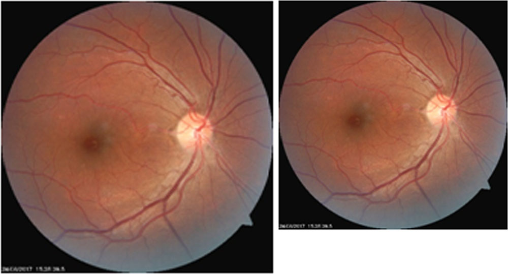
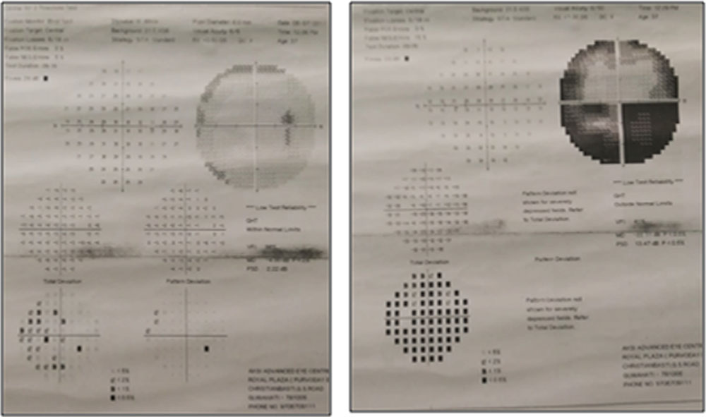
Figure 2. HFA(30-2) shows altitudinal field defect in the left eye due to AION (patient no.2)
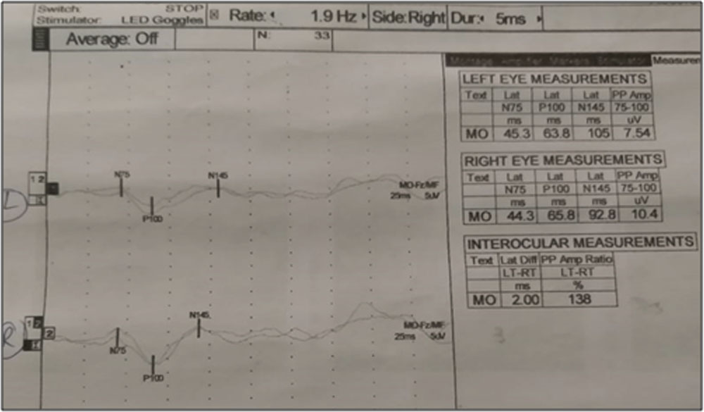
Figure 3. VECP shows Prolonged P100 latency in both eyes (patient no. 3)
Discussion:
Visual changes are experienced by many women during and immediately after pregnancy. Pregnancy can also cause refractive changes. It is suggested that refractive changes are partly due to progesterone-mediated fluid shifts in the corneal stroma and corneal edema may lead to blurred vision.2,3 Dizziness,diplopia,photophobia,glare,eye strain, occasional blurring of vision, flashes of light or temporary loss of vision are some commonly reported problems in postpartum period. There may be associated mild to moderate headache orintermittent frontal headaches.
Visual complaints encountered after the childbirth usually persists for six to eight months. However, in a few patients postpartum vision loss may be a permanent complication.
After 20 weeks of pregnancy if blood pressure remains at 140/80 mm of Hg or higher it is termed as pregnancy induced hypertension(PIH). Preeclampsia is a pregnancy complication.It is characterized by high blood pressure and signs of damage to organs like liver and kidneys. Preeclampsia resolves with delivery.4,5 But, the condition can worsens in 48 hours after delivery.4,6
Acute vision loss in the post-partum period may occur due to severe preeclampsia or eclampsia. Vision loss could be due to exudative retinal detachment, hypertensive retinopathy and cortical blindness7.Cortical blindness may occur up to 15% of preeclampsia and eclampsia patients 8,9,10.In patients with cortical blindness both vasogenic and cytotoxic edema have been observed10,11.Other causes are PRES which can cause reversible acute vision loss 12, pituitary apoplexy 13, posterior ischemic optic neuropathy 14, anterior ischemic optic neuropathy15, cortical venous sinus thrombosis 16, and central serous choroidopathyand choroidal infarction.
Management of preeclamptic patients who develop cortical blindness is the same as for women without this visual complication.8.9,10 Aggressive antihypertensive management in the ICU is necessary in PIH, postpartum aggravation of preeclampsia and eclampsia.Eclampsia is the development of convulsions in a pre-eclamptic patient.Signs of eclampsia include headaches,visual disturbances and epigastric abdominal pain17,18.
Corticosteroids are safe and effective in reducing neonatal complications in women with severe preeclampsia before week 34 of pregnancy.
Any visual complaint that develops during and after pregnancy should be promptlymanaged. The physiologic changes in pregnancy make vascular conditions more frequent, including intracranial arteriovenous malformation, aneurysm, retinal artery occlusion, spontaneous orbital hemorrhage, and pituitary apoplexy. Alsotumorssuch as pituitary adenomas, meningiomas, and orbital schwannomas may grow during pregnancy because of hormonal shifts. Multiple sclerosis(MS) and other autoimmune conditions tend to improve during pregnancy, but a demyelinating attack postpartum are more likely.
In our series defective pupillary reflex (RAPD/APD) was present in 10 eyes. Fundus changes at presentation included mild to severe disc edema, macular edema(focal or diffuse), macular striae, exudative retinal detachment, soft and hard exudates, arterial tortuosity and dilation of the veins.
Of 12 study eyesvision in 6 eyes had improved after correction of systemic causes like PIH and preeclampsia without any sequelae. 1 patient with anterior ischaemic optic neuropathy was treated with IVMP (1gm/day for 3 days), however, after 1 month disc pallor developed(final BCVA 6/18p). 1multigravida patients had severe nutritional anaemia. Fundus examinations revealed retinal haemorrhage, soft exudates and retinal edema, Roth’s spots(anaemic retinopathy). Retinopathy had improved with systemic treatment with vision improvement after 4 months.Two patient who had vision loss due to preeclampsia responded to oral steroid(final BCVA OU- 6/6). One of four patientswherecause of vision loss could not be determined aftercomplete ocular and neurological workupcould have been suffering from postpartum depression(PPD).
PPD is a very common problem where alongwith other symptoms patient may complain headaches, stomach ache and blurred vision. The condition develop within 4-6 weeks after delivery.However, sometimes it can take several months to appear19.
A multidisciplinary approach involving ophthalmologist,neurologist,obstetrician and psychologist is required to arrive at a definitive diagnosis in patients with postpartum loss of vision.
Conclusion:
Although rare, postpartum acute unilateral or bilateral loss of vision may occurin both primy and multigravida following vaginal and caesarean delivery. In our series vision improvedspontaneously(completely or partially) or after treatment of systemic causesin 8 patients. However, irreversible profound vision loss had occurred in 2 patients.Profound bilateral vision loss at presentation, exudative detachment,optic atrophy, RPE atrophy and pigment hypertrophy in the macula carried poor prognosis.Pregnancy induced hypertension, eclampsia, anaemia, PPD were few identified cause of loss of vision. Early detection and prompt management lead to good visual recovery in majority of the patients.
References:
- Aniruddha More et al: Acute vision loss in post-partum period as presenting symptom of HIV-associated cryptococcal meningitis–an unusual case report;BMC Infectious Diseases BMC series 201616:582, https://doi.org
- Digre KB. J Neuroophthalmol. 2011;31(4):381-387.
- Birkholz ES et al. SurvOphthalmol. 2010;55(2):162-168.
- Sibai B, Dekker G, Kupferminc M. Pre-eclampsia. Lancet. 2005 Feb-Mar;365(9461):785–99. [PubMed]
- Henry CS, Biedermann SA, Campbell MF, Guntupalli JS. Spectrum of hypertensive emergencies in pregnancy. Crit Care Clin. 2004;20:697–712. ix. [PubMed]
- Magee AL, Moutquin LM, Dadelszen P. The Society of obstetricians and gynaecologist of Canada. Diagnosis, Evaluation and Management of the Hipertensive Disorders of Pregnancy. J ObstetGynaecol Can. 2008;30:S1–49. [PubMed]
- Servillo G, Apicella E, Striano P. Posterior reversible encephalopathy syndrome (PRES) in the parturient with preeclampsia after inadvertent dural puncture. Int J ObstetAnesth. 2008;17:88–9. [PubMed]
- Swende TZ, Abwa T. Reversible blindness in fulminating preeclampsia. Ann Afr Med. 2009;8:189–91.[PubMed]
- Waziri-Erameh MJ, Omoti AE, Edema OT. Bilateral total loss of vision following eclampsia–a case report. Afr J Reprod Health. 2003;7:106–8. [PubMed]
- Bona M. The eyes in pregnancy. Ophthalmology Rounds [serial on the Internet] 2007. May, [Last accesed on Apr 2011]. Published online, May 2007. Available from: https://ophthalmologyrounds.ca.
- Hladunewich M, Karumanchi SA, Lafayette R. Pathophysiology of the clinical manifestations of preeclampsia. Clin J Am SocNephrol. 2007;2:543–9. [PubMed]
- Magee AL, Moutquin LM, Dadelszen P. The Society of obstetricians and gynaecologist of Canada. Diagnosis, Evaluation and Management of the Hipertensive Disorders of Pregnancy. J ObstetGynaecol Can. 2008;30:S1–49. [PubMed]
- Henry CS, Biedermann SA, Campbell MF, Guntupalli JS. Spectrum of hypertensive emergencies in pregnancy. Crit Care Clin. 2004;20:697–712. ix. [PubMed]
- Fugate JE, Claassen DO, Cloft HJ, Kallmes DF, Kozak OS, Rabinstein AA. Posterior reversible encephalopathy syndrome: associated clinical and radiologic findings. Mayo Clin Proc. 2010;85:427–32.[PMC free article] [PubMed]
- Garg RK. Posterior leukoencephalopathy syndrome. Postgrad Med J. 2001;77:24–8. [PubMed]
- Kahana A, Rowley HA, Weinstein JM. Cortical blindness: clinical and radiologic findings in reversible posterior leukoencephalopathy syndrome: case report and review of the literature. Ophthalmology. 2005;112:e7–11. [PubMed]
- Steegers,Eric AP,von Dadelszen et al:”Preeclampsia”.The Lancet.376(9741):631-644.doi:10.1016/S0140-6736(10)60279-6PMID 20598363
- F Gary Cunningham,et al.(editors)(2010).Williams obstetrics(23rded).New York:McGraw-Hill Medical.ISBN 978-0071497015.
- Post partum depression:symptoms, causes and diagnosis.https://www.medicalnewstoday.com
Leave a Comment