

Dr.Satyam Gupta,Dr.Shreya Shah,Dr.SIDDHARTH RAJENDRAGIRI GOSAI,Dr.VIVEK KUMAR
ABSTRACT:
INTRODUCTION: Dacryoliths are concretions consisting of cellular debris and proteins, found in the lacrimal sac. Dacryolithiasis is often an under-diagnosed condition.
MATERIALS: A 67 yrs old female presented to our OPD with complaints of epiphora and discharge in left eye. After complete evaluation she was diagnosed with left upper canalicular abscess and hard medial canthal mass. The case was managed by incision and drainage with broad spectrum topical and systemic antibiotics. The patient remain asymptomatic for 4 months after which she presented with watering, discharge and was ROPLAS +. This prompt for an external DCR surgery during which we found multiple stones in the sac. The sac was explored for complete removal of stones. The patient is asymptomatic since then.
CONCLUSION: Dacryolithiasis may present as canalicular abscess, which should raise a high index of suspicion. DCR surgery with removal of dacryoliths remains the most effective treatment.
Key words: Dacryoliths, Dacryolithiasis
INTRODUCTION
Dacrolithiasis derives its meaning from the Greek word “dakry” meaning “a tear” and “lithiasis” meaning formation of concretions (calculi). Dacryoliths, sometimes referred to as mucoliths, are calculi of proteins and cellular debris, formed in the lacrimal system. Their color varies from yellowish white or light brown.
Etiopathogenesis is unclear, but some form of lacrimal drainage obstruction potentially triggers numerous mechanisms leading to epithelial metaplasia and alteration of the mucin and trefoil factor peptides.[1]
The incidence of dacryocystitis occurs in a bimodal distribution, with greater frequency in neonates and in people more than 40 years of age.[2]
Clinical features vary from epiphora, punctal discharge, localized swelling (usually a firm mass lesion at medial cantus) to dacryocystitis. Calcification of dacryoliths causes further obstruction so they are usually found in conjunction with intermittent or chronic dacryocystitis. In suspected cases dacryo-endoscopy may be employed for direct visualization of the dacryolith. However, in large number of cases definitive diagnosis is made during a dacryocystorhinostomy.
CASE REPORT
A 67 years old female presented to our OPD with complaint of painful swelling over the medial side of the upper lid in the left eye, along with watering and discharge (Figure 1a & 1b). She was being treated elsewhere for these complaints but was not relived, and was referred by the primary treating ophthalmologist to the Oculoplasty Department of our hospital, as a case of recurrent dacryocystitis. On examination tender swelling was found in the left upper canalicular area with pouting of upper punctum and discharge. She was diagnosed as a case of chronic canaliculitis with superimposed secondary infection leading to formation of localized abscess. On inspection lower punctum appears normal. There is no history of any systemic illness and any systemic stone formation (renal/biliary).
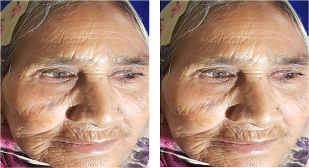
Figure 1a & 1b. A 67 years old female presenting with painful swelling over the medial side of the upper lid in the left eye.
After routine investigations, and physician clearence surgical exploration was planned. Under all aseptic precautions and local anesthesia, incision and drainage was done.(Figure 2a & 2b)
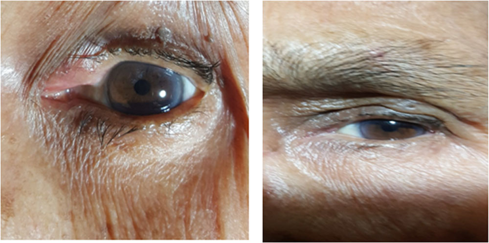
Figure 2a & 2b- Post-operative photograph following Incision and drainage.
She was started on systemic antibiotic and anti inflammatory drugs along with topical antibiotic.
The patient remained asymptomatic for 4 months after the procedure after which she presented to us with epiphora and discharge. After examination she was diagnosed as a case of NLD obstruction. For this an external DCR surgery is planned as per the protocol. To our surprise we found multiple stones of different sizes, shapes, consistency and appearance (small, big, rough, smooth, lobulated like mulberry, opaque and translucent) in the lacrimal sac and the sac was explored for complete removal of stones.(Figure 3)
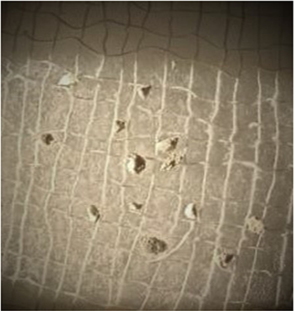
Figure 3-Dacryoliths(small-big, rough-smooth, opaque-translucent, lobulated)
Patient was kept on systemic antibiotics under regular follow-up and was asymptomatic since then (Figure 4a & 4b).
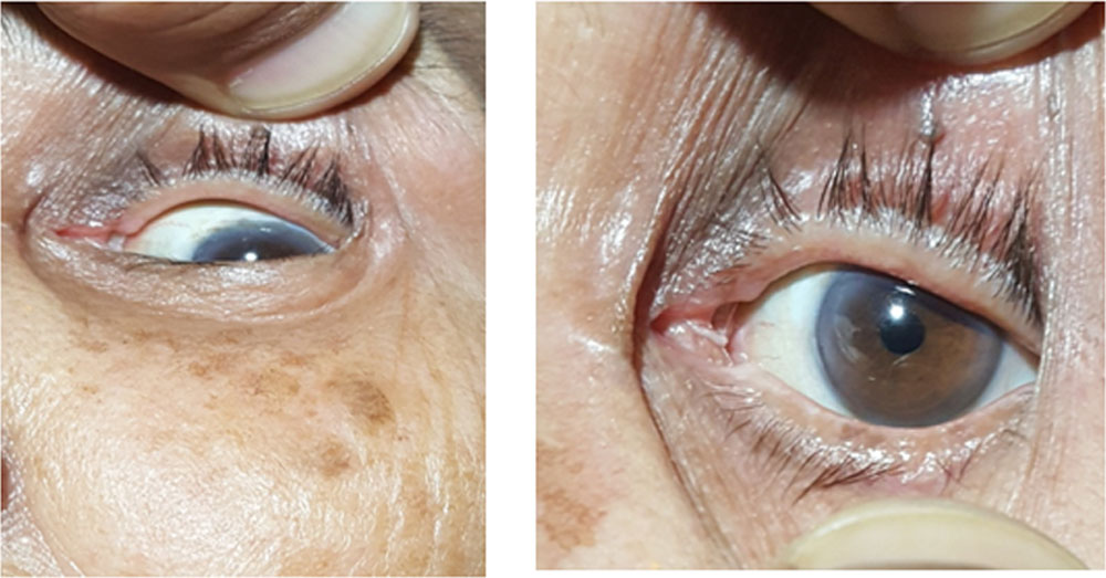
Figure 4a & 4b- Post-operative photograph after DCR and dacroliths removal.
DISCUSSION
Dacryolithiasis is a frequent disease of the lacrimal system, and middle-aged women are commonly affected [3-10].
Dacryolithiasis most commonly effects lacrimal sac, rarely canaliculus and hardly NLD.
Dacryolithiasis often presents with epiphora along with swelling overlying the area of involvement; canalicular dacryolith will present with swelling of the medial end of the lids while lacrimal sac dacryolithiasis will cause swelling over medial canthal area. Discharge and pouting of the punctum may also be present. Most of these patients suffer from recurrent episodes of NLD obstruction. Rarely one can palpate this mass in the region of the lacrimal system [11-12]. Lacrimal sac syringing reveals resistance or regurgitation. Investigation modalities include dacryocystography, B-scan or CT scan [13].
The clinical presentation of these patients is similar to chronic dacryocystitis and hence poses a diagnostic challenge. A detailed history, thorough examination and specific investigations can confirm the diagnosis, so that appropriate treatment can be given to these patients of dacryolithiasis.
The association of infection with dacryolithiasis was reported in the literature [3-10].They can be broadly classified as infectious and non-infectious dacryoliths [14-15].
We have presented a case of canalicular and lacrimal sac dacrolithiasis which was initially diagnosed as an abscess in the left upper canalicular area and definitive diagnosis was made only during the curative surgery (external DCR).
Since dacryolithiasis is a relatively common disorder that is often under diagnosed [16], so in majority of cases it is incidentally found during DCR, and the procedure itself is the cure.
Due to unavailability and unaffordability of the required investigations, proper history and detailed clinical examination is the key to diagnosis.
CONCLUSION
Although in most of the cases, dacryoliths are an incidental finding during DCR surgery, certain clinical features such as epiphora with swelling over canalicular or medial canthal area should arouse a high index of suspicion.
REFERENCES
1.Paulsen FP, Schaudig U, Fabian A, et al. TFF peptides and mucins are major compone components of dacryoliths. Graefes Arch Clin Exp Ophthalmol. 2006;244:1160–70.
2.Gilliland G. Dacryocystitis. e-Medicine. February 22,2005 .
3.L. J. Perry, F. A. Jakobiec, and F. R. Zakka, “Bacterial and mucopeptide concretions of the lacrimal drainage system: an analysis of 30 cases,” Ophthalmic Plastic and Reconstructive Surgery, vol. 28, no. 2, pp. 126–133, 2012.
4.L. T. Jones, “Tear-sac foreign bodies,” The American Journal of Ophthalmology, vol. 60, no. 1, pp. 111–113, 1965.
5.A. J. Berlin, R. Rath, and L. Rich, “Lacrimal system dacryoliths,” Ophthalmic Surgery, vol. 11, no. 7, pp. 435–436, 1980.
6.R. B. Wilkins and J. P. Pressly, “Diagnosis and incidence of lacrimal calculi,” Ophthalmic Surgery, vol. 11, no. 11, pp. 787–789, 1980.\
7.M. J. Hawes, “The dacryolithiasis syndrome,” Ophthalmic Plastic and Reconstructive Surgery, vol. 4, no. 2, pp. 87–90, 1988. \
8.B. Yazici, A. M. Hammad, and D. R. Meyer, “Lacrimal sac dacryoliths: predictive factors and clinical characteristics,” Ophthalmology, vol. 108, no. 7, pp. 1308–1312, 2001.
9.J. K. Marthin, J. Lindegaard, J. U. Prause, and S. Heegaard, “Lesions of the lacrimal drainage system: a clinicopathological study of 643 biopsy specimens of the lacrimal drainage system in Denmark 1910–1999,” Acta Ophthalmologica Scandinavica, vol. 83, no. 1, pp. 94–99, 2005.
10.D. J. Repp, C. N. Burkat, and M. J. Lucarelli, “Lacrimal excretory system concretions: canalicular and lacrimal sac,” Ophthalmology, vol. 116, no. 11, pp. 2230–2235, 2009.
11.Orbit, eyelids, and lacrimal system: 2003–2004 basic and clinical science course, Vol. 7. American Academy of Ophthalmology, USA, 2003, p 28o.
12.Som PMM, Curtin HD. Head and neck imaging (3rd ed). St. Louis, MO: Mosby Inc 1996, pp 1153–1155.
13.Dutton JJ. Standardized echography in diagnosis of lacrimal drainage dysfunction. Arch Ophthalmol 1989; 107(7):1010-2.
14.Perry LJ, Jakobiec FA, Zakka FR. Bacterial and mucopeptide concretions of the lacrimal drainage system. An analysis of 30 cases. Ophthal Plast Reconstr Surg. 2012;28:126–33.
15.Paulsen FP, Schaudig U, Fabian A, et al. TFF peptides and mucins are major components of dacryoliths. Graefes Arch Clin Exp Ophthalmol. 2006;244:1160–70.
16.Wilkins RB, Pressly JP. Diagnosis and incidence of lacrimal calculi.Ophthalmic Surg1980.
Leave a Comment