

Dr.Madhusudan Mandal, Dr.Maheswari Rakesh,Prof.Rizvi Syed Ali Raza
Abstract:
Objective:
Evaluation of the relationship between Interleukin-4 (IL-4) level in tears and disease severity in vernal keratoconjunctivitis (VKC) patients to understand the applied role of IL-4 in clinical assays and management.
Materials and Methods:
Tears samples were collected from 32 VKC patients (mild=18, moderate=8, severe=6) and 32 controls. IL-4 level was measured in tears using ELISA. Patients with other inflammatory diseases were excluded.
Results:
IL-4 level in tears was significantly higher in VKC than controls (p=0.011). Statistically significantly increased IL-4 levels were determined in moderate VKC group as compared to mild (p<0.001) and in severe group as compared to moderate (p=0.011). The ROC curve analysis also revealed that tears IL-4 measurement would be an excellent test to discriminate mild, moderate and severe VKC.
Conclusions:
Tears IL-4 may serve as a potential bio-marker of disease activity and progression in VKC reflecting its role in future therapeutics.
FuLL TEXT:
IntroductioN
Vernal keratoconjunctivitis (VKC) is a chronic, bilateral, seasonally exacerbated, allergic inflammation of the ocular surface, involving tarsal and/or bulbar conjunctiva.[1] The clinical presentation of VKC can vary greatly from mild, intermittent symptoms with few signs to severe, vision-threatening conditions. Converting clinical observations into clinical scores is useful when evaluating the clinical severity of allergic conjunctivitis objectively. The effective severity score may make it possible to objectively determine the indication or cancellation stage for therapeutic drugs such as anti-allergic drugs, immunosuppressive drugs, or corticosteroids. Therefore, in clinical practice with allergic conjunctivitis patients, an accurate diagnosis of the severity of the disease is essential in selecting the most effective therapy.
Several cytokines are involved in the recruitment and activation of inflammatory cells in VKC. The pleiotropic impact of interleukin-4 (IL-4) on immune system cells and its association with T helper type 2 (Th2) cells suggest that IL-4 might be involved in the pathogenesis of VKC.[2]However, this biomarker needs to be validated along with various stages of disease progression to enable the possibility of targeted pharmacological interventions in VKC.In this study, we had evaluated the level of IL-4 in tears in patients of VKC using Enzyme-linked Immunosorbent Assay (ELISA) and compared among the patients of different severity groupsto understand the applied role of IL-4 in clinical assays and management.
MATERIAL AND METHODS
Thisobservational, cohort study was conducted in accordance with the ethical standards formulated in the Helsinki Declaration and informed written consent was taken from all the patients or their guardians (in case of minor).This study involved 32 patients of VKC and 32 age- and sex- matched healthy controls. The VKC patients were categorised asmild (n = 18), moderate (n = 8) and severe form (n = 6). Patients having any non-ocular allergy, dry eye and other ocular inflammatory disease or any autoimmune inflammatory disease were excluded from the study.
Diagnosis of VKC was based on very severe clinical signs and symptoms.The clinical severity of VKC was evaluated according to the 5-5-5 exacerbation grading scale [3] (Table1) where the clinical observations were converted into clinical scores (Table2).
Table1: Exacerbation Grading Scale in VKC
| Grading of clinical sign | 100 points grade | 10 points grade | 1 points grade |
| Clinical sign | 1. Active giant papillae 2. Gelatinous infiltrate of the limbus 3. Exfoliativeepithelial keratopathy4. Shield ulcer 5. Papillary proliferation at lower palpebral conjunctiva |
1. Blepharitis 2. Papillary proliferation with velvety appearance3. Horner-Trantas dots 4. Oedema of bulbar conjunctiva5. Superficial punctate keratopathy |
1. Papillae at upper palpebral conjunctiva
2. Follicular lesion at lower palpebral conjunctiva 3. Hyperaemia of palpebral conjunctiva 4. Hyperaemia of bulbar conjunctiva 5. Lacrimal effusion # |
| Score | 100 points x number of positive sign | 10 points x number of positive sign | 1 points x number of positive sign |
| Range | 0-500 points | 0-50 points | 0-5 points |
# Lacrimal effusion refers to epiphora or tear meniscus increase caused by an eye irritation symptom
Table2: Criteria of severity classification in VKC
| Mild | Moderate | Severe |
| Clinical observation classified in 1- or 10-point-grade is present but 100-point-grade is absent. | Only one clinical observation classified in 100-point-grade is observed along with 1- and / or 10-point-grade. | More than two clinical observations classified in 100-point-grade are observed |
Sample collection and storage:
A volume of tears exceeding 100 µl was collected from single eyes of the subjects with a micropipette (Thermo Fischer Scientific) and disposable microtips (Tarsons) and transferred into Eppendrof tubes -1.5ml (Future Bio Science). Tears samples were stored at -20º C until assayed.
Measurement of IL-4:
IL-4 levels were measured by the ELISA method (Diaclone Human IL-4 ELISA kit, France) according to the manufacturer’s instructions. The absorbance value (OD value) of each well was read on a spectrophotometer at a wavelength of 450 nm. We then drew the standard curve which was almost linear and calculated the concentrations of IL-4 by extrapolating OD values against IL-4 standard concentrations using the standard curve.
Statistical analysis:
We analysed the data in our study using SPSS statistical software (version 20). Demographic characteristics were presented as mean ± standard deviation (SD) for continuous variables and as frequencies and percentages for categorical variables. IL-4 levelsin tears were reported as mean ± SD pg/ml.IL-4 levels were compared between VKC and control group using unpaired two-tailed Student’s t-test and among the severity groups of VKC using One-way analysis of variance (ANOVA) test and Post Hock (Tukey HSD) analysis.All analyses in this study employed a significance level of p < 0.05 (two-tailed).
A receiver-operating characteristic (ROC) curve was used to describe the performance of continuous variable. The predictive accuracy of IL-4 levels in patients with VKC was examined using the area under the receiver-operating characteristic curve (AUC). Optimal cut-off values were determined from ROC curve analyses by calculating the minimum distances from graph coordinates with maximum sensitivity and specificity.
RESULTS
The mean age of the patients in VKC group was 11.6±3.09 years (range 8 to 17) and in control group 11.1±2.87 years (range 8 to16). 24 patients (75%) in VKC and 22 (68.7%) in control group were male. Out of 32VKC patients, 18 (56.25%) were in mild grade while 8 (25%) and 6 (18.75%) were in moderate and severe grade respectively
The mean level of IL-4 in tears of control group was 1.02 ± 0.40 pg/ml (median = 1.04 pg/ml and range being 0.94 pg/ml to 1.06 pg/ml) and in VKC group it was 3.53 ± 2.87 pg/ml (median 1.86 pg/ml and range being 0.93 pg/ml to 10.38 pg/ml) (Figure 1). However it wassignificantly higher in VKC patients than controls (p = 0.011).
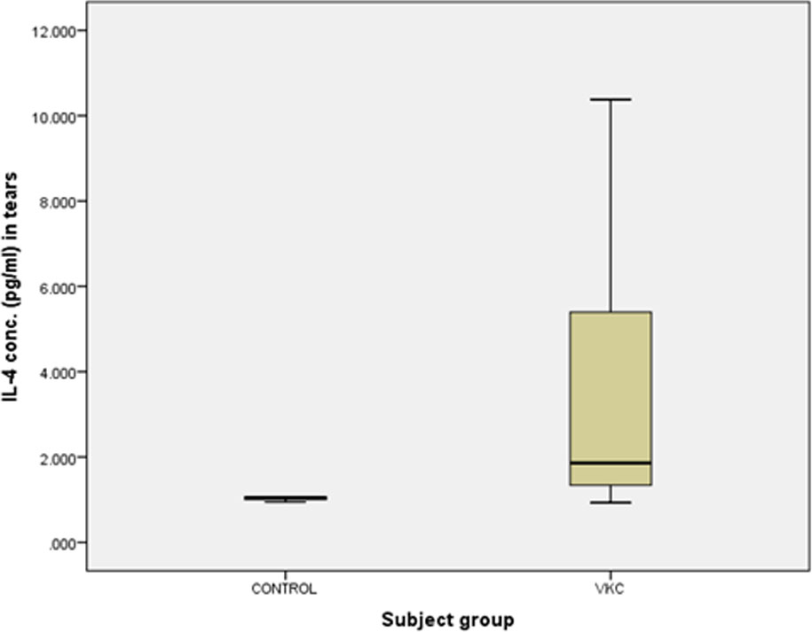
Figure1. IL-4 level in tears of VKC patients and control
The mean level of IL-4 in tears of mild VKC patients was 1.62 ± 1.00 pg/ml (median = 1.38 pg/ml and range being 0.93 pg/ml to 4.26 pg/ml)whereas those in moderate and severe groups were 4.88 ± 2.16 pg/ml (median = 4.97 pg/ml and range being 2.28 pg/ml to 7.29 pg/ml) and 7.48 ± 2.72 pg/ml (median = 7.11 pg/ml and range being 4.96 pg/ml to 10.38 pg/ml) respectively (Figure 2).
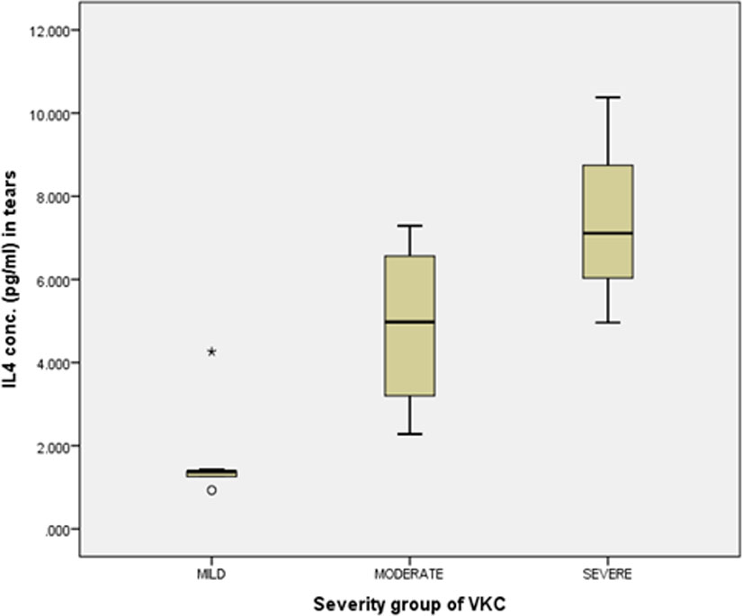
Figure2. IL-4 level in tears of different severity groups of VKC patients
One-way ANOVA showed that there was statistically significant difference in the mean IL-4 levels in tears in between or within the severity groups of VKC (p < 0.001). However the Post Hock (Tukey HSD) analysis showed that the mean IL-4 level in tears was significantly higher in severe VKC as compared to mildand moderate VKC (mild,P <0.001 and moderate,p = 0.011) and in moderate VKC as compared to mild VKC (p < 0.001).
The ROC curve analysis revealed that IL-4 measurement in tears would be an excellent test to discriminate mild VKC patients from controls (AUC = 0.889), moderate VKC from mild (AUC = 0.944) and severe group from moderate group (AUC = 0.750) (Figure3).
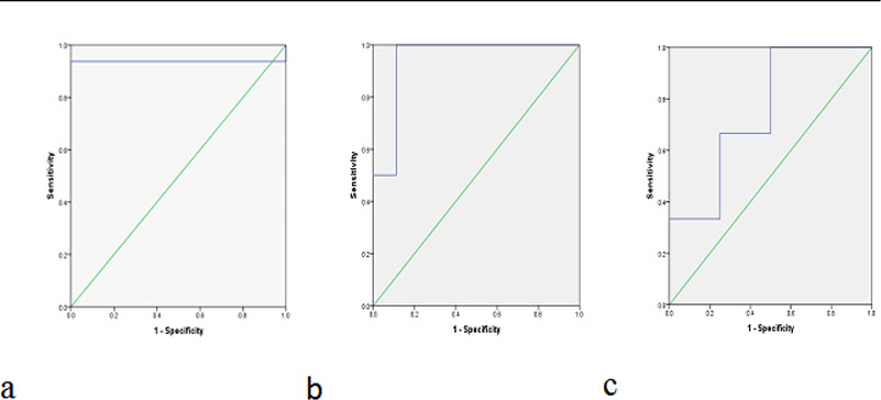
Figure3. ROC curve for IL-4 concentration in tears of a) mild VKC patients as compared to
controls, b)moderate VKC patients as compared to mild group, c) severe VKC
patients as compared to moderate group.
The cut off value of IL-4 in tears to diagnose mild VKC would be 1.162 pg/ml (sensitivity: 88.9% and specificity: 100%), 1.86 pg/ml (sensitivity: 100% and specificity: 99%) for moderate VKC and 6.47pg/ml (sensitivity: 66.7% and specificity: 75%) for severe VKC.
DISCUSSION
Over the last decade, the prevalence of allergic conjunctivitis is increasing, probably through environmental factors. VKC is a severe form of allergic conjunctivitis. The clinical and histopathological changes associated with VKC have been demonstrated to be both an IgE- (type I hypersensitivity) and TH2-mediated disease (type IV hypersensitivity). In the past several years, many clinical and experimental studies about the cells and mediators involved in initiating and perpetuating the ocular allergic inflammation have shown that T helper type 2 cells and their cytokines, corneal fibroblasts and epithelium along with various growth factors play an important role in the pathogenesis of VKC.[4,5]
The previous studies showed the expression of the interleukin-4 receptor α in human conjunctival epithelial cells. Matsuura N et al.(2004) showed that the frequency of IL-4-producing conjunctival CD4+ T cells in patients with VKC were significantly higher than normal control. [6]Fujishima H et al.observed the significantly elevated levels of IL-4 in the conjunctival cells supernatants in allergic conjunctivitis. [7]
In our study, the quantitative assessment of IL-4 in tears of VKC patients revealed that IL-4 level in tears was significantly higher in VKC patients than controls (p = 0.011). H Fujishimaet al. (1995)[8] and Leonardi A, et al. (2006)[9] andUchio E et al. (2000) [10] also found the significantly elevated level of IL-4 in tears of patients of VKC.
Although there have been many studies evaluating IL-4 in patients with VKC, the present study is, to the best of our knowledge and PubMed search results, the first to evaluate thecorrelation of IL-4 level in tears with severity of VKC. The mean level of IL-4 in tears of mild VKC patients was 1.62 ± 1.00 pg/ml whereas those in moderate and severe groups were 4.88 ± 2.16 pg/ml and 7.48 ± 2.72 pg/ml respectively. These results showed that the level of IL-4 in tears was significantly related to the severity of VKC (p = < 0.001). The ROC curve analysis also showed that IL-4 measurement in tears would be a good test to discriminate moderate group from mild group (AUC = 0.944) and severe group from moderate group of VKC patients (AUC = 0.750). The cut off value of IL-4 in tears for diagnosis of moderate group would be 1.86 pg/ml (sensitivity: 100% and specificity: 99%) whereas in severe group that would be 6.47 pg/ml (sensitivity: 66.7% and specificity: 75%).
CONCLUSIONS
These resultsindicate that IL-4 may serve as a potential bio-marker of disease activity and progression in VKC. So this cytokine would be having specific diagnostic and prognostic value in the future therapeutics andmight be a therapeutic target in VKC specially in severe and resistant cases.
SUGGETIONS
The study was limited due to its sample size. Another replicative study in a larger cohort is required.
Financial support and sponsorship
Nil.
Conflicts of interest
There are no conflicts of interest.
References:
- Kumar S: Vernal keratoconjunctivitis: a major review. ActaOphthalmol 2009, 87:133–147.
- Leonardi A, De Dominicis C, Motterle L: Immunopathogenesis of ocularallergy: a schematic approach to different clinical entities. CurrOpin Allergy ClinImmunol 2007.
- Jun Shoji, Noriko Inada and Mitsuru Sawa. Evaluation of Novel Scoring System Named 5-5-5 Exacerbation Grading Scale for Allergic Conjunctivitis Disease .Allergology International. 2009;58:591-597.
- VKC Leonardi A, Secchi AG: Vernal keratoconjunctivitis. IntOphthalmolClin 2003, 43(1):41–58.
- Bonini S, Coassin M, Aronni S, Lambiase A: Vernal keratoconjunctivitis. Eye (Lond) 2004, 18(4):345–351.
- Matsuura N, Uchio E, Nakazawa M, Yago T, Matsumoto S, Ohno S, Minami M. Predominance of infiltrating IL-4-producing T cells in conjunctiva of patients with allergic conjunctival disease. Curr Eye Res. 2004 Oct-Nov;29(4-5):235-43.
- Fujishima H, Saito I, Takeuchi T, Shinozaki N, Tsubota K. Measurement of interleukin-4 and histamine in superficial cells of conjunctiva in patients with allergic conjunctivitis. Curr Eye Res. 1996 Feb;15(2):209-13.
- H Fujishima, T Takeuchi, N Shinozaki, I Saito, and K Tsubota.Measurement of IL-4 in tears of patients with seasonal allergic conjunctivitis and vernal keratoconjunctivitis. ClinExpImmunol. 1995 November; 102(2): 395–398.
- Leonardi A, Curnow SJ, Zhan H, Calder VL . Multiple cytokines in human tear specimens in seasonal and chronic allergic eye disease and in conjunctival fibroblast cultures. ClinExp Allergy. 2006 Jun;36(6):777-84.
- Uchio E, Ono SY, Ikezawa Z, OhnoS.Tear levels of interferon-gamma, interleukin (IL) -2, IL-4 and IL-5 in patients with vernal keratoconjunctivitis, atopic keratoconjunctivitis and allergic conjunctivitis. ClinExp Allergy. 2000 Jan;30(1):103-9.
Leave a Comment