

Dr.Bhramaramba Banagar,
Introduction
- Meibography is a specialised imaging technique used to visualise the morphology of the meibomian glands.
- Meibography provides an in vivomethod of assessing the morphology of the glands.
Purpose of study
- Meibography is a specialised imaging technique used to visualise the morphology of the meibomian glands.
- Meibography provides an in vivomethod of assessing the morphology of the glands.
Study Design
Subjects recruited in this study
・ 100 subjects attending clinic for general health check up were included in the study.
(The data used in this study were obtained from the right eye in each subject.)
・ Males: 62 , females: 38
・ Mean age 46.5 ±15.4 years (range 19~75 years)
Exclusion criteria
- Is a current contact lens wearer
- Has any ocular disease
- Is using any topical medications that may affect ocular health
- Has known sensitivity to the diagnostic pharmaceuticals to be used in the study
Ethical committee clearance taken from the institite.
Subjects were informed about the test and consent taken .
OSDI questionnariewas provided to patients and their respone was analysed and OSDI Score given.
Following tear function tests conducted .
Tear function tests
Schirmer test Ⅰ
Break-up time of tear film (BUT)
Fluorescein staining score (FS)
Meibography
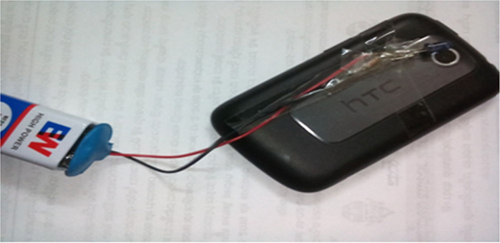
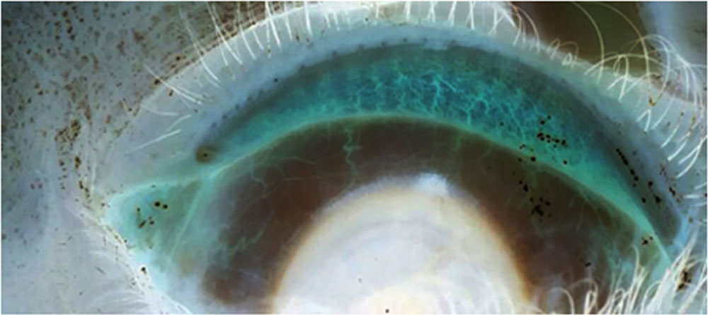
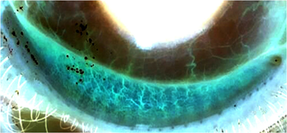
The meibography images taken in infrared mode in high definition resolution with maximum magnification in a Smartphone and infrared bulb for illumination .
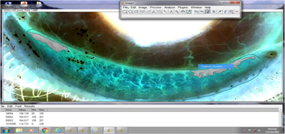
The digital images obtained by meibographywere recorded and analyzed using image J software.
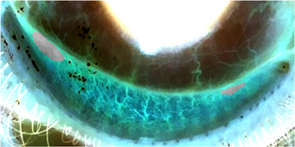
Morphometric indices like percentage area of meibomian gland aciniNumber of gland drop-outs was calculated.
Smart phone attached with infrared bulb with the battery
Meibography image taken with the everted upper eyelid
Meibography image taken with the everted lower eyelid
Gray area showing gland dropout
Statistical analysis
The Pearson and Spearman tests were performed to determine correlations between meibomaingland morphologic values and tear function test parameters.
RESULTS
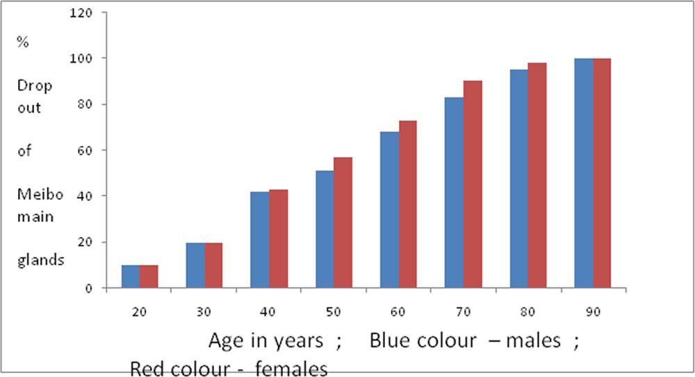
Graph showing the correlation between age and sex and percentage of meibomian gland dropout
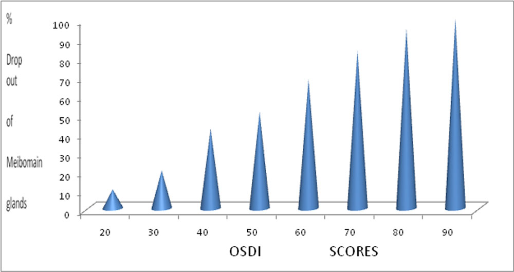
Graph showing the correlation OSDI questionnaire score and percentage of meibomian gland dropout
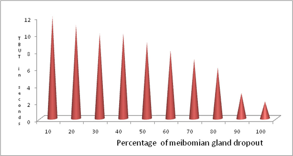
Graph showing the correlation between TBUT (sec ) and percentage of meibomian gland dropout
Correlations of meiboscore with dry eye parameters
| R ( correlation ) | P value | Mean | |
| OSDI | 0.052 | 0.484 | 37.92+18.27 |
| Fluorescein score | 0.402 | 0.005 | 3.67+2.87 |
| BUT (s) | 0.382 | 0.005 | 4.73+1.78 |
| Schirmer I (mm) | 0.021 | 0.776 | 11.53±8.32 mm |
| Age | 0.412 | 0.001 | 46.5 ±15.4 years |
| Sex | 0.032 | 0.654 |
DISCUSSION
The result of our study in respect to morphological changes in meibomianglandswith age and sex correlatedwith the results of studies by Nichols et al , Foulks et al, Dogru M, et al.
Our study results in respect to Schirmers test , TBUT , Fluorescein score correlated with results of Miller KL, Walt JG, Mink DR.
Conclusion
Infrared meibography using smart phone offers a useful tool for obtaining a morphological analysis of the meibomian glands in ocular surface evaluation.
References
- Arita R, et al. Noncontact infrared meibography to document age-related changes of themeibomian glands in a normal population. Ophthalmology.2008 ;115:911-5.
- Arita R, et al. Contact lens wear is associated with decrease of meibomian glands. Ophthalmology.2009 ;116:379-84.
- Arita R, et al. Proposed diagnostic criteria for obstructive meibomian gland dysfunction. Ophthalmology.2009 ;116:2058-63.
- Den S, et al. Association between meibomian gland changes and aging, sex, or tear function. Cornea.2006 ;25:651-5.
- Obata H. Anatomy and histopathology of human meibomian gland. Cornea.2002 ;21::S70-4
Leave a Comment