

Dr.Anubhav Goyal,Dr.Giridhar Anantharaman,Dr.Mahesh G,Dr.Thomas Thachil
Abstract
Purpose: To study the incidence, risk factors, and anatomical outcomes after laser treatment in retinopathy of prematurity.
Methods: Retrospective cohort interventional study. Infants admitted to neonatal intensive care unit (NICU) of 12 referral hospitals between April 2016 and March 2017 were screened according to latest Indian guidelines of international classification of retinopathy of prematurity (ICROP).
Results: The incidence of ROP in 1,648 eyes screened was 25.36% (418 eyes), out of which high-risk pre-threshold ROP (type 1) ROP was observed in 9.95% (164 eyes). Decreased hemoglobin(p<0.001), oxygen requirement(p=0.008) and number of blood transfusions(p=0.037) were significant with type 1 than type 2 (low-risk pre-threshold) ROP. Stages 1, 2, and 3 were observed in 82 (32.28%), 154 (60.62%), and 18 (7.08%) eyes, respectively. APROP was observed in 20.73% eyes with type 1 ROP. Ten eyes showing APROP were treated at an early gestational age of 29 weeks. All infants with type 1 ROP were treated with laser photocoagulation only. Two eyes showed falciform fold over macula and 1 eye was blind from stage 5 ROP.
Conclusion: One-fourth of the infants showed ROP and one-tenth needed laser photocoagulation, the outcome of which was excellent. Risk factors predisposing to ROP were low hemoglobin, high oxygen supplementation, increased number of blood transfusions, more hours on ventilation and septecaemia.
Retinopathy of prematurity (ROP) is a disease mostly reported in preterm neonates with a wide spectrum, ranging from mild, transient changes in the retina with regression to severe progressive vasoproliferation, fibrosis, and retinal detachment finally leading to blindness. ROP-related vision loss is also called “third epidemic” in developed countries, and many of these countries are organizing screening programs for its better management. The development of retina is incomplete during the course of gestation and depends mainly on the severity of prematurity of retina at birth. In 1942, Terry1 was first to describe retrolental fibroplasia with implication of oxygen therapy as the causative agent. Hence, administration of oxygen therapy in premature infants was severely curtailed, resulting in increased mortality. Now, because of improved neonatal survival rate,
The incidence of ROP is increasing in India between 38% and 51.9% in preterm infants.2 Today, it is well known that oxygen therapy is not the single causative factor, but several other risk factors also play a major role in the pathogenesis of ROP.3 Although current ablation treatments can reduce the incidence of blindness by ~25% in infants with advanced ROP, the patients often still have poor visual acuity even after treatment, and the life-long impact of the disease on eye and vision development remains significant.4 Early identification and successful treatment can reduce final visual morbidity.
The aim of this retrospective study was to find the incidence and risk factors predisposing to ROP and to assess the outcome after laser photocoagulation for ROP done in neonatal intensive care unit (NICU) of multiple referral hospitals to a tertiary eye center of a developing country.
Materials and Methods
Latest Indian screening guidelines5
- Birth weight <1700 g
- Gestational age at birth <34–35 weeks
- Exposed to oxygen >30 days
- Infants born at <28 weeks and weighing <1200 g are particularly at high risk of developing severe form of ROP.
- Presence of other factors such as respiratory distress syndrome, sepsis, multiple blood transfusions, multiple births (twins/triplets), apneic episodes, and intraventricular hemorrhage increase the risk of ROP. In these cases, screening should be considered even for babies >37 weeks gestation or >1700 g birth weight.
All neonates admitted to NICU of 12 referral hospitals in Kochi, Kerala, India, were routinely screened for ROP between April 2015 and June 2016 (15 months) according to the current Indian guidelines. The initial examination was carried out at 4 weeks after birth or 31 weeks postconceptional age, whichever was later. All the infants were screened by the same ophthalmologist (AG).
A detailed history including birth weight, gestational age at birth, and adverse events during NICU stay and its management were recorded. The screening was done with a binocular indirect ophthalmoscope and +28 diopter (D) lens. Eyes were examined with an infant Barraquer wire speculum and a Kreissig scleral depressor, under topical anesthesia using 2% proparacaine drops. The pupils were dilated using 0.4% tropicamide + 2.5% phenylephrine eye drops three times, till full dilatation occurred. ROP was graded into stages and zones as per the international classification of ROP (ICROP) classification.6
Type 1 or threshold ROP is defined as zone I any stage ROP with plus disease, zone I stage 3 ROP without plus disease, zone II stage 2 or 3 ROP with plus disease. Type 2 or pre-threshold ROP is defined as zone I stage 1 or 2 ROP without plus disease, zone II stage 3 ROP without plus disease. Aggressive posterior ROP (APROP) is defined as severe plus disease, flat neovascularization in zone I or posterior zone II, intraretinal shunting, hemorrhages, and a rapid progression to retinal detachment.
Those with ROP were examined periodically or every week till completely regressed or till they reached threshold for laser treatment. Any stage 3 ROP with plus disease with 5 contiguous or 8 cumulative clock hours in zone I or II was considered as threshold for treatment.7 MII RET CAM (courtesy Dr. Ashish Sharma),8 a naive innovation of smartphone (with built-in camera and flash)-based fundus camera device and +20D lens was used to capture fundus images in preterm infants during ROP screening (Figure 3).
Laser Treatment
Laser photocoagulation was advised for infants who developed threshold disease as per ICROP classification6 or if APROP was seen in the disease. Laser was done using 810-nm diode laser (Iridex, Iris Medical) with laser indirect ophthalmoscope as early as possible, at least within 1–3 days of diagnosis of threshold plus disease. Laser treatment was done under topical anesthesia, using an infant wire speculum and sclera indentation under neonatologist supervision in the respective NICUs only. The avascular retina beyond the ridge was ablated using confluent medium intensity burns over one session. Invariably, both eyes were treated in all infants in single sitting. Topical treatment with tobramycin and dexamethasone was given for 14 days posttreatment. If regression was found to be inadequate or skip areas were seen on subsequent examination, laser was repeated after 1 week or more.
Follow-up
All children who had undergone laser therapy were reviewed periodically until all signs of threshold disease were regressed. In general, follow-up for all babies terminated once retinal vascularization stabilized or was completed till periphery.
Statistical analysis was done using SPSS version 16.
Results
A total of 1,648 eyes of 824 infants were screened for ROP in NICUs of 12 referral hospitals in Kochi, Kerala, from April 2016 to September 2017 (18 months). Postmenstrual age ranged from 24 to 38 weeks with a mean of 31.76 (SD ± 2.837) weeks. The birth weight ranged from 495 to 3000 g with a mean of 1468.37 (SD ± 454.50) g. There were 472 males and 352 females. ROP was observed in 418 eyes (209 infants) with an incidence of 25.36%. Out of 418 eyes, type 1 ROP was found in 164 eyes with an incidence of 9.95%. Of 418 eyes, 254 eyes showed type 2 ROP. Of these 254 eyes, stages 1, 2, and 3 were observed in 82 (32.28%), 154 (60.62%), and 18 (7.08%) eyes, respectively. APROP or “rush disease” was diagnosed in 34 (20.73%) of 164 eyes with type 1 ROP. No ROP was found in infants with birth weight >2000 g and gestational age >36 weeks. And, no type 1 ROP was seen in infants with gestational age >32 weeks and birth weight >2000 g. Incidence of type 1 ROP decreases with increase in postmenstrual age and birth weight (Figures 1 and 2; Table 1). None of the studied neonates initially presented with stage 4 or 5 ROP.
Laser treatment was performed in 164 (9.95%) eyes showing type 1 ROP. Table 1 shows the number of infants who received laser treatment according to gestational age and birth weight. More than one laser treatment was performed in twelve eyes. Infant demographics and course of care correlated with severity of ROP. However, even after appropriate laser treatment, 3 (0.18%) eyes progressed to falciform fold over macula and 1 (0.06%) eye developed blindness due to retinal detachment. These four eyes were having APROP in zone I and were treated with only laser treatment. All babies withstood laser. Five infants were born at gestational age between 24 and 25 weeks and were given laser treatment for APROP at a still early postmenstrual age of 29 weeks. All infants were screened till vascularization has proceeded to the retinal periphery in all quadrants.
Infants with type 1 ROP had statistically significant lower mean gestational age (P < 0.001), lower mean birth weight (P < 0.01), less mean hemoglobin (P < 0.001), higher mean oxygen requirement (P < 0.001), more mean hours on ventilation (P < 0.001), and higher mean number of blood transfusions (P < 0.001) compared to those ROP (Table 2). There were 5 infants having birth weight >1700gram developed ROP dueto presence of risk factors like respiratory distress syndrome, septicemia, multiple blood transfusions, multiple births (twins/triplets), apneic episodes, and intraventricular hemorrhage (Table 3).
Figure 1: Incidence of ROP according to gestational age
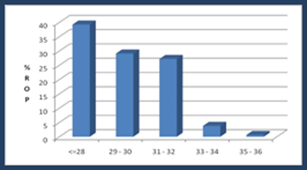
Figure 2: Incidence of ROP according to birth weight
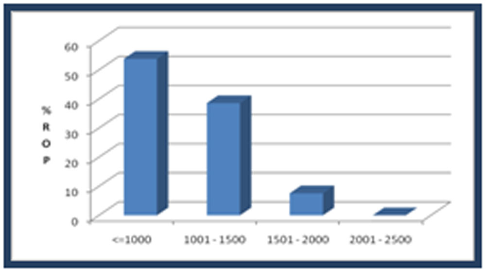
Table 1: Proportion of type 1 ROP eyes treated with laser monotherapy according to gestational age and birth weight
| Gestational Age (weeks) | Total, n | Laser, n (%) | Birth weight (g) | Total, n | Laser, n (%) |
| ≤28 | 168 | 86 (52.43) | ≤ 1000 | 224 | 108 (65.85) |
| 29–30 | 124 | 52 (31.70) | 1001–1500 | 164 | 48 (29.26) |
| 31–32 | 110 | 26 (15.85) | 1501–2000 | 30 | 8 (4.87) |
| 33–34 | 12 | 0 | 2001–2500 | 0 | 0 |
| 35–36 | 4 | 0 |
Table 2: Correlation of gestational age, birth weight, hemoglobin, oxygen requirement, hours on ventilator, number of blood transfusion between type 1 and type 2 ROP
| Type 1 ROP | Type 2 ROP | p-Value | |||
| Mean | SD | Mean | SD | ||
| Gestational age | 28.3 | 2 | 29.6 | 2.1 | <0.001* |
| Birth weight | 996.5 | 256.2 | 1110.8 | 290.2 | 0.01* |
| Hemoglobin (g/dl) | 9 | 1.8 | 12.4 | 2.8 | <0.001* |
| Oxygen requirement | 40.8 | 13.5 | 31.2 | 9.7 | <0.001* |
| Hours on ventilator | 258.1 | 215.1 | 125.3 | 160.8 | <0.001* |
| Number of blood transfusions | 4.3 | 2.6 | 1.7 | 2.3 | <0.001*
|
| Septicemia | 0.23 | 0.11 | 0.09 | 0.10 | <0.001* |
*Mann–Whitney U test
Table 3:- Systemic associations in infants ≥1700 grams
| S No. | Birth weight (grams) | Gestational age (weeks) | Stage of ROP | Associated risk factors |
| 1 | 1700 | 32 | 1 | Septicemia, RDS, apneia |
| 2 | 1710 | 31 | 1 | Septicemia, Rh incompatibility |
| 3 | 1700 | 29 | 2 | Septicemia, intraventricular hemorrhage, RDS, twins birth, apneia |
| 4 | 1900 | 32 | 2 | RDS, apneia |
| 5 | 1710 | 30 | 3+ | Septicemia, RDS, apneia |
Discussion
We screened all babies admitted to NICUs according to recent Indian guidelines of screening Type1 & 2 ROP with birth weight <1700gms and gestational age <34 weeks. The AAP recommends screening of all eligible babies at four to six weeks chronologic age or 31-33 weeks postconceptional age, whichever is later.8 Infants with birth weight >1700gms and gestational age >34 weeks were screened only if they had additional risk factors and/or on neonatologist discretion. While old Indian screening guidelines for ROP suggested screening in birthweight <1500gms and gestational age <32 weeks.4,8,10,11 Vinekar,et al.12 suggested different scenario of ROP screening in developing countries like India. Larger and gestationally ‘older’ infants in India can also develop ROP as compared to their counterparts in Western countries. Praveen Sen et al2 and Jalali et al6 suggested all infants in India with birth weight <1700gms and gestational age <34-35 weeks should be screened regularly. The rate of favourable outcome and a posterior location of the disease are inversely related.
In our study we would have missed 82(25.94%) eyes with ROP if we had used <30 weeks criteria, as per American Academy of Paediatrics(AAP) updated recommendations8 and missed 12(3.79%) eyes with ROP if we had used <32 weeks criteria, as per older Indian Screening Guidelines.4,10,11 So, we suggest that all babies with birth weight <1700gms and gestation <34-35 weeks should be routinely screened. Infants with birth weight >1700gms and gestational age >35 weeks should be screened at the discretion of the neonatologist, depending on other risk factors during the course of stay in the NICU.
Chaudhari et al4 treated only 1 affected eye in 7 infants but we aggressively treated both eyes of all infants diagnosed to have threshold ROP. In accordance to other studies2,4,6 we also found incidence and severity of ROP was closely related to lower birth weight and lower gestational age. Incidence of APROP in our study was 20.73%, which is less than 25% documented by Jalali et al.22 Improved neonatal services and better extreme preterm survival seen in our study may contribute to higher incidence of APROP in Cochin, India. We didn’t find any neonate presented with stage 4 or 5 ROP in this study duration.
Many risk factors have been reported to predispose to the development of ROP. Oxygen therapy, anaemia, exchange transfusion, packed cell volume transfusion, septicaemia, enhanced ventilator support, apnoea and clinical sepsis are important risk factors4,12,23. In our study, anaemia, oxygen administration, hours on ventilation and number of blood transfusions were found to be significant risk factors for ROP, more in Type 1 ROP as compared to Type 2 ROP.
Shift of treatment paradigm from CRYO-ROP study14 to ETROP study15 suggested that by ablating peripheral avascular retina, laser therapy significantly allows more precision of treatment as well as reduces the unfavorable side effects of the cryotherapy and has successful results of 90% more than cryotherapy15. BEAT ROP study compared bevacizumab monotherapy with conventional laser therapy and showed promising results for APROP or stage 3+ ROP in zone 1 but not in zone 2 disease16. Wei-Chi Wu et al19 documented ocular complications associated with bevacizumab intravitreal injection were vitreous or pre-retinal hemorrhage in 8% and transient vascular sheathing in 4% eyes. Other studies showed that anti-VEGF systemic absorption may cause vascular development delay in other organs in these premature babies especially with already persisting subnormal growth. Moreover, followup period after Anti-VEGF monotherapy is unpredictable as there can be a recurrence of neovascularisation even beyond 54 weeks of post-gestational age17. Therefore, anti-VEGF treatment is not recommended by many as the first-line therapy.
Though only laser treatment results are poorer and cause permanent ablation of peripheral avascular retina resulting in permanent peripheral visual field loss16 and very high myopia20 seen in 36.4% as compared to 1.7% eyes treated in Bevacizumab monotherapy treatment group. The rate of recurrence (primary outcome) for zone I and zone II combined was significantly higher with conventional laser therapy than with intravitreal Bevacizumab which was 26% vs. 6%.16 Even than laser treatment is still a gold standard treatment for threshold ROP and practised in most of the places. Treatment with anti-VEGF followed by a 4–5 days later with laser treatment in these cases has improved the efficacy of laser along with a reduced need for extensive laser especially in Zone 1 ROP.18 Still larger sample studies needed to rule out any systemic or local side effects of anti-VEGF treatment in ROP.16
With the advent of new light weight, portable, handy and inexpensive (costing only 19,999 Rs. in Indian currency or 380$ US currency) smartphone-based fundus camera (MII RET CAM – a noble invention by Dr. Ashish Sharma) 5 attached with +20D lens, we were able to capture high-quality fundus videos and images in preterm infants documenting threshold ROP and improvement after laser treatment (figure 3). Though this smartphone-based fundus camera has only approximately 30 degree field of view, still we are able to capture both central and peripheral retinal images which can be used for mass screening, demonstration and better understanding for treating ophthalmologist, neonatologist and counselling parents especially in case of Type 1 or threshold ROP.
As a track changer we found that 3 infants had gestational age between 24-25 weeks and were given laser treatment for APROP at a still early post conceptional age of 29 weeks. We had treated all infants with laser treatment alone which has its own limitations of peripheral retinal ablation resulting in permanent peripheral visual field loss and laser induced very high myopia. We found that the results of laser are extremely satisfactory and out of all the infants who completed followup till complete retinal stabilisation, only 2 (3.27%) infants, having APROP had poor outcome. In accordance to our study Sanghi G et al21 observed falciform fold in macula in 2 eyes and stage 5 ROP in 1 eye treated with laser therapy. The limitation of our study was the study design, as we performed a retrospective, observation study of data extracted from medical records and databases. Strength of this study is large database. We didn’t use imaging modality like RETCAM to screen ROP and none of the preterm infant was treated with Anti-VEGF therapy. Still, we achieved excellent results even with laser monotherapy. Strength of our study is good sample size in small period of time and excellent results even with laser treatment alone.
Conclusion
25.4% of the infants showed ROP out of which only 9.8% infants needed laser photocoagulation treatment, the outcome of which was excellent. Earlier the pre-term and lower the gestational age, the higher is the risk of developing ROP. Birth weight and gestational age are directly proportional to haemoglobin% while these are inversely proportional to oxygen requirement, number of blood transfusions and hours on ventilator. The current treatment of laser ablation therapy has limitations with regard to acute and long term complications. Novel treatment approach of anti-VEGF therapies have not yet been sufficiently evaluated to be broadly recommended for clinical treatment. In ROP management timing is critical in any medical or surgical intervention, since both Type 1 and Type 2 ROP require different approaches13. Despite not using anti-VEGF treatment in any of the treated infants, we managed to get excellent outcomes. We restrict use of anti-VEGF treatment especially for zone 1 disease or APROP and it may not be necessarily given for all stages of ROP. Instead of 31 weeks8 we suggest starting ROP screening at a still early gestational age ≤29 weeks or 4 weeks postgestational age whichever is later. Laser monotherapy can itself give excellent results in treatment of ROP. It has to be noted that in a fragile neonate, careful monitoring and assessing advantages and risks of any treatment/intervention must be weighed very carefully.
Bibliography
- Terry TL et al. Extreme prematurity and fibroblastic overgrowth of persistent vascular sheath behind each crystalline lens. Am J Ophthalmol 1942; 25: 203-204.
- Parveen Sen, Chetan Rao and Nishat Bansal et al. Retinopathy of Prematurity: An Update. Sci J Med & Vis Res Foun June 2015 | volume XXXIII | number 2: 93-96.
- Seiberth V, Linderkamp O et al. Risk factors in retinopathy of prematurity. A multivariate statistical analysis. Ophthalmologica 2000; 214: 131-135.
- Chaudhari S, Patwardhan V, Vaidhya U et al. Retinopathy of Prematurity in a Tertiary Care Center –Incidence, Risk Factors and Outcome. Indian Paediatrics Volume 46, March 17, 2009: 219-224.
- Sharma, Subramaniam, S. Devi, Ramachandran et al. “Smartphone-based fundus camera device (MII Ret Cam) and technique with ability to image peripheral retina.”, Eur J Ophthalmol, vol. 26, no. 2, pp. 142-4, 2016.
- Jalali S, Anand R, Kumar H, Dogra MR, Azad R, Gopal L et al. Programme planning and screening strategy in retinopathy of prematurity. Indian J Ophthalmol. 2003;51(1):89–99.
- International Committee for the Classification of the Late Stages of Retinopathy of Prematurity. An international classification of retinopathy of prematurity II. The classification of retinal detachment. Arch Ophthalmol 1987; 105: 906-912.
- American Academy of Pediatrics. Screening examination of premature infants for retinopathy of prematurity. Pediatrics 2001; 108: 809-811.
- Vasumathy Vedantham et al. Retinopathy of prematurity screening in the Indian population: It′s time to set our own guidelines!. Indian J 2007 SepOct;55(5): 329–330.
- Gupta VP, Dhaliwal U, Sharma R, Gupta P, Rohtagi J et al. Retinopathy of prematurity – risk factors. Indian J Pediatr 2004; 71: 887- 892.
- Maheshwasri R, Kumar H, Paul VK, Singh M, Deorari AK, Tiwari AK et al. Incidence and risk factors of retinopathy of prematurity in a tertiary newborn unit in New Delhi. Natl Med J India 1996; 92: 211-214.
- Vinekar A, Dogra M, Sangtam T, Narang A, Gupta A et al. Retinopathy of prematurity in Asian Indian babies weighing greater than 1250 grams at birth: ten year data from a tertiary care center in a developing country. Indian J Ophthalmol 2007; 55: 331-336.
- Jing Chen, Andreas Stahl et al. Current update on retinopathy of prematurity: screening and treatment. Curr Opin Pediatr. 2011 April ; 23(2): 173–178.
- Cryotherapy for retinopathy of prematurity cooperative group. Multicentric trial of retinopathy of prematurity: 3 months outcome. Arch Ophthalmol. 1990;108:195–204.8.
- Early Treatment of Retinopathy of Prematurity Cooperative Group. Revised indications for treatment of retinopathy of prematurity: results of early treatment of retinopathy of prematurity randomized trial. Arch Ophthalmol. 2003;121:1684–96.
- Mintz-Hittner HA, Kennedy KA, Chuang AZ et al. BEAT-ROP Cooperative Group. Efficacy of intravitreal bevacizumab for stage 3+ retinopathy of prematurity. N Engl J Med. 2011;364 (7):603–15.
- Fierson WM et al. Screening examination of premature infants for retinopathy of prematurity. Pediatrics. 2013;131:189–95.
- Mota A, Carneiro A, Breda J, Rosas V et al. Combination of intravitreal ranibizumab and laser photocoagulation for aggressive posterior retinopathy of prematurity. Case Rep Ophthalmol. 2012;3(1):136–41.
- Wei-Chi Wu, Po-Ting Yeh et al. Effects and Complications of Bevacizumab Use in Patients with Retinopathy of prematurity:A Multicenter Study in Taiwan. Ophthalmology Volume 118, Number 1, January 2011.
- Megan M. Geloneck, MD1; Alice Z. Chuang et al. Refractive Outcomes Following Bevacizumab Monotherapy Compared With Conventional Laser Treatment. : A Randomized Clinical Trial. JAMA Ophthalmol. 2014;132(11):1327-1333.
- Sanghi G, Dogra MR, Das P, Vinekar A, Gupta A, Dutta S. Aggressive posterior retinopathy of prematurity in Asian Indian babies: spectrum of disease and outcome after laser treatment. Retina 2009;29(9):1335–1339.
- Jalali S, Kesarwani S et al. Outcomes of a Protocol-Based Management for Zone 1 Retinopathy of Prematurity: The Indian Twin Cities ROP Screening Program Report Number 2. Am J Ophthalmol 2011;151:719–724.
- Hakeem AHAA, Mohamed GB, et al. Retinopathy of Prematurity: A Study of Prevalence and Risk Factors. Middle East Afr J Ophthalmol. 2012 Jul-Sep; 19(3): 289–294.
Leave a Comment