

Dr.Krithika Santhi Selvan, K21484, Dr.Santhi Selvan
Case report
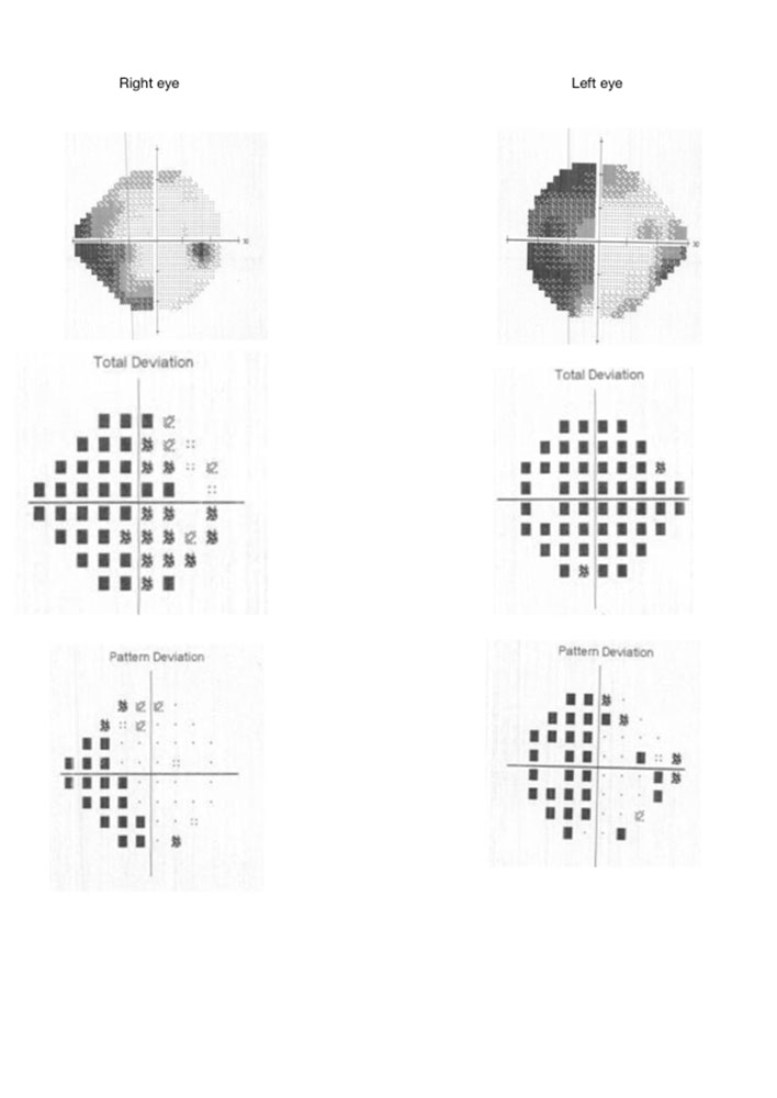
A 12 year old female child presented to the clinic with the complaint of throbbing , right sided headache associated with sleeplessness for the past 2 days which was not getting relieved with oral NSAID’s . She gave no past history such episode and there was no family history . Her uncorrected visual acuity was 6/6 in both eyes . Ocular examination showed no abnormalities in the anterior segment with normal sized pupils reacting to both direct & consensual light and no relative afferent pupillary defect . Ocular motility was normal with normal fundus appearance . Colour vision testing with Ishihara’s pseudoisochromatic plates was normal . In office visual field examination by confrontation method revealed a left homonymous hemianopia which was confirmed with HFA 24-2 . CT Brain and MRI brain were normal . The patient was treated with systemic beta blockers (propronalol) and cerebro-selective calcium channel blockers ( flunarizine ) . She was monitored with periodical review in the ophthalmologist clinic . The patient reported progressive clearing of vision in the peripheries with complete resolution at around the 6th day after the headache episode . HFA done on the 10the day after headache episode revealed no visual field deficits and the patient has remained symptom free since then with no recurrences. This led to the diagnosis of transient incongruous left homonymous hemianopia presenting as visual symptom during a migraine episode .
Discussion
The Second Edition of The International Classification of Headache Disorders (ICHD-II) divides migraine into migraine without aura and migraine with aura. According to the ICHD-II, migraine headaches are recurrent and typically last from 4 to 72 hours. They should have at least 2 of the following characteristics: unilateral, pulsating, moderate to severe intensity and worsening with physical activity. Nausea/vomiting or photophobia and phonophobia should be present during the headache.Visual aura associated with migraine accounts for about 36% of patients. Common descriptions of visual aura include bright spots (42%), flashes of light (39%), blind spots (32%), foggy vision (27%), and fortification spectra (20%) .The neurologic symptoms typically develop gradually over 5 to 20 minutes and last less than 60 minutes.Visual phenomenon associated with migraine can be negative , such as scotomas, or positive, such as photopsias or scintillating scotomas. Frequently, scintillating scotomas accompany fortification spectrum, a zigzag design that starts near fixation and spreads peripherally to form a C-shaped figure with a shimmering border and an accompanying absolute or relative scotoma.
Migraine is postulated to be caused by two mechanisms.The first suggests that migraines are primarily a vascular disorder, with vasoconstriction responsible for the aura and subsequent vasodilation resulting in the headache. It is thought the dilation of cranial arteries in the scalp, dura, and pia triggers the trigeminal pain fibers that innervate these vessels and causes the headache. The second major theory for the pathogenesis of migraines is based upon cortical spreading depression (CSD). It is thought that slowly propagating waves of depolarization in the brain followed by inhibition of the affected areas is what is responsible for the headache as well as the aura. This is thought to be caused by altered cellular excitability which triggers the CSD.
Various visual field abnormalities have been reported with migraine headaches such as monocular vision loss , hemianopia , quadrantopia , sectronopia ,etc. That aura of migraine is considered as a cortical phenomenon whilst these field abnormalities could be presumed to be precortical in nature .Migraine associated cerebral ischemia tends to occur readily in the occipital lobe . The most prominent clinical sign of migraine associated cerebral ischemia is a homonymous field defect such as hemianopsia or hemiopic scotoma, due to a posterior cerebral artery infarct.Because our patient also presented with a homonymous field defect, pathological mechanisms same as those of migraine-associated cerebral ischemia may have been responsible for symptom expression in this case.
CT scan done in patients presenting with migrainous aura have shown hypoperfusion , increased median transit time and decreased cerebral blood flow which readily reversed after the resolution of symptoms . This is likely to be secondary to vasodilation, supportive of the vascular theory. MRI and MRA images are typically normal in migraine cases, subclinical infarcts, deep white mater lesions and changes in perfusion imaging, represented by decreased blood flow in the cortex contralateral to the hemifield affected by the aura, have been documented before and after the aura.
Standard automated perimetry (SAP) is the standard perimetric technique and is most used in clinical practice.SAP was performed with a Humphrey Visual Field Analyzer (Carl Zeiss Meditech, Inc., Dublin, CA) with a 4-mm2 Goldmann size III stimulus (0.43° diameter) on a dim background (31.5 apostilb). The SITA Standard strategy was used for the 24-2 test presentation pattern. Visual field defects have also been picked up by Temporal modulation perimetry (TMP) and short-wavelength automated perimetry (SWAP) . Standard automated perimeter is non pathway specific and these different forms of perimetry test different aspects of visual processing, with flicker perimetry preferentially assessing magnocellular pathways and SWAP assessing the blue-on-yellow (or koniocellular) system .
Differential diagnoses
The most common causes of visual field defects in children include trauma (34%) , tumour (27%), infarction (23%) and cerebral hemorrhagic (7%) in decreasing order of frequency . Migraine is typically a diagnosis of exclusion because many conditions that mimic migraines have potentially devastating consequences and must be ruled out.Retinal migraine affects the anterior visual pathway resulting in monocular visual loss that lasts less than 30 minutes. Transient constriction of retinal arteries may also be observed during a retinal migraine.. A typical aura without a headache can be difficult to distinguish from a stroke or a transient ischemic attack (TIA). Stroke or TIA can be considered if the patient is older than 40 years or if there is risk factors. TIA typically has a more abrupt onset and shorter duration (3 to 10 minutes) static visual symptoms. Diagnosis of stroke is confirmed with Neuroimaging.
Some medications have been reported to cause transient homonymous hemianopia such as cisplatinum, vinblastine, and bleomycin used for testicular carcinoma and incongruous homonymous heminopia after topiramate therapy. The visual field defects slowly improved after discontinuation of the medications. Tumours of the occipital lobe can cause photopsias that resemble an aura. However, these are typically persistent and stationary. Although occipital seizures can result in both positive and negative visual symptoms, visual patterns differ between this and migrainous episodes. Occipital seizures tend to have more centrally located positive phenomenon and diffuse negative symptoms, whereas the symptoms with migraine patients are more peripherally located.
Conclusion
This case report really emphasises the need for visual field testing to be a baseline evaluation in patient presenting to the clinic with a headache or a migrainuous episode It is possible that some of the dysfunction measured the day after a migraine can be explained due to effects of fatigue or poor concentration after migraine..It is evident that not all healthy young migraineurs with interictal visual field abnormalities will progress to having glaucoma. Indeed, we do not know whether the presence of visual field deficits between migraines is predictive of future eye disease. It is not known whether migraine events themselves affect visual processing, possibly in a cumulative fashion, or whether there are underlying vascular, metabolic, or neurologic differences in some migraineurs that predispose them to both migraine and to eye disease.Studying the time course of visual field deficits relative to migraine events may provide information essential to understanding their relevance, if any, to eye disease, and for enabling correct differential diagnosis of visual field loss due to treatable cause (such as glaucoma).Even if migrainous visual field defects have no long-term cumulative significance, knowing whether duration after migraine is likely to affect visual field performance is essential for accurate perimetric interpretation for the management of other eye disease in migraineurs and periodic prolonged decreased sensitivity after migraine has implications for both differential diagnosis in a clinical setting, and clinical research studies using perimetry.
References
1)Rich WM. Permanent Homonymous Quadrantanopia after Migraine. British Medical Journal. 1948;1(4551):592-594.
2)Goodwin D. Homonymous hemianopia: challenges and solutions. Clinical Ophthalmology (Auckland, NZ). 2014;8:1919-1927.
3)Gupta SN, Gupta VS, Fields DM. Spectrum of complicated migraine in children: A common profile in aid to clinical diagnosis. World Journal of Clinical Pediatrics. 2015;4(1):1-12.
4) Spiri D, Rinaldi VE, Titomanlio L. Pediatric migraine and episodic syndromes that may be associated with migraine. Italian Journal of Pediatrics. 2014;40:9
5)Kapinos G, Fischbein NJ, Zaharchuk G, Venkatasubramanian C. MIGRAINE-LIKE HEADACHE WITH VISUAL DEFICIT AND PERFUSION ABNORMALITY ON MRI. Neurology. 2010;74(21):1743-1745.
Leave a Comment