

Dr.Shreya Shah,
Dr.Mehul Ashvin Kumar Shah,Dr.Romi Singh,Dr.SIDDHARTH RAJENDRAGIRI GOSAI
Abstract:
Purpose
To explore a new technique to find out the proximal end of lacerated canaliculi and a new material for the stent.
Methods
Surgery was performed on 10 eyes of 10 patients using a 5/0 prolene suture needle as amodified probe.Prolene suture was inserted as a stent and left in place for two months. All the data were analyzed.
Results
The surgery was successful in all cases, and the tubes were removed after two months. The mean follow-up time after the tube removal was 3.8 months (range 3–6 months). No other complications associated with the prolene sutures were noticed except for epiphora and corneal irritation in three cases. All the tubes were removed successfully without any difficulty. No iatrogenic injuries occurred during prolene removal.
Conclusions
The reported surgical technique is a very cost-effective option for lacrimal canalicular laceration repair.
Key words: Lacrimal canalicular laceration; Prolene suture; Stent; Identification of proximal end
INTRODUCTION:
Lid lacerations are common with ocular injuries[1–3], while an association with lacrimal passage injuries is less common[1].
Canalicular injuries are relatively common, but controversy persists regarding repair and the surgical methods to be employed. In this review, various techniques have been analyzed in a historical context. Recent studies of lacrimal drainage in systems with monocanalicular obstruction are cited. Various surgical techniques in the repair of canalicular injuries are reviewed, including methods of identifying the medial lacerated lacrimal passages and the various lacrimal stents that have been used. Various methods of stent placement and fixation are systematically categorized. Case series of canalicular repair reported in the literature is reviewed, and the results and complications are compared.
Methods: Our study was approved by the Hospital Ethical Committee. We have developed a surgical technique to manage lid laceration, and have enrolled patients for this program.
The eyelid was repaired surgically in layers. The lacrimal canal was repaired using a very novel technique. A new method to identify the proximal and distal ends of lacerated canaliculi has been identified along with a newer material of stent.
The choice of general or local anesthesia will depend on the age of the patient, the location and extent of the injury, the type of stent, and the surgeon’s experience(4). General anesthesia is mandatory for virtually all children and for many adults. The techniques of “local” anesthesia include topical intranasal anesthetics and vasoconstrictors, a regional block of theintratracheal and anterior ethmoidal nerves, as well as local subcutaneous infiltration.
We have penetrated the Bowmen lacrimal probe from the lower punctum (Fig-1) and identified the distal end of the laceration.
We have utilized the 5/0 prolene suture with spatula needle to identify the proximal end of laceration, and broke the sharp tip of the needle (Figure-2).
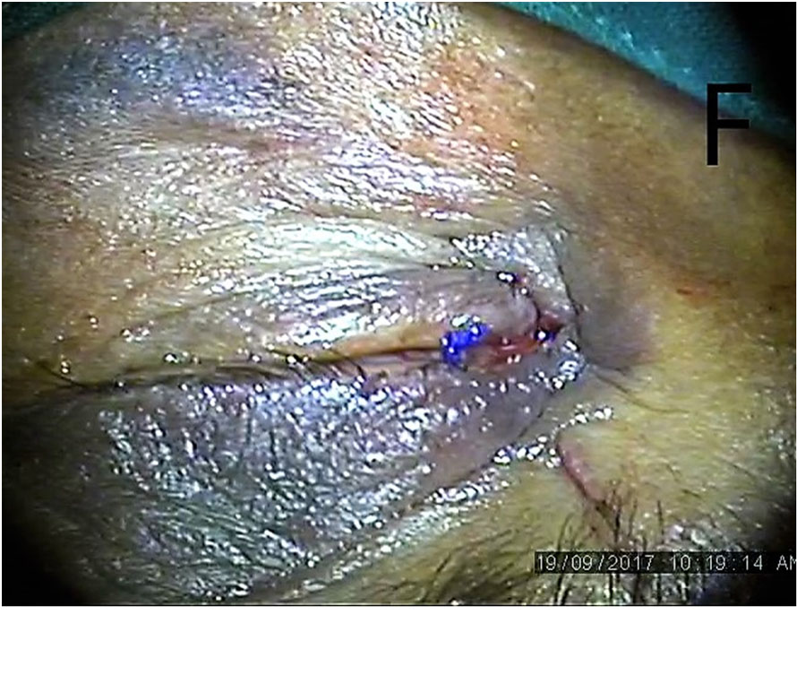
The blunt end of the needle inserted in the upper punctum which is intact was passed through the canaliculi and brought out of the proximal torn end (Fig-3). The blunt end was followed up in the distal end with railroad technique with lacrimal probe (Fig-4). The prolene suture was left in place as a stent for six weeks (Fig-5,6) and the rest of the lid was sutured in layers.
We have removed the prolene suture after eight weeks.
Results: We have enrolled nine cases with a mean follow up of 98 days. The causes of canalicular laceration include fighting, dog bites, fall or collisions while running, and sticks or cattle horns. Laceration of the inferior canaliculus is more common than that of the superior canaliculus. We have found all patients to be doing well anatomically in relation to integrity. We have found lacrimal passages in all cases and patients were symptom-free.
Discussion:
Although canalicular injuries are relatively common, controversy persists regarding repair and the surgical methods to be employed. Methods of treating canalicular laceration include a laissez-faire approach, exteriorization, or anastomosis with the placement of a canalicular stent. Canalicular stents may be placed with monocanalicular, bicanalicular-annular or -nasal fixation techniques. Although a wide variety of lacrimal stents have been used in the past, most reported case series of canalicular laceration have emphasized one or more of these techniques with the use of silicone tubing as the stent material. Although iatrogenic injury of the uninvolved canaliculus and premature loss of a stent have been cited, high rates of success have been reported using these techniques and complications have been infrequent or minor (1).
Many authors have suggested the use of pig tailto detect the proximal end (4-8); similar mechanism is used in the current technique but in a more cost-effective way.
Naturally occurring organic and metal stents have generally been used in a monocanalicular fashion. The relative inflexibility of metal limits its application as a simple monocanalicular stent. More flexible, synthetic stents of nylon, polyethylene, and silicone make have been placed in either a mono- or bicanalicular fashion. Although metal canalicular stents are still in use, silicone tubing has become more popular and is viewed as the canalicular stent material of choice. This is particularly true while considering medial lacerations or tissue loss involving the common canaliculus or lacrimal sac (1).Bicanalicular silicon tube insertion is reported to be most the accepted material and method (9–15).
Mini monoka is a monocanalicular silicone stent reported to have a high success rate and ease of insertion (16–18).
Teflon is also reported as an option but is unlikely to be usedin the long term (19).
Studies suggest stent removal time interval of two-six months; we have also removed the prolene suture after two months (1).
Conclusion: The reported method is a unique technique to repair, which is very cost effective, easy to adopt, and assures of excellent results without much discomfort to the patient.
Funding: This study not funded by anybody
Conflict of Interest: Author A declares that he/she has no conflict of interest. Author B declares that he/she has no conflict of interest. …
Ethical approval: All procedures performed in studies involving human participants were in accordance with the ethical standards of the institutional and/or national research committee and with the 1964 Helsinki declaration and its later amendments or comparable ethical standards.
Informed consent: Informed consent was obtained from all individual participants included in the study.
Refrences:
- Lima-Gómez V, Mora-Pérez EOcular lesions associated with lid wounds with or without tear duct affectionCir Cir.2006 ;74:11-4.[Article in Spanish]
- Walter WL. Ophthalmic Surg.The use of the pigtail probe for silicone intubation of the injured canaliculus.1982; 13:488-92.
- Naik MN, Kelapure A, Rath S, Honavar SGManagement of canalicular lacerations: epidemiological aspects and experience with Mini-Monoka monocanalicular stent.Am J Ophthalmol.2008; 145:375-380.
- Reifler DM. Management of canalicular laceration. Surv Ophthalmol. 1991; 36(2):113–132. [PubMed]
- Brian J. Forbes, William R. Katowitz, Gil Binenbaum, Pediatric canalicular tear repairs—revisiting the pigtail probe J AAPOS. 2008 Oct; 12(5): 518–520.
- Saunders DH, Shannon GM, Flanagan JC. The effectiveness of the pigtail probe method of repairing canalicular lacerations. Ophthalmic Surg. 1978;9 (3):33–40. [PubMed]
- Pecora JL. Pediatric nasolacrimal pigtail probes. Ophthalmic Surg. 1980; 11 (4):249. [PubMed
- Cho SH, Hyun DW, Kang HJ, Ha MS. A simple new method for identifying the proximal cut end in lower canalicular laceration. Korean J Ophthalmol. 2008; 22 (2):73–76.[PubMed
- Baylis HI, Axelrod R: Repair of the lacerated canaliculus. Ophthalmology 1978; 85:1271-1276.
- Beyer-Machule CK: Lacrimal stents, in Bosniak SL (ed): Advances in Ophthalmic Plastic and Reconstructive Surgery; The Lacrimal Syslem, Vol 3. New York, Pergamon Press, 1984, 171-173
- Corin SM, Hurwitz JJ, Corin WJ, Kazdan MS: Lacrimal catheterization. Ophthalmic Surg 1989; 20:202-204,
- Fox SA: Ophthalmic Plastic Surgery. New York, Grune& Stratton, 1976, ed 5, 584-607
- Adams AD: Silicone-loop repair of the torn canalicrllus. .Arrh Ophtha/mol1976; 94: 1958-1960
- X Liang, Y Lin, Z Wang, L Lin et ala modified bicanalicular intubation procedure to repair canalicular lacerations using silicone tubes Eye (Lond) 2012; 26(12): 1542–1547.
- Wang L, Chen D, Wang Z. New technique for lacrimal system intubation. Am J Ophthalmol. 2006; 142 (2):252–258. [PubMed]
- Gonnering R: Simplified monocanalicular silicone intubation. Arch Ophthalmol1987; 105:1024
- Jeganathan VSE, Gao Z, Verma N Mini Monoka Stent Insertion for the Management of Epiphora Secondary to Canalicular Obstruction or Stenosis. Optom Open Access 2016;1: 112.
- Lee H, Chi M, Park M, Baek S. Effectiveness of canalicular laceration repair using monocanalicular intubation with Monoka tubes. Acta Ophthalmol. 2009;87 (7):793–796. [PubMed]
- Hurwitz JJ: Teflon tubes for stenting and bypassing the lacrimal drainage pathways. Ophthalmic Surg1889; 20;855-859
Legands:
Fig-1 Lacrimal Probe passed thru lower punctum to distal end
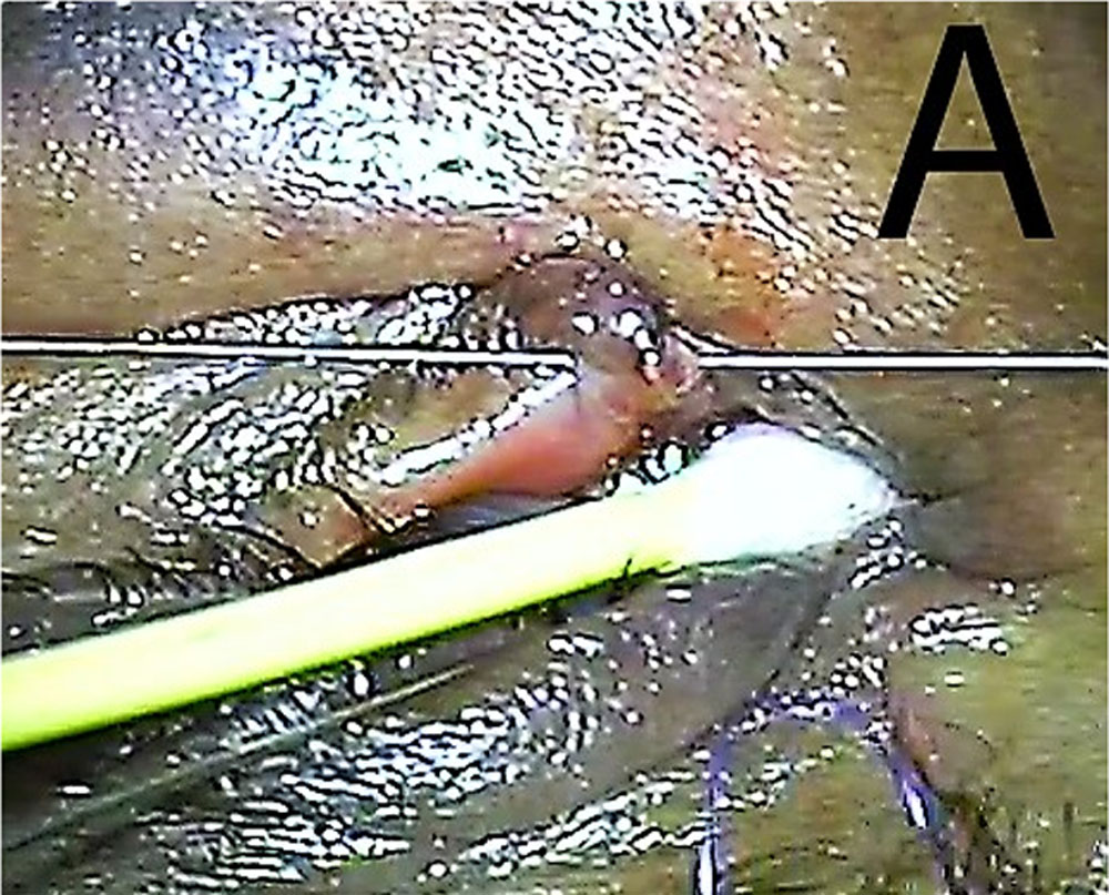
Fig-2 Blunt prolene needle passed thru upper punctum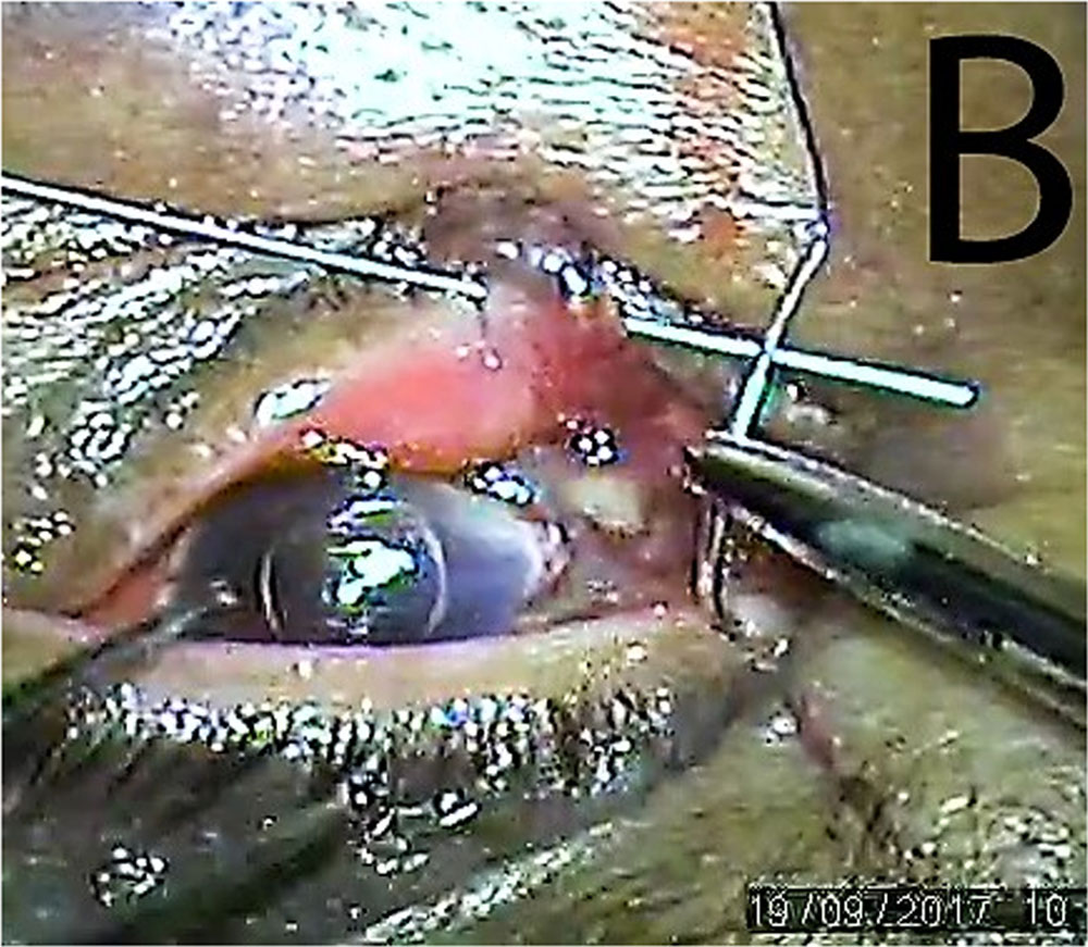
Fig-3 Blunt end of needle brought out of proximal lacerated end
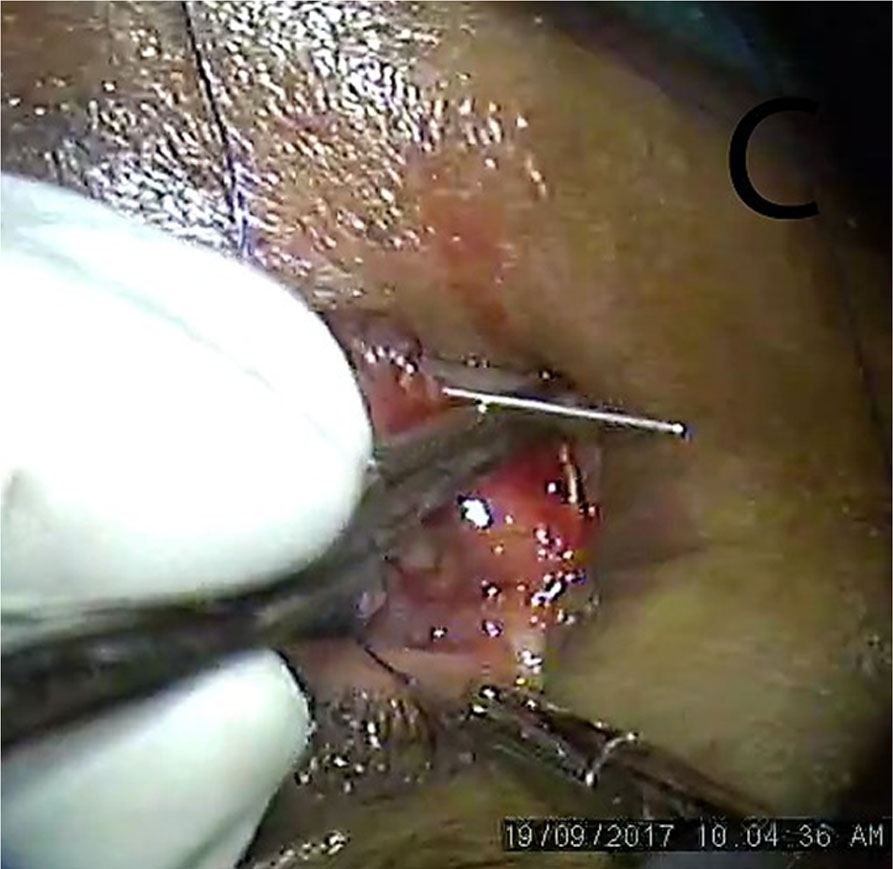
Fig-4 Blunt Prolene needle passed thru distal lacerated end using rail road technique
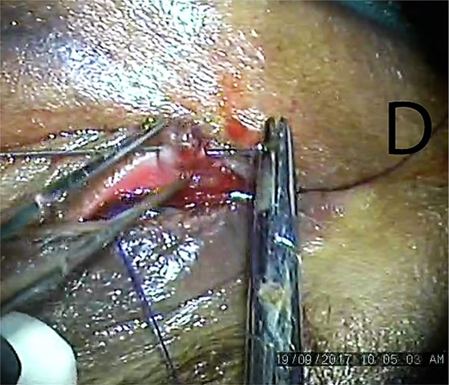
Fig-5 Prolene suture Passed and loop created
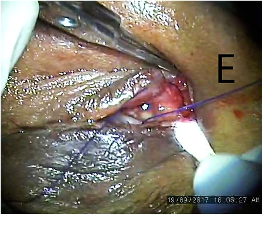
Fig-6 Prolene suture left in place as stent
Leave a Comment