

Dr.Durga Priyadarshini, D15392, Dr.Ambika
Abstract :
Aim: To analyse clinical profile, etiology and treatment outcome of Recurrent optic neuritis (RON) patients.
Methods : 86 Recurrent optic neuritis patients (110 eyes) who presented to neuro ophthalmic clinic in a tertiary eye institution between January2011 to November2016 were analysed.
Results : Bilateral involvement was noted in 27.9%. 31.4% had painless attacks .Mean age of presentation was 35.9 +-12yrs with female preponderance .Mean follow up period was 2.7 yrs. Common etiologies of RON noted in our cohort were NMOSD in 63.95% (n=55pts) out which 46.5% were IgG Ab positive (n=40 ) , , MS 12.8% (n= 11), Tuberculosis 8.1% (n=7) ,Autoimmune 3.5%(n=3), Sarcoid 2.3% (n=2) and CRION 9.3% (n=8). CRION patients had maximum number of relapses.
Conclusion: Recurrent optic neuritis is challenging in terms of diagnosis and treatment. Patients with NMO IgG Ab had poor visual recovery.
Introduction:
Optic Neuritis is an inflammatory disorder of the optic nerve of various etiology, the most common being reported as Multiple sclerosis in western literature and other causes include idiopathic, granulomatous like tuberculosis, syphilis, lyme disease or cryptococcal meningitis, viral infections and autoimmune [1-3] .Optic neuritis can have recurrence [4].The aim of this study was to analyse the clinical profile, etiology and treatment outcome of the recurrent optic neuritis (RON) patients.
Materials and Methods:
The study included 86 patients of recurrent optic neuritis who had presented to department of neuro ophthalmology in a tertiary eye centre in South India between January 2011 and November 2016 and they were analysed retrospectively .Patients with ischemic ,hereditary, compressive ,toxic and traumatic optic neuropathy were excluded .
Optic neuritis was classified as recurrent following unilateral or bilateral attack occurring after an interval of more than or equal to 4 weeks. The demographics, vision on presentation, associated pain, laterality, number of relapses, associated neurological attacks, associated diseases ,neuro imaging characteristics, serological workup which included granulomatous ,vasculitis and aquaporin 4 antibody ,csf analysis ,diagnosis , the type of treatment ,follow up period and visual outcome were assessed .
Multiple sclerosis diagnosis was made as per the 2017 McDonald Multiple sclerosis (MS) Diagnostic criteria [6]. Neuromyelitis optica spectrum disorder (NMOSD) diagnosis was suspected according to International consensus diagnostic criteria for neuromyelitis optica spectrum disorders. [5] Granulomatous and autoimmune optic neuropathy were presumed based on positive serological assay, imaging characteristics and response to treatment [7].Chronic relapsing inflammatory optic neuropathy (CRION) was suspected based on the characteristic pain and prompt response to steroids after excluding other causes [8] .
Statistical Tools
All statistical analysis was performed using SPSS V14.0 .Descriptive statistics was used to find the distribution of continuous variables and frequency distribution was used to assess the distribution of categorical variables. Independent sample t test was used to test the mean difference between two groups and paired t test was used to test the mean difference in visual acuity between vision at presentation and last follow – up. Any p – value less than 0.05 was considered as statistically significant.
Results :
In our study 86 patients (110 eyes ) with recurrent optic neuritis who met our inclusion criteria were analysed .The mean age of presentation was 35.9+-12 years with female preponderance (5:1 ) ,however there was no statistical difference in mean age between the male and female . 27.09% patients ( 24 patients) had bilateral presentation .The attacks were painless in 31.4% (27 patients) .The mean follow up period was 2.7 years (Range 1 week to 16 years ) .The mean visual acuity on presentation was logmar 1.1173+-0.986 .The mean number of relapse was 3.2+-1.6.
The etiologies of RON noted in our cohort is listed in Table 1 .Neurological symptoms like paresthesia,transverse myelitis ,quadriparesis were noted more frequently in the NMOSD with aquaporin 4 antibody (AqP4 ab ) group (N=16) than MS and NMOSD without AqP4ab group and never in the other group of patients .The clinical characteristics of RON in different etiologies are shown in Table 2 .
All patients underwent MRI Brain and orbit and 29 patients (33%) had brain signals .Table 3 explains the neuroimaging abnormality noted in RON patients. Long optic nerve segment and posterior optic nerve involvement was noted in 24 patients (27.9%) and out of which 15 patients (62.5%) were NMOSD .MRI spine involvement was noted in 13 patients (37%) of NMOSD with AqP4 ab group out of the 35 patients who underwent.2 patients who were initially AqP4 ab negative on subsequent repetition of the test were AqP4ab positive on relapse .2 patients of NMOSD with AqP4 ab had associated systemic lupus erythmatosis.18 patients among our cohort underwent CSF analysis which was normal in 15 patients, 2 patients had oligoclonal bands ,and 1 patient had raised proteins .Refer Table 4 for the serological assay of the patients in the study.
Patients who presented with an acute attack received intravenous methylprednisalone 250mg 6th hourly per day for 5 days and followed by oral 1mg/Kg/body weight weekly taper .In the maintenance therapy 6 patients had received oral ,long and slow tapering of steroids ,28 patients received immunosuppressant therapy ,10 patients received immunomodulator therapy and 4 patients anti tuberculosis treatment.2 patients who were noted have NMOSD with aquaporin 4 ab after relapse and 1 patient of NMOSD without AqP4 ab who was clinically worsening was shifted to immunosuppressant therapy from immunomodulator therapy .The mean visual outcome of 51 eyes of RON patients in the last follow up was Log Mar 0.6786+-0.888 which is statistically significant on comparison with the mean vision at presentation of the 110 eyes LogMar 1.1173+-0.986 (P value 0.000427) .
Discussion:
Recurrent optic neuritis is diagnosed in patients who had two disseminated episodes of optic neuritis with an interval of more than or equal to 4 weeks . [9] It can be manifestation of the central nervous system demyelinating disorders or autoimmune pathology .Recurrent optic neuritis increases the risk of MS significantly [10] , though in our study the incidence was only 12.8%. Strong female preponderance was noted in our MS group as in literature .90.09% of them had associated ocular pain during the attack as in Optic neuritis treatment trial (ONTT), where pain usually worsened by eye movement, accompanied the visual loss in 92.2% of cases and actually preceded the onset of visual symptoms in 39.5%. [11] In our study the mean vision on presentation in the MS group was Log mar 0.4933+-0.9260 and the visual outcome was good in follow up (Log mar 0.095+-0.13).
Neuromyelitis optica is the other demyelinating central nervous system disorder which has been noted to present with bilateral simultaneous or relapsing optic neuritis with transverse myelitis [12] . But not always cases are fitting in the classical definition and due to such expanding clinical spectrum of NMO , new international criteria for NMO spectrum disorder has been reported [5] . In our cohort we had NMOSD with AqP4 ab (N=40 patients) and NMOSD without AqP4 ab(N=15patients) and we analysed them separately . NMOSD with AqP4 ab was the commonest etiology noted among our cohort .There was female preponderance and lower incidence of pain on ocular movements was noted in NMOSD group. One of the strict criteria for diagnosing NMOSD [5] , bilateral simultaneous presentation was noted in 18 patients of the NMOSD with AqP4 ab .The mean vision on presentation was noted to be profound vision loss in the NMOSD group.
The incidence of brain abnormalities in NMOSD as per the revised 2006 NMO diagnostic criteria was noted to be 50 to 85 % and a trend to posterior involvement of the optic nerve is more specific for NMOSD [13] . Cranial MRI among our patients of NMOSD with AqP4 ab had signals in areas specific to NMOSD in 15 patients and 10 patients had long segment and posterior optic nerve involvement .Spine signals were noted in 13 patients of NMOSD with AqP4 ab . Among the NMOSD without AqP4 ab group, brain signals in areas specific to NMOSD were noted in 4 patients and posterior optic nerve involvement was noted in 5 patients, which lead to suspicion of NMOSD in those patients [Table3].
The novel biomarker and antibody specific to NMOSD, anti aquaporin 4 antibody was positive in 40 patients. Immunosuppressant’s can reduce the levels of aquaporin 4 antibody and so the testing has to be done prior to any therapy .Retesting is important in relapsing optic neuritis in such scenarios [14] .In our cohort repeat aquaporin 4 antibody during relapse was positive in 2 patients who were negative initially . There has been reported association between NMOSD and several autoimmune pathologies such as systemic lupus erythematosis, Sjogren’s, Behcets and Hashimoto’s thyroiditis though the real pathophysiological association remains unknown [15] . In our study, 2 patients of NMOSD with AQP4 ab had associated systemic lupus erythematosis . The visual outcome of the NMOSD with and without AqP4 ab was noted worse among the different etiologies of RON in our study .
The other etiologies noted in our cohort of recurrent optic neuritis were presumed tuberculosis, sarcoidosis and autoimmune optic neuropathy based on the serological assay ,neuroimaging and response to treatment .The mean vision on presentation was profound in tuberculosis .In our study ,4 patients received antituberculosis treatment in addition to systemic steroids . Early identification and initiation of antituberculosis treatment in suspected tuberculous optic neuropathy results in better visual outcome (Logmar 0.488+-0.67) and reduction of chances of relapse in future [16].Sarcoid optic neuropathy was suspected in view of raised angiotensin converting enzyme and a good response to steroids (Logmar 0.000) .3 patients of RON in our cohort with positive antinuclear antibody were suspected autoimmune etiology , despite the lack of systemic vasculitis and 2 among them was on immunosuppressant therapy .The visual outcome was good post treatment (Logmar 0.369+-0.626).
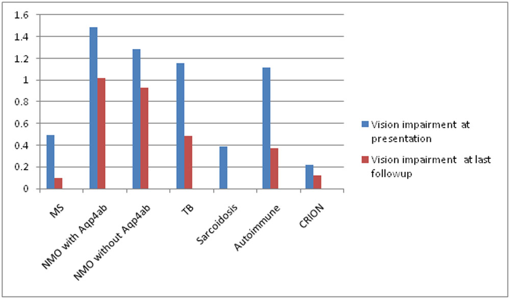
Steroid responsive recurrent optic neuropathy is one of the important etiology of recurrent optic neuritis and recently termed as Chronic relapsing inflammatory optic neuropathy [8] .It is described as a unilateral or bilateral isolated recurrent optic neuropathy without any signs of underlying demyelinating ,granulomatous or autoimmune pathology ,characterised by painful optic neuropathy with immediate response to steroids and relapse on withdrawal of steroids [17] .In our cohort of patients 8 were diagnosed to have CRION ,The mean vision at presentation among them was Logmar 0.2176+-0.4001 with 87.5 % of them having associated pain .The mean number of relapse was noted to be highest in the CRION group (mean relapse = 4+-1.5 ) .The mean visual outcome at last follow up was Logmar 0.125 +-0.186.Out of the 8 patients ,3 patients received immunosuppressant’s in addition to systemic steroids .Table 5 gives the comparison of the vision at presentation and at last follow-up in different etiologist noted in our cohort.
Our study has limitations because of its retrospective nature and varied and short follow up. But the study provides useful data regarding the different etiologies associated with recurrent optic neuritis and thereby early identification and management.
Conclusion :
Recurrent optic neuritis is challenging in terms of diagnosis and treatment. NMOSD is one of the commonest recurrent optic neuropathy with worse visual outcome in our cohort . Identification of patients who are not classical of MS and clinically behaving or progressing towards NMOSD is very essential as treatment with immunomodulators in NMOSD can cause worsening of the condition. Always rule out tuberculosis in endemic country like India before initiating immunosuppressant’s .CRION though is a diagnosis by exclusion can be suspected based on the characteristic pain and prompt response to steroids. Thus signifying, etiological identification of RON patients is vital for their management.
Table 1 :Etiologies of RON in our cohort
| Diagnosis | Number of patients ,n= 86 | Percentage |
| MS | 11 | 12.5 |
| NMOSD with
AqP4 Ab
|
40 |
46.5 |
| NMOSD without Aqp4Ab
|
15 |
17.4 |
| TB | 7 | 8.1 |
| Sarcoidosis | 2 | 2.3 |
| Autoimmune | 3 | 3.5 |
| CRION | 8 | 9.3 |
Table 2:Clinical characteristics of RON in different etiologies
| Clinical Characteristics | MS
N=11 |
NMOSD with
AqP4 Ab N=40 |
NMOSD without Aqp4Ab
N=15 |
TB
N=7 |
Sarcoidosis
N=2 |
Autoimmune
N=3 |
CRION
N=8 |
| Mean age (yrs) | 35.72+-9.6 | 34.45+-12.9 | 34.85+-11.3 | 42.85+-9.8 | 47.5+-27.5 | 34 +-15.6 | 37.37+-13.7 |
| F:M | 10:1 | 7:1 | 14:1 | 6:1 | 0:2 | 2:! | 1.6:1 |
| Mean Vision at presentation (Logmar) | 0.4933+-
0.9260 |
1.4839+-
0.9315 |
1.2818+-
0.9705 |
1.1543+-
0.8115 |
0.3891+-
0.3012 |
0.1193+-
0.1468 |
0.2176+-
0.4001 |
| Pain | 90.9% | 50% | 66.6% | 71.4% | 100% | 100% | 87.5% |
| Bilateral simultaneous | 1 | 18 | 4 | 0 | 1 | 1 | 1 |
| No of relapse | 3.18+-1.07 | 2.94+-
1.47 |
3.93+-
2.08 |
3.2+-0.95 | 2 | 3.33+-1.5 | 4+-1.5 |
| Neurological attacks | + | +++ | ++ | – | – | – | – |
Note : ‘+’ -2 Patients had neurological attacks ; ‘++’-4 patients had neurological attacks ; ‘+++’-11 ppatients had neurological attacks
Table 3: Imaging characteristics in RON
| Diagnosis | Brain signal | Long segment/Posterior optic nerve involvement | Spine Signals * |
| MS ,N=11 | 9 | 3 | 1(OSMS) |
| NMOSD with
AqP4 Ab N=40 |
15 | 10 | 13 |
| NMOSD without Aqp4Ab
N=15 |
4 | 5 | 1 |
| TB,N=7 | 1 | 3 | 0 |
| Sarcoidosis,N=2 | 0 | 0 | 0 |
| Autoimmune ,N=3 | 0 | 0 | 0 |
| CRION,N=8 | 0 | 3 | 0 |
*MRI spine was done in 35 patients out of 86 patients
Table 4: Serological assay
| Blood workup | Positive | Total Sample |
| Aquaporin 4 antibody | 40 | 849 |
| RA | 1 | 57 |
| ANA | 5 | 59 |
| ANCA | 2 | 57 |
| DsDNA | 1 | 2 |
| APL AB | 2 | 3 |
| Sarcoid/Riased ACE | 4 | 48 |
| Mantoux/Quantiferon | 6 | 46 |
| CSF analysis
Normal OCB Raised proteins Pleocytosis PCR MTB
|
15 2 1 – – |
18 |
Table 5 : Comparison of vision at presentation and at last followup in RON patients
References:
- Ebers GC. Optic neuritis and multiple sclerosis. Arch Neurol . 1985; 42-702-704.
- Froozon et al.Acute Demyelinating Optic neuritis.Current Opinion ophthalmology . 2002;13( 6): 375-80.
- Rohit Saxena et al.Clinical Profile and short term outcomes of optic neuritis patients in India .Indian J Ophthalmology 2014 ;62(3):265-67.
- Zeid NA, Bhatti MT. Acute inflammatory demyelinating optic neuritis:evidence-based visual and neurological considerations. Neurologist.2008;14:207–223
- DM Wingerchuck et al. International consensus diagnostic criteria for neuromyelitis optica spectrum disorders .Neurology 2015 July 14 ; 85 (2) :177-89.
- Thompson et al.Diagnosis of multiple sclerosis :2017 revisions of McDonalds criteria .The Lancet Neurology 2018 Feb ,Vol 17 (2) :162-173 .
- MJ Kuppersmith ,RM Burde,FA Warren et al. Autoimmune optic neuropathy: evaluation and treatment. Journal ofNeurology, Neurosurgery, and Psychiatry 1988;51:1381-1386
- Kidd D, Burton B, Plant GT, Graham EM. Chronic relapsing inflammatory optic neuropathy (CRION). Brain 2003; 126(Pt 2): 276-84
- A Kurne et al. Recurrent optic neuritis: clues from a long-term follow up study of recurrent and bilateral optic neuritis patients.Eye and Brain 2010;2:15-20
- Nilsson P, Larsson EM, Maly-Sundgren P, Perfekt R, Sandberg-Wollheim M. Predicting the outcome of optic neuritis: evaluation of risk factors after 30 years of follow-up. J Neurol. 2005;252:396–402.
- Optic Neuritis Study Group. The Clinical Profile of Optic Neuritis.Experience of the Optic Neuritis Treatment Trial.Arch Ophthalmol. 1991; 109:1673-78.
- Wingerchuk DM. Diagnosis and treatment of neuromyelitis optica.Neurologist. 2007;13:2–11.
- HJ Kim et al.MRI characteristics of neuromyelitis optica spectrum disorder An international update .Neurology 2015.March 17;84(1) :1165-1173.
- L Pandit et al.Neuromyelitis optica spectrum disorders: An update.Ann Indian Acad Neurol. 2015 Sep ; 18 (Suppl 1) :S11-S15.
- M Adawi et al. Systemic Lupus Erythematosus (SLE) Complicated by Neuromyelitis Optica (NMO – Devic’s Disease): Clinic-Pathological Report and Review of the Literature.Clin Med Insights Case Rep .2014;7:41-47.
- Davis EJ, Rathinam SR, Okada AA, et al. Clinical spectrum of tuberculous optic neuropathy. Journal of Ophthalmic Inflammation and Infection. 2012;2(4):183-189. doi:10.1007/s12348-012-0079-5.
- Arzani M ,Sahraian MA,Rezaei H,Naser Moghadasi A.Recurrent isolated optic neuritis :A study on 22 patients .Iran J of Neurol 2017 ;16(3):130-5
Leave a Comment