

Dr.Srishti Ramamurthy,Dr.Rathinam Sivakumar R
ABSTRACT
Aim: To report visual outcomes & complications in Pediatric uveitis
Methods: Retrospective case series conducted in Uvea Services of tertiary eye care hospital in South India. Consecutive patients of age < 16 years were included in the study. Detailed systemic evaluation was performed.
Results: 98 patients (152 eyes) with a mean age of 12.07 were included. Mean BCVA at presentation was 0.45(0.59)LogMar. 55% had bilateral presentation & 70.8 % had granulomatous uveitis. The most common diagnoses were pars planitis, toxoplasmosis, sarcoidosis, tuberculosis, JIA & idiopathic. 93% received systemic steroids& 32% received immunotherapy. Common complications were cataract, band keratopathy & glaucoma. Mean BCVA at last follow up was LogMar 0.32(0.55);p value=0.0007
Conclusion: The challenges in pediatric uveitis lie in diagnosis, need for long term follow up and management of complications caused by both disease sequelae and treatment modalities.
Introduction:
Uveitis in children contributes to 5-10% of all cases.1 Children tend to present late in the course of the disease due to its chronic nature; the early phase being predominantly asymptomatic.2 Childhood uveitis encompasses a challenging diagnostic entity. Preventing complications such as amblyopia is critical at this age. Heterogeneity is noted in the etiology of childhood uveitis in different populations being impacted by demographic, geographic, genetic and sociocultural factors.3 Studying the epidemiology of the disease in pediatric age group is important in aiding the ophthalmologist and paediatrician in choosing appropriate treatment modalities and managing ocular and systemic complications.4
Materials and Methods:
The medical records of children under the age of 16 years presenting to the Uvea clinic, Aravind Eye Hospital, Madurai between January 2017 and December 2017 was retrospectively reviewed. Visual acuity at presentation and after treatment,age, gender, laterality of involvement, anatomical, morphological types, clinical course, number of relapses, systemic associations, patients requiring immunosuppressive therapy; type and duration, surgical intervention and complications of disease and therapeutic strategies were collected. The clinical diagnosis was made using the criteria proposed by the International Uveitis Study Group (IUSG). Each patient underwent thorough ophthalmic examination; visual acuity, intraocular pressure and dilated fundus exam in every visit.
Complete blood count, ESR, C reactive protein, Mantoux, VDRL was done in all patients. Other systemic investigations such as ANA, Rheumatoid factor, HLA B27, Serum Angiotensin converting enzyme (ACE) levels, Toxoplasma serology, Interferon gamma release assay (IGRA) for TB ,imaging (Chest X-ray, CT, MRI- chest/abdomen/sacroiliac joints) were performed whenever required. Ophthalmic ancillary tests such as B scan, fundus fluorescein angiography, optical coherence tomography, vitreous biopsy and PCR were done on the clinician’s discretion. Statistical analysis was done using SPSS Statistics and Wilcoxan sign rank test was done to find significant difference between baseline and final visual acuity.
Results:
Mean (SD) of the age of 98 study subjects is 12.07 years and it ranges from 4 years to 16 years. The study included 55% boys and 45% girls. Involvement was unilateral in 45% and bilateral in 55%. Intermediate uveitis was found to be the most common anatomical type. Granulomatous uveitis occurred in 70.8% and Non granulomatous in 29.2%. Non infectiousetiology 63.3% appeared to be more common than infectious causes.
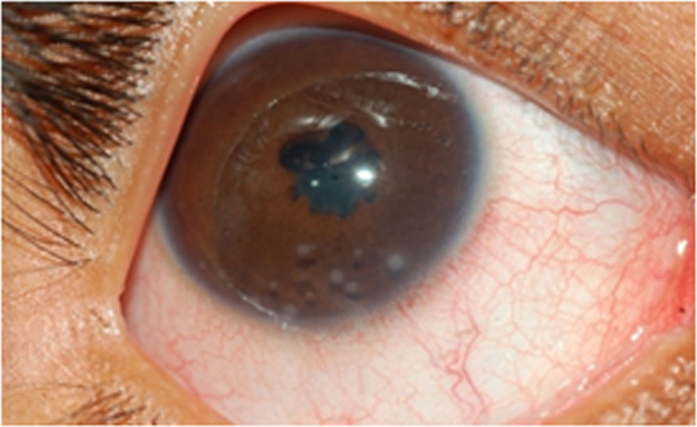
Among Non-infectious etiology, pars planitis(15.30%) and sarcoidosis(9.4%) were most common. Infectious etiology showed a wide range of agents, most common being Tuberculosis and Toxoplasmosis (10.6% each)
Non infectious:
| Diagnosis | % |
| Pars planitis | 15.30 |
| Sarcoidosis | 9.4 |
| JIA | 7.1 |
| Vogt Koyanagi Harada | 2.3 |
| Behcet’s | 2.3 |
| Acute zonal outer retinal necrosis | 1 |
| Idiopathic | 36.73 |
Infectious:
| Diagnosis | % |
| Tuberculosis | 10.6 |
| Toxoplasmosis | 10.6 |
| Toxocariasis | 7.1 |
| Acute retinal necrosis | 3 |
| Diffuse Unilateral Subacute Neuroretinitis | 3 |
| Endogenous endophthalmitis | 3 |
| Herpes | 2.3 |
| Leptospirosis | 2.3 |
| Viral | 2.3 |
| River water granuloma | 1.2 |
| HIV | 1.2 |
Steroids were required in the management of 93.87% (92 patients) in topical/periocular/systemic forms. Commonly used additional immunomodulatory agents included Methotrexate and Mycophenolate Mofetil.
Most common complication of chronic uveitis was Cataract (24%), band shaped keratopathy (11%) and glaucoma(7%).
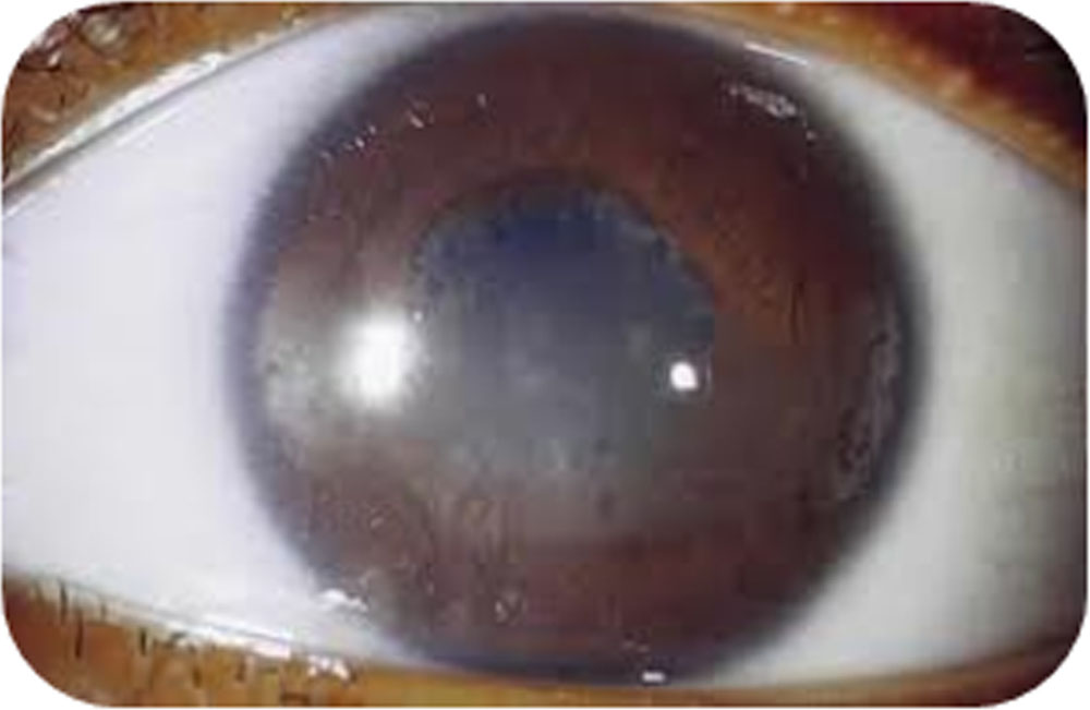
Surgical intervention was warranted in 19 patients including cataract surgery, pars plana vitrectomy and trabeculectomy. Other interventions such as Intravitreal steroids (triamcinolone injections, Ozurdex implant), Intravitreal anti-VEGF agents, PRP laser, YAG Peripheral iridectomy were needed in the management of complications of Pediatric uveitis.
83.3% achieved a final visual acuity of 6/18 or better. Severe visual impairment occurred in 1.3%and blindness (<3/60) in 6%.
Wilcoxon sign rank test was used to find out the significant difference between the baseline and final visual acuity. P-value less than 0.05 considered as statistically significant. p-value 0.0007 shows there is a significant difference between the baseline (median logMAR = 0.24, IQR (0 to 0.60)) and final (median logMAR = 0.0, IQR (0 to 0.48)) BCVA.
Discussion:
Diagnoses were found to be similar to those reported by Tugal-tutkun et al but there was a higher percentage of infectious etiology than previously reported in this study. De Boer et al in their study of 123 patients had a final visual outcome >6/18 in 72% compared to the 83% which achieved the same outcome in the current study which could be attributed to the recent trend of early institution of immunosuppressive therapy.
References:
- Paivonsalo-Hietanen T, Tuominen J, Saari KM. Uveitis in children: population-based study in Finland. ActaOphthalmolScand 2000;78:84–88.
- Rosenbeg KD, Feuer WJ, Davis JL. Ocular complications of pediatric uveitis. Ophthalmology 2004;111:2299–306
- Kadayifcilar S, Eldem B, Tumer B. Uveitis in childhood. J PediatrOphthalmol Strabismus. 2003; 40:335-340
- Cuningham ET., Jr Uveitis in children. OculImmunolInflamm. 2000;8;251-261
- Tugal-Tutkun I, Havrlikova K, Power WJ, Foster CS. Changing patterns in uveitis of childhood. Ophthalmology 1996;103:375–383.
- Kimura SJ, Hogan MJ. Uveitis in children: analysis of 274 cases. Trans Am OphthalmolSoc 1964;62:173–192.
- Kanski JJ, Shun-Shin GA. Systemic uveitis syndromes in childhood: an analysis of 340 cases. Ophthalmology 1984; 91:1247–1252.
Leave a Comment