Dr.Hiteshi Saini, H21173
Type of article: Original Article
Design: Case control study.
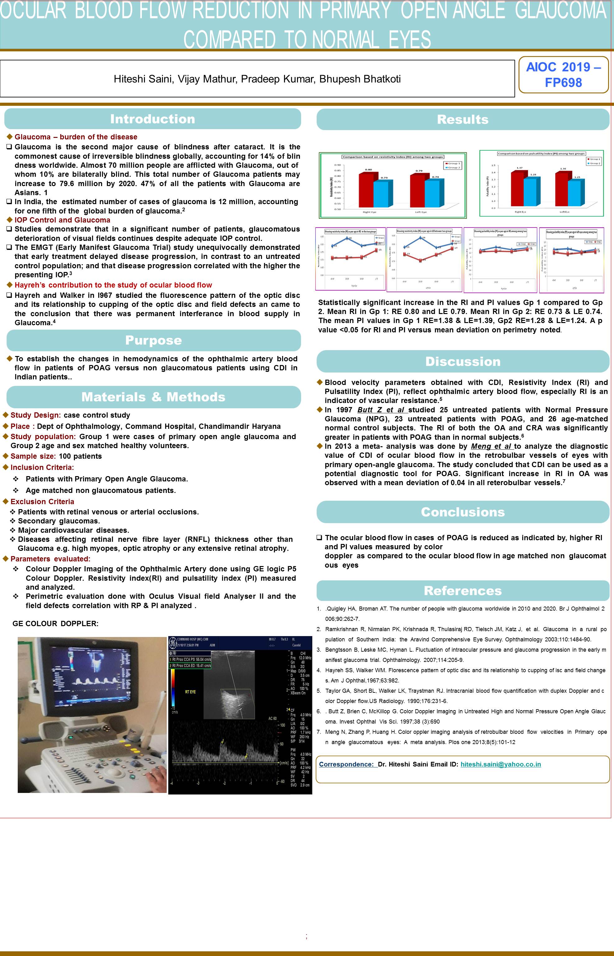
Material and Methods: N= 100. Age range: 40-80 years. Group 1 were cases of primary open angle glaucoma and Group 2 age and sex matched healthy volunteers. Colour Doppler Imaging of the Ophthalmic Artery done using GE logic P5 Colour Doppler. Resistivity index(RI) and pulsatility index (PI) measured and analyzed. Perimetric evaluation done with Oculus Visual field Analyser II and the field defects correlation with RP & PI analyzed .
Result: Statistically significant increase in the RI and PI values Gp 1 compared to Gp 2. Mean RI in Gp 1: RE 0.80 and LE 0.79. Mean RI in Gp 2: RE 0.73 & LE 0.74. The mean PI values in Gp 1 RE=1.38 & LE=1.39, Gp2 RE=1.28 & LE=1.24. A p value <0.05 for RI and PI versus mean deviation on perimetry noted.
Conclusion: The ocular blood flow was reduced in cases of POAG compared to the non glaucomatous eyes. The more the visual field loss, more was the reduction in ocular blood flow.
Leave a Comment