

Dr.SHAH CHINTAN MUKESHKUMAR,
Dr.Pradhnya Wasule,Dr.Amit Mohan,Dr.Elesh Jain
Introduction
Ocular injury is the leading cause of visual impairment1 and responsible for almost 10% cases of unilateral cataract in children.2 Ocular trauma is commoner in children due to unprotected and unattended playing. Mechanical ocular injury is broadly classified in open and closed globe injuries.3 Cataract is the commonest cause of reduction of vision in open globe injuries.4 Timely management of cataract is of utmost importance in children to avoid loss of vision and amblyopia.
Primary intraocular lens (IOL) implantation is possible in almost all cases of closed globe injuries. However in open globe injuries it is doubtful due to many complicating factors. Secondary IOL implantation is not without disadvantages like second intervention, repeat general anaesthesia and delay in visual rehabilitation in amblyogenic age.
This study was done to compare visual and refractive outcomes and complications of primary versus secondary IOL implantation.
Materials and methods
We obtained approval from the Institutional Review Board at our institution to conduct this study. A retrospective review of files was done in paediatric patients from January 2014 to July 2017 with open globe injury and traumatic cataract. Patients were divided in two groups. Group A included the patients with primary IOL implantation. While group B included patients in whom injury was repaired and subsequent secondary IOL implantation was done later. The data collected from files included: age, sex, eye involved, initial and final uncorrected and best corrected visual acuity, mode of injury, wound details, associated anterior segment complicating factors, time of primary and secondary surgery, type and location of IOL implanted and posterior segment details. Refractive errors were noted and spherical equivalent was counted at 3 months post IOL implantation. Spherical equivalent was considered desirable if within + two dioptres (D).
At every visit the unaided and best corrected visual acuity were assessed by either Snellen visual acuity chart or Lea symbols. For the statistical analysis, the visual acuity obtained by any of these two methods was converted to LogMAR. Visual acuity was grouped into three grades: 20/40 or greater, 20/50 to 20/100 and less than 20/100.
The wound location was defined according to an Ocular trauma classification group as follows: zone I (isolated to cornea, including corneoscleral limbus), zone II (corneoscleral limbus to a point 5 mm posterior into the sclera), and zone III (posterior to anterior 5 mm of sclera).If the injury involved multiple zones, it was defined by the most posterior extent. 3
Written and informed consent was taken for interventions from all patients’ guardians. All the procedures were performed under general anaesthesia. Keratometry was done using hand held automated keratometer. Axial length was measured using A scan ultrasound contact technique. IOL power was calculated using SRK 2 formula. In children less than 2 years and 2-8 years, IOL power was reduced as recommended by Dahanet al.5
Wound size and extent were measured in millimetre (mm) using Castroviejo Caliper. Cornel and scleral lacerations were repaired with 10-0 nylon and 8-0 polyglactin sutures respectively. Cataractous lens matter aspiration was done if anterior capsule was ruptured and status of posterior capsule was noted. Primary posterior capsulorhexis (PPC) with anterior vitrectomy was done in children younger than 8 years and in the presence of posterior capsule opacification (PCO). Primary IOL implantation was considered by the surgeon if visualisation was adequate. However IOL implantation was deferred in any complicating situations.
Results
Total 139 patients were found during the study period that underwent ocular trauma repair with cataract surgery and IOL implantation. After globe injury repair, primary IOL implantation was done in 61(44%) patients, while rest 78(56%) patients underwent secondary IOL implantation. Statistical analysis was done using SPSS software version 24.0.0.0. Trauma by wooden objects was most common in both the groups followed by metal objects.
Table 1 shows preoperative assessment in patients. 77% patients were males. Greater outdoor activities and more playful nature in boys are considered responsible for male preponderance.6 In group A, 43(70.5%) patients had smaller wounds (less than 5 mm); mean wound size was 4.2 mm. While in group B, 35 (44.9%) had smaller wound and mean wound size was 6.3 mm. Ocular injury most commonly involved zone 1. Zone 3 was not involved in any patients. Central corneal oedema/ tissue defect was found in 15(24.6%) patients in group A compared to 34(43.6%) patients in group B. Anterior and posterior capsule tears were very common in both groups. Intraocular foreign body was seen in 4 and 8 patients in group A and B respectively; however they were limited to anterior segment. None of the patients in group A had endophthalmitis or posterior segment related complications as these were considered contraindications for primary IOL implantation. Mean time between trauma and IOL implantation was 6.6 days (range 1-22 days) in group A compared to 150 days (range 20-320 days) in group B. (p= <0.00001)
Post operative commonest complication was fibrinous uveitis in both groups (p >0.05). Optic capture and lens decentration were seen in 8 cases in group A compared to 2 cases in group B (p= 0.02). Posterior capsule opacification (PCO) was seen in 8 cases in group A, 4 cases in group B (p >0.05). Endophthalmitis at the time of presentation was present in 7 cases in group B. 5 patients (8.2%) developed squint in group A compared to 16 (20.5%) in group B (p= 0.04). Mean visual acuity, spherical and cylindrical error were comparable in both group. [Table 2]
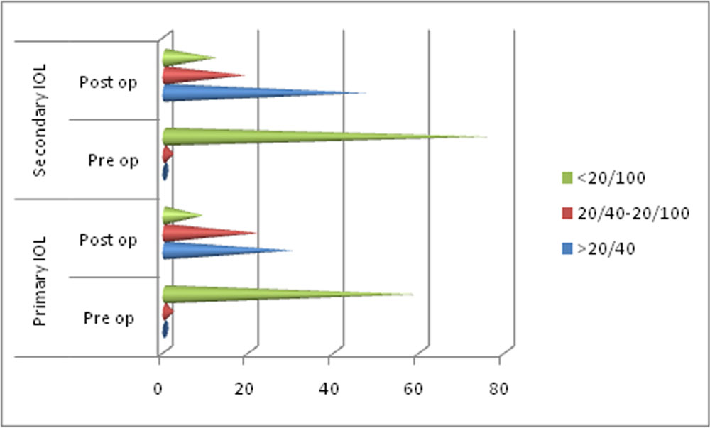
Figure 1 shows visual acuity in pre and post operative states.
Discussion
Management of ocular trauma is very challenging in young children due to risk of amblyopia. Trauma to lens and subsequent cataract in commonly seen after injury. Treatment of cataract is very challenging due to many complicating factors as stated below. Visualisation is often poor. IOL power calculation is another challenge in hypotonous eyes. So in many cases IOL is implanted according to other eye measurement which may result in more refractive surprises postoperatively.
Removal of traumatic lens matter is usually recommended along with primary trauma repair particularly if anterior capsule is breached. 7 Some studies have reported increased risk of infection, inflammation, raised intraocular pressure and retinal detachment if IOL is implanted with primary repair.8, 9, 10 some suggested that secondary IOL implantation allowed for more accurate determination of IOL power.11, 12However visual rehabilitation is delayed affecting binocular functioning of eyes. Two most controversial issues are timing of cataract removal and IOL implantation.
Primary IOL implantation benefits the patient by shortening rehabilitation time for those patients who may not or cannot followup.8, 9, 11, 13, 14, 15We usually try to implant an IOL in one step approach if feasible.
When we retrospectively evaluated the data, cases in which primary IOL implanted were better cases than group B. Mean time between trauma and IOL implantation was significantly greater in secondary IOL group (150 days versus 6.6 days, p= <0.00001).
IOL power calculation is a major hurdle in cases with ocular trauma. In 27(44%) cases from group A reliable keratometry readings were possible from the involved eye itself due to peripheral corneal involvement. Even for secondary IOL implantation, keratometry may not be possible due to irregular astigmatism. Biometry from same eye was possible in 46(59%) patients in group B. The fear of getting refractive surprises when biometry is done from other eye is often exaggerated. As in one study mean refractive anisometropia was only 0.37+0.57 dioptres.16 In our study when IOL power was calculated from other eye, 59% from group A and 66% from group B had desirable refractive error less than two spherical equivalents. However refractive surprise was extremely uncommon in both the groups.
Post operatively fibrinous uveitis was the common complication seen in 19(31%) and 25(32%) cases of group A and B respectively. Bowman et al also noted 29(40%) out of 72 cases of fibrinous uveitis as the common postoperative complication.15 Other notable complications included pupillary optic capture and lens decentration, seen in 13% cases in group A compared to 2.5% in group B (p=0.02). Posterior capsule opacification was seen in 13.1% cases of group A and in 5.1% cases of group B. This was much lower than a rate of 57% of Nd:YAG capsulotomy reported by Moisseiev et al.13 Primary posterior capsulorhexis was performed in 92 (66.2%) cases combined which lowered the PCO rate significantly in our study. Zone 3 was not involved in any case which may explain none developed retinal detachment.
Maximum vision gain following trauma was achieved much later in secondary IOL (214 days versus 84 days, p<0.0001) group. This may be the cause of 20.5% incidence of squint in group B at last visit. Furthermore, amblyopia was responsible for vision less than 20/40 in 32% cases of group B compared to 16% of group A.
Visual and refractive outcome in both groups were good and comparable. Mean best corrected visual acuity (logMAR) in group A and B was 0.42 and 0.47 respectively. In group A, 30(49%) patients achieved 20/40 or greater vision compared to 47(60%) in group B (p=0.19). BenEzra et al17 did primary IOL implantation and globe repair in 23 eyes and obtained >20/40 vision in 15(65%) eyes which is slightly better than present study. Mean spherical error 3 month post operatively was 1.39 and 1.18, while cylindrical error was 2.2 and 2.3 in group A and B respectively.
Single surgeon didn’t perform all the surgeries and patients were not randomised for the timing of IOL implantation. Also primary IOL implantations were done in relatively less complicating cases. These are the some limitations of this study. Timing of IOL implantation in our study was considered by the operating surgeon only while doing primary repair. Hence further study is warranted in similar groups.
In conclusion, both primary IOL and secondary IOL implantation after open glob injury can have good and comparable visual outcomes. However due to early visual rehabilitation primary IOL implantation should be considered whenever possible particularly in less complicating smaller lacerations and in rural areas where compliance for followup is poor.Secondary IOL implantation should be considered for larger extensive lacerations particularly involving central corneal region.
References
- Thylefors B. Epidemiological patterns of ocular trauma. Aust N Z J Ophthalmol. 1992;20:95–8
- Kenneth W. Wright, textbook of Pediatric Ophthalmology and Strabismus Second Edition, table 27-1, page no.458.
- Pieramici DJ, Sternberg P, Jr, Aaberg TM, Sr, Bridges WZ, Jr, Capone A, Jr, Cardillo JA, et al. The ocular trauma classification group. A system for classifying mechanical injuries of the eye (globe) Am J Ophthalmol. 1997;123:820–31.
- Thakker MM, Ray S. Vision-limiting complications in open-globe injuries. [Internet]. Can J Ophthalmol 2006;41:86–92.
- Dahan E., Drusedau M.U.H. Choice of lens and dioptric power in pediatricpseudophakia. J Cataract Refract Surg. 1997;23:618–623.
- Koo L, Kapadia MK, et al. Gender differences in etiology and outcome of open globe injuries. J Trauma 2005; 59:175-78
- Muga R, Maul E. The management of lens damage in perforating corneal lacerations. Br J Ophthalmol. 1978;62:784–787
- Lamkin JC, Azar DT, Mead MD, et al. Simultaneous corneal laceration repair, cataract removal, and posterior chamber intraocular lens implantation. Am J Ophthalmol. 1992;113:626–631
- Chan TK, Mackintosh G, Yeoh R, et al. Primary posterior chamber IOL implantation in penetrating ocular trauma. IntOphthalmol. 1993;17:137–141
- Andreoli CM, Andreoli MT, Kloek CE, et al. Low rate of endophthalmitis in a large series of open globe injuries. Am J Ophthalmol. 2009;147:601–608
- Rubsamen PE, Irvin WD, McCuen BW II, et al. Primary intraocular lens implantation in the setting of penetrating ocular trauma. Ophthalmology. 1995;102:101–107
- Cohen KL. Inaccuracy of intraocular lens power calculation after traumatic corneal laceration and cataract. J Cataract Refract Surg. 2001;27:1519–1522
- Moisseiev J, Segev F, Harizman N, et al. Primary cataract extraction and intraocular lens implantation in penetrating ocular trauma. Ophthalmology. 2001;108:1099–1103.
- Ng JS, Leung HT, Lam DS. Cataract extraction and IOL implantation. Ophthalmology. 2002;109:1197
- Bowman RJ, Yorston D, Wood M, et al. Primary intraocular lens implantation for penetrating lens trauma in Africa. Ophthalmology. 1998;105:1770–1774
- Hu YY et al. Prevalence and Associations of anisometropia in children.Invest Ophthalmol Vis Sci.2016 Mar;57(3):979-88.
- BenEzra D, Cohen E, Rose L. Traumatic cataract in children: correction of aphakia by contact lens or intraocular lens. Am J Ophthalmol. 1997;123:773–782
| Parameter | Group A | Group B | p value |
| Male/ Female | 47/14 (77%/ 23%) | 60/18 (77%/ 23%) | 1 |
| Mean age (in years) | 10.3 + 4.7 (range 2 – 18) | 9.2 +3.4 (range 3 – 17) | >0.05
|
| Corneal wound size
Small <5 mm Large >5 mm |
43 (70%) 18 (30%) |
35 (45%) 43 (55%) |
0.002
|
| Mean wound size (millimetre) | 4.2 + 1.8 (range 2 to 10) | 6.3 + 2.8 (range 2 to 18) | <0.0001
|
| Wound involvement
central 3 mm cornea (pupillary zone) peripheral part of cornea |
30 (49%) 31 (51%) |
35 (45%) 43 (55%) |
>0.05 |
| Zone of injury
Zone 1 Zone 2 |
53 (87%) 8 (13%) |
67 (86%) 11 (14%) |
>0.05 |
| Associated ocular complications
Uveal tissue prolapse Vitreous prolapse/ loss Central corneal edema (3 mm) Anterior & posterior capsular tear Intraocular foreign body Endophthalmitis (pre existing) |
29 (47.5%) 7 (11.5%) 5 (8.2%) 47 (77%) 4 (6.6%) 0 |
51 (65.4%) 12 (15.4%) 34 (43.6%) 50 (64.1%) 8 (10.3%) 7 (9%) |
<0.05 0.5 <0.00001 >0.05 0.5 – |
Table 1- demographic data, preoperative evaluation and associated complicating factors
| Parameter | Group A | Group B | p value |
| IOL power calculation
Keratometry (same eye) Keratometry (other eye) |
27 (44%) 34 (56%) |
46 (59%) 32 (41%) |
0.08
|
| Complications
Immediate fibrinous uveitis Pupillary optic capture+ lens decentration Posterior capsule opacification |
19 8 8 |
25 2 4 |
0.90 0.02 >0.05 |
| Post operative strabismus | 5 (8.2%) | 16 (20.5%) | 0.04 |
| Mean best corrected visual acuity(logMAR) | 0.42 | 0.47 | 0.32 |
| Time required for maximum vision gain following trauma (days) | 84 | 214
|
<0.0001 |
| Mean spherical error | 1.39 (-5 to +5) | 1.18 (-4.5 to +3) | >0.05 |
| Mean cylindrical error | 2.2 (-5 to +5.5) | 2.3 (-6 to +5) | >0.05 |
| Mean spherical equivalent | 1.81 | 1.55 | 0.15 |
Table 2- intra operative and post operative comparison between group A & B
Disclosure: the authors have no financial or proprietary interest in any material or method mentioned. The article has been sent elsewhere for consideration of publication.
Leave a Comment