



Dr.MEGHAL GAGRANI, Dr.Ramanjit Sihota,Dr.Dada Tanuj
ABSTRACT
Purpose: To evaluate effect of Mindfulness based stress reduction (MBSR) in Primary open angle glaucoma (POAG) patients
Methods:Sixty patients were randomized into two groups. Group 1 underwent 45 minutes of MBSR daily for 6 weeks along with standard medical treatment while Group 2 received only standard medical treatment. Inclusion criteria were patients with POAG, age>45 years, best corrected visual acuity >6/60. Patients with other ocular co-morbid conditions were excluded. An assessment of IOP, brain oxygenation in prefrontal cortex using functional near infrared spectroscopy (fNIRS), QOL (WHO-BREF QOL) and stress markers in serum was made at baseline and at 6 weeks.
Results:21 female and 39 male patients were enrolled with a mean age of 57.28±9.37 years. At 6 weeks mean level of IOP decreased significantly in group 1 (15.9±1.8 mmHg to 14.4±1.21mm Hg, p-value 0.0001) as compared to group 2 (15.7±1.4 mmHg to 15.65±1.41, p-value 0.41). fNIRS showed significant improvement in oxygenated hemoglobin change (ΔHbO) in group 1(p-value <0.0001) as compared to group 2 (p-value 0.52). WHO-BREF QOL score increased significantly in group 1(p-value 0.0001) as compared to group 2(p-value 0.74). Biochemical markers like serum cortisol, reactive oxygen species decreased significantly (p-value<0.5) and brain derived neurotrophic factor, beta endorphin and total antioxidant capacity increased significantly (p-value <0.5) in the group 1as compared to group 2.
Conclusion:A short term course of MBSR was associated with a significant reduction in IOP and stress markers with improvement in brain oxygenation and QOL.
KEY WORDS
Glaucoma, intraocular pressure, mindfulness meditation, optic nerve head, quality of life, relaxation, stress, fNIRS, brain oxygenation, prefrontal cortex
INTRODUCTION
Glaucoma is a form of neurodegenerative disorder characterized by selective loss of retinal ganglion cells (RGCs)(1) and progressive damage to optic nerve. It is the second leading cause of blindness after cataract(2) and affects approximately 68 million people worldwide, 10% of whom are blind(3). Primary open angle glaucoma (POAG) is the most common form of all glaucoma types(4). Raised IOP is the most important modifiable risk factor for glaucoma. Decreased cerebral and optic nerve head (ONH) blood flow have been shown to be associated with glaucoma etiopathogenesis indicating that Glaucoma may be one of the manifestations of a generalized vascular insufficiency(5).Decreased cerebral blood flow autoregulation has also been shown to be associated with visual field loss.
Glaucoma patients require strict compliance to medications and lifelong follow-ups. Association between stress and glaucoma has been established by some studies (6)suggesting that stress in glaucoma patients leads to adverse quality of life (QOL)(7) .A patient with glaucoma lives under psychological stress which may progress to depression and the stress may further accelerate glaucoma progression(8).Various stress markers have been identified which can objectively quantify both physiological/psychological and cellular stress like cortisol. β- endorphins, interleukin-6 (IL 6), reactive oxygen species (ROS), total antioxidant capacity (TAC)(9). Studies have found that brain derived neurotrophic factor (BDNF) to have a role in RGC escape from death in mice with raised IOP. Since glaucoma patients need lifelong medications, live under anxiety and constant fear of losing sight and consequently poor QOL; this has led to the search of complementary forms of treatment which in addition to decreasing the progression of glaucoma would address these additional issues.
Mindfulness/meditation refers to a physiological state of reduced metabolic activity reported to enhance psychological balance and emotional stability (10). It is a set of attentional practices leading to an altered state of consciousness characterized by improved awareness, greater presence, and a more integrated sense of self. Meditation is a well-accepted modality in reducing stress and anxiety and to bring a sense of well-being. Mindfulness based stress reduction (MBSR) is a type of meditation wherein breath is used as an object of awareness(11).Practice of MBSR has proven to be associated with a number of significant physiological changes in the body including changes in the balance of sympathetic- parasympathetic activity, heart rate, respiratory and pulse rate, blood pressure, oxygen metabolism, skin resistance, changes in secretion of endocrine hormones and central neurophysiologic changes(12,13). Mindfulness meditation improves cerebral blood flow(5,14) and oxygenation levels of many areas of brain. MBSR has been used as an adjunct in the treatment and prevention of many chronic diseases like depression, diabetes, hypertension, coronary heart disease and in cancer patients. MBSR has also shown to increase cerebral blood flow, improve cognition in a variety of diseases which correlates with improved quality of life and well-being(12,15).
Studies have shown changes in various parts of the brain beyond the visual cortex in primary open angle glaucoma (POAG) (16,17) including various areas of the prefrontal cortex. The anxiety and depression associated with POAG, also affects the prefrontal cortex. Also, MBSR has been shown to increase activity in visual attention related occipital brain centers (18). Although, no direct increase in activity in the visual cortex has been studied in literature, we believe that a functional connectivity operates between these areas for visual processing and, therefore, could lead to an increase in activity in the visual cortex as well.
MATERIAL AND METHODS
Study Design: The present study is a prospective randomized, controlled trial with POAG patients aimed at investigating the effect of MBSR on the blood flow and oxygenation status of the brain. The study was approved by the Institute Ethics Committee of the All India Institute of Medical Sciences, New Delhi (No. IECPG-362/29.06.2016, Dated:28/10/2016) and registered with the Clinical Trial Registry of India (Ref: CTRI/2018/04/013397) prior to patient enrolment observing the Declaration of Helsinki and Good Clinical and Laboratory Practice Guidelines. This study was designed and carried in compliance with the Consolidated Standards of Reporting Trials (CONSORT) and the study protocol is shown in Figure 1.
Participants: Patients from follow up glaucoma clinic (minimum 3 months follow up) of Dr. Rajendra Prasad Centre for Ophthalmic Sciences were screened for the inclusion/exclusion criteria.
Inclusion criteria: Patients with moderate and severe POAG with intraocular pressure (IOP) range 15-21 mm Hg, age > 45 years, best corrected visual acuity of the better eye ≥ 6/60, conversant in Hindi or English and consent to attend the complete course of MBSR.
Exclusion criteria: Patients having co-morbid ocular conditions other than glaucoma contributing to visual loss, chronic systemic diseases that could affect QOL, history of any surgery in previous 3 months, medical therapy for any other illnesses, already practicing yoga or meditation in any form were excluded from the study. Patients who were deaf or communication impaired or with subnormal mentation were also excluded.
Sample size calculation: There is no previous study reported that evaluated effect of MBSR on patients with POAG. A convenient number of 60 patients was taken and randomized into 2 groups – 30 cases and 30 controls to conduct a pilot study. The primary objective was change in IOP.
Randomization: Randomization into intervention and control group was done by computer based randomization using permuted blocks. The generated random sequence was transferred to sealed envelope to conceal the randomization till the actual allocation.
Intervention: In the intervention group, the patients underwent a 6 weeks course of MBSR by a trained instructor. MBSR was carried out at the Integral Health Clinic, Department of Physiology, All India Institute of Medical Sciences. No change was done to the standard treatment of any patient. The session was for 45 minutes every day in the morning 9 a.m. to 10 a.m.. Patients were asked to sit comfortably on the floor where there was no disturbance and to focus on breathing. Patients were instructed to refocus on breathing whenever other thoughts enter the mind(11).
IOP: Morning IOP at 7.00 am was measured by Goldmannapplanation tonometry for all patients and at all visits.
Visual assessment: Visual acuity was recorded using Snellen visual acuity chart and converted to logMAR. Optic disc changes were documented on a 90 D slit lamp biomicroscopy and Humphrey visual field SITA STD 30-2.
Brain oxygenation: A 16-channel continuous wave fNIRS imager system (FNIR1000- ACK-W, BIOPAC Systems, Inc., U.S.A) was employed to map changes in HbO over bilateral prefrontal cortex (PFC). After cleaning the forehead, the band was applied to the forehead stretched from the hairline to the eyebrow in a sagittal manner and was firmly held with a velcro band[Figure 2]. The patient was given a cognitive task to perform in the form of n-back test which tests visual memory. A 1-back test was explained to the patient which consisted of 36 test images and 36 blank images spanning a duration of 145 seconds. After explaining the test, recording was started and patient was asked to perform the task. COBI software was used to analyse the waveform and get values of maximum change in oxygenated hemoglobin (ΔHbO) during the task across all channels.
Quality of life: Quality of Life assessment was assessed using the World Health Organization Quality of Life (WHOQOL-BREF) questionnaire. The WHOQOL-BREFinstrument assesses the individual’s perceptions in the context of their culture and value systems, and their personal goals, standards and concerns. The WHOQOL-BREF instrument comprises 26 items, which measure the following broad domains: physical health, psychological health, social relationships, and environment.
Biochemical markers: A 5 ml Blood samplewas taken from the antecubital vein at 8.00 am in the morning to avoid any effect of diurnal variation. The assessment of serum cortisol, β-endorphins, BDNF, levels was done using commercially available kits. Reactive oxygen species (ROS) were measured using chemiluminiscence and total antioxidant capacity (TAC) was assessed by colorimetric assay.
Statistics: Statistical analysiswas done using STATA 11.2 software for windows. Paired t-test was used to compare within group parameters and independent t-test was used to compare parameters between two groups for parametric data. Wilcoxan sign rank test (within group) and Mann-Whitney test (intergroup) was used to analyze change in oxygenated haemoglobin. A p-value of <0.05 was considered to be statistically significant.
RESULTS
Sixty patients (21 females, 39 males) were enrolled in the study with a mean age of
57.28±9.37 years. The baseline clinical characteristics were comparable in both groups.At 6 weeks mean level of IOP decreased significantly in intervention group (15.9±1.8 mmHg to 14.4±1.21mm Hg, p-value 0.0001) as compared to controls (15.7±1.4 mmHg to 15.65±1.41, p-value 0.41). The percentage reduction in IOP was compared between the two group and 10-15% reduction in IOP from baseline was seen in 40% of participants who underwent MBSR (Table 1).
The change in oxygenated hemoglobin was comparable in both groups at baseline in all channels except channel 5 and 9 as assessed by Mann-Whitney test (Table 2). Channels 5 and 9 had artefacts and noise, therefore were not fit for statistical analysis and were excluded from analysis.All subjects had >80% correct responses in the n-back test at baseline and at 6 weeks. Data from fNIRS showed significant increase in ΔHbO in intervention group (as compared to control group) in left PFC, right PFC and medial PFC over several channels. Significant changes over time was observed in channels 1,3,4 and 6 overlying the left PFC in the intervention group but not in control group. The difference in ΔHbO values at 6 weeks was observed only at channel 1,2,4 and 6 among the groups. While there was uniformly an increase in ΔHbO values observed among intervention group, the control group participants had no or slight increase and even a reduction of ΔHbO values over time. A significant increase in ΔHbO over time was also seen at right PFC in channels 11, 13 and 15 among the intervention group as compared to controls, although difference among the groups at 6 weeks was noted only in channel 12, 14 and 15. Medial PFC (channels 7,8 and 10) also showed a significant increase in ΔHbO in the intervention group as compared to controls, although the difference was non-significant among the groups at 6 weeks. The median values of ΔHbO in various channels is shown in Table 2.
The mean quality of life was comparable in both groups at baseline (p-value 0.06). It increased significantly in intervention group (86.6±6.16 to 93.3±5.66, p-value 0.0001) as compared to controls (89±7.25 to 89.07±3.24, p-value 0.74].
The mean serum cortisol was comparable at baseline in both groups (p-value 0.07). It decreased significantly in intervention group (497±46.37ng/ml to 447±53.78ng/ml, p-value 0.01) as compared to control group (519.75±24.5ng/ml to 522.58±26.63ng/ml, p-value 0.64) at 6 weeks.
The mean serum beta endorphin was comparable at baseline in both groups (p-value 0.27). It increased significantly in the intervention group (33±5.52pg/ml to 43.27pg/ml, p-value<0.0001) as compared to controls (34.78±4.1pg/ml to 36.33pg±4.07pg/ml, p-value 0.27) at 6 weeks.
Mean ROS were comparable at baseline in both groups (p-value 0.74). ROS decreased significantly in intervention group (1596.19±179.14 to 1261±244.31 RLU/min/104 neutrophils p-value 0.0001) as compared to controls (1577.5±172.02 to 1662.5±84.75 RLU/min/104 neutrophils p-value 0.16) at 6 weeks. The mean TAC was comparable at baseline in both groups (p-value 0.83). It increased significantly in the intervention group (6.15mM to 9.18 mM, p-value <0.0001) as compared to controls ( 6.23mM to 6.02mM, p-value 0.61) at 6 weeks.
Mean serum BDNF was comparable in both groups (p-value0.63). It increased significantly in intervention group (52.24±6.71ng/ml to 63.25±13.48 ng/ml, p-value 0.004) as compared to control arm (53.23±5.82ng/ml to 54.42±5.66ng/ml, p-value 0.54).
DISCUSSIONS
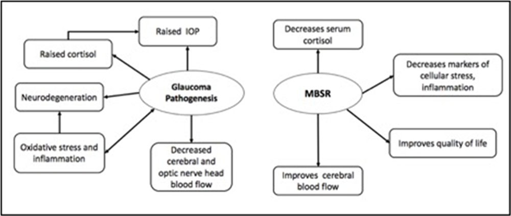
Glaucoma is the second leading cause of blindness in the world and is associated with severe compromise in visual faculties(19) and cognitive decline (20). It leads to a compromise in the QOL consequently ensuing stress and anxiety. Stress and glaucoma have a two way relationship each exacerbating the effect of the other. MBSR is, in this context, a suitable intervention aimed at breaking this reciprocal amplification and provide psychological and physiological relief to the patients [Figure 3].
Intraocular pressure is the only modifiable risk factors for POAG(21). Almost all of the current treatment options available for glaucoma are aimed at lowering IOP by pharmacological (involving lifelong use of topical medications and a financial burden in addition to various side effects and compromise in QOL) and/or surgical means. Evaluation of changes in IOP following MBSR was pivotal in evaluating the role of mindfulness based practices in POAG and assessing its candidature as a complementary form of treatment. 1 mm Hg decrease in IOP has been shown to decrease the risk of glaucoma progression by 10%(21), so MBSR may also reduce glaucoma progression by decreasing IOP(22). Moreover, mental and psychological stress has been found to be associated with elevation in IOP(8), a process mediated by cortisol (an important stress marker). It is evident from our results that MBSR may be a good candidate therapy to decrease IOP in POAG in addition to standard medical treatment with added benefits of decreasing the psychological stress.
MBSR has been shown to enhance oxygenation of the cerebral cortex(23) and improve cognitive functioning. Mindfulness increases the global cerebral blood flow including the occipital cortex(24). An increase in the regional blood flow and oxygenated hemoglobin is mediated by changes in neuronal activity in that part of the brain(25). Mindfulness improves cerebral blood flow and decreases cerebrovascular resistance suggesting an underlying vascular mechanism which reflects cerebral activation(24). fNIRS has been used to show the increase in oxygenation in prefrontal cortex after practicing mindfulness(26). Techniques such as fMRI and PET have also been used for functional brain studies and have good spatial resolution but they are expensive, highly susceptible to motion artefacts and require exposing the subjects to foreign substance (PET) or loud noise (fMRI). EEG and magnetoencephalography provide good temporal resolution. But fNIRS is non-invasive, cost effective, realtime functional imaging system with good temporal resolution, easy and safe to operate.Regional brain communication can be quantified using similarities in oscillation frequencies of hemodynamic signals asserted through fNIRS (27). The frontal cortex, and more specifically, the dorsolateral prefrontal cortex (DLPFC), is critically involved during working memory processes and, therefore, was included in this study (28). Based on this premise, we studied the effect of MBSR on brain oxygenation in the prefrontal cortex using fNIRS.
We found an increase in the change in oxygenated hemoglobin while performing a n-back test in participants who underwent MBSR as compared to controls. An increase in ΔHbO values at all parts of the prefrontal cortex was observed in the intervention group as compared to controls suggesting a possible role of the beneficial effect of MBSR on cognitive functioning, in particular working memory. Meta-analysis suggests consistent activation at bilateral rostral prefrontal cortex or frontal pole, bilateral dorsolateral prefrontal cortex and bilateral mid-ventrolateral prefrontal cortex during performance of working memory tasks such as n-back task (28)(46).The current study also reiterates the findings of these previous varied functional studies of activation of this cortical area while performing a working memory task. MBSR possibly enhanced the cortical activation in this region leading to significant changes after 6 weeks’ duration which was not observed in the control group participants. Literature suggests that even single bout of exercise can improve the performance on cognitive tasks (29).
The difference among the groups was observable at 6 weeks in channels overlying the right and left DLPFC but not in medial PFC although there was an increasing trend observed. Consensus in literature supports major role of the DLPFC in working memory tasks particularly in those utilizing fNIRS (28,30,31).We also observed no change and even a reduction of ΔHbO values over time in the control group. The inadequate increase in cerebral oxygenation during cognitive tasks may be an indicator of cerebral fatigue or due to the challenge of source allocation between the areas responsible for physical and cognitive workload (32–34). It can be suggested that the participants receiving intervention did not suffer cognitive overload due to the performance of MBSR and thus had an increasing trend of values while the control participants were not able to compensate the increasing workload and thus a no or negative change was observed.
Apart from degeneration in the visual pathway, higher prevalence of white matter changes and small vessel ischemic changes have been found in patients with glaucoma(1), suggesting a generalized vascular insufficiency and a role of altered blood flow in the pathogenesis of glaucoma(5) rather than just a localized vascular deficit of the visual pathway. In the light of these findings, MBSR could be used as a modality to prevent the progression of glaucoma by improving cerebral blood flow in different parts of the brain. Using fMRI, studies have shown bidirectional functional connection between the prefrontal cortex and the occipital cortex during tasks like visual search(35) . So an activation of the prefrontal cortex would cause a functional activation of the visual cortex and subsequent increase in the regional blood flow. Exact characterization of these changes would require studying activity of the occipital cortex, which was a limitation of our study.
As already mentioned, POAG has been associated with psychological stress, anxiety and a poor QOL. In a parallel context mindfulness meditation improves subjective well-being by instilling behavioral modification and mental discipline(36) and has shown benefits in normal as well as diseased subjects(37). In our study also POAG patients who underwent MBSR showed significant improvement in QOL as compared to controls (as assessed by WHOQOL-BREF questionnaire).
Cortisol is released in acute stress and is also found elevated in chronic stressful conditions. Increased levels of cortisol have been found in patents with ocular hypertension and glaucoma(38). Serum cortisol has been found to decrease in healthy individuals after a short term course of mindfulness meditation(39). In our study a short term course of MBSR was associated with significant reduction in serum cortisol providing an objective indicator of decrease in stress and also additional association with a decrease in IOP. β-endorphins are endogenous neuropeptides produced by arcuate nucleus and the pituitary gland and play a defensive role in stressful situations(40). Studies on rabbit have shown that stimulation of arcuate nuclei and consequent release of endorphins lead to reduction in IOP(41). Mindfulness meditation has been demonstrated to increase β-endorphin level(42). indicating that β-endorphins may also play a role in the stress reduction mediated lowering of IOP. In our study, participants undergoing MBSR had a significant increase in β-endorphin levels which further bolsters our proposition of employing MBSR as an adjunct to the routine treatment of POAG.
Oxidative stress
is associated with widespread release of free radicals and ROS affects cellularity of human trabecular meshwork (TM) (especially its endothelial cells) (47,48)and, hence, plays an important role in pathogenesis of POAG. Decreased levels of TAC have been found in patients of major depressive disorder(47,49). A negative correlation is found between serum TAC and severity of depression. Decrease in antioxidant defense and increase in oxidative stress plays a key role in pathogenesis of POAG. Oxidative stress will cause mitochondrial damage and apoptotic cell death in the trabecular meshwork as well as retinal ganglion cell loss. Several studies have documented the beneficial effect of mindfulness and meditation on the oxidative status of the body(49). This study for the first time documents the role of short term practise of MBSR in decreasing the oxidative stress and increasing total antioxidant capacity in POAG patients.
According to current literature, glaucoma is a neurodegenerative disorder with progressive loss of neurons, RGCs in this case. Changes are seen all along the visual pathway upto the occipital cortex, which is known to occur by means of transynaptic degeneration(50). BDNF, a neurotrophic factor, has been seen to play an imperative role in RGC rescue in rat eyes with elevated IOP. Also lower BDNF levels have been found to be present in patients with POAG as compared to healthy controls (51). BDNF expression has also been found to dramatically decrease in the hippocampus in response to acute stress (52). In the light of these findings, we decided to evaluate BDNF levels in response to MBSR in patients with POAG. There is only one study published till date showing increase in BDNF levels with MBSR. In our study, we found a significant increase in BDNF levels in participants who underwent MBSR as compared to controls suggesting a possible role MBSR in preventing glaucoma progression by upregulating BDNF and preventing retinal ganglion cell death.
LIMITATIONS AND RECOMMENDATIONS OF THE STUDY
The present study reports changes after six weeks, but we suggest long term follow-ups to see if the effects of MBSR sustain for longer periods. We analyzed changes in the prefrontal cortex in the present study, for better correlation, imaging of the occipital cortex would be a usefulcomplementing marker and of obvious advantage.
COMPETING INTERESTS
All the authors declare “NO CONFLICT OF INTEREST”.
FIGURE LEGENDS
Figure 1: Flowchart showing the study framework in compliance with the CONSORT guidelines
Figure 2: fNIRS head band showing sensor with 4 light sources and 10 detectors and the 16 channel measurement locations registered on the sensor(left).Representative image showing corresponding locations of the 16 channels on the prefrontal cortex (right)
Figure 3: Concept of the study: various factors involved in the pathogenesis of glaucoma and the effects of MBSR on the risk factors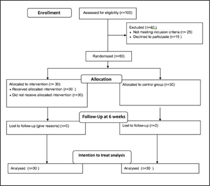
Figure 2
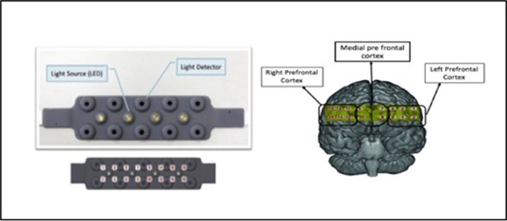
FIGURE 3
Tables:
TABLE 1Comparison of percentage reduction in IOP
| Decrease in IOP
At 6 weeks |
MBSR
(n=60) |
No MBSR
(n=60) |
| <5 % | 25 (41.6%) | 56 (93.3%) |
| 5 % to 10 % | 0(0) | 0(0) |
| 10 % to 15 % | 24 (40%) | 4(6.66%) |
| 15% to 20 % | 4 (6.66%) | 0(0) |
| >20% | 7 (11.66%) | 0(0) |
TABLE 2Median of change in oxygenated Hb(ΔHBO) in prefrontal cortex
| Cortical area | Baseline (mM)
Median (Min, Max) |
At 6 weeks (mM)
Median (Min, Max) |
P value
(Intragroup) |
|
| Left prefrontal cortex | ||||
| Channel 1 | MBSR | 0.92(-54.56,153.6) | 3.13(.167,7.20) | 0.02 |
| No MBSR | 1.86(-53.56,15.07) | 2.01(.12,7.03) | 0.49 | |
| P value (Intergroup) | 0.10 | 0.01 | ||
| Channel 2 | MBSR | 1.9(-48.6,162.12) | 3.01(.18,7.64) | 0.08 |
| No MBSR | 2.52(-49.9,14.71) | 2.06(.22,4.63) | 0.89 | |
| P value (Intergroup) | 0.87 | 0.04 | ||
| Channel 3 | MBSR | 1.62(-50.72,172.73) | 2.82(-28.7,6.5) | 0.01 |
| No MBSR | 2.46(-50.72,16.09) | 3.05(-.38,4.43) | 0.68 | |
| P value (Intergroup) | 0.44 | 0.8 | ||
| Channel 4 | MBSR | 1.43(-47.02,172.74) | 2.85(.17,7.62) | 0.05 |
| No MBSR | 2.33(-57.26,16.58) | 1.9(-.81,3.63) | 0.23 | |
| P value (Intergroup) | 0.4 | 0.06 | ||
| Channel 6 | MBSR | 0.33(-58.77,11.78) | 4.4(.06,11.78) | 0.003 |
| No MBSR | 0.46(-56.6,5.26) | 0.47(-1.61,9.96) | 0.15 | |
| P value (intergroup) | 0.45 | 0.001 | ||
| Right prefrontal cortex | ||||
| Channel 11 | MBSR | 1.33(-68.54,5.77) | 2.88(.09,7) | 0.008 |
| No MBSR | 2.37(-68.54,3.96) | 3.29(-1.98,3.96) | 0.5 | |
| P value (intergroup) | 0.26 | 0.7 | ||
| Channel 12 | MBSR | 0.99(-54.53,9.31) | 1.97(.12,6.63) | 0.35 |
| No MBSR | 0.54(-14.9,4.46) | 0.54(-1.72,15.2) | 0.17 | |
| P value (intergroup) | 0.36 | 0.002 | ||
| Channel 13 | MBSR | 1.85(-64.5,3.69) | 2.85(-.15,11.53) | 0.0007 |
| No MBSR | 2.71(-64.5,4.55) | 2.74(1.47,5.92) | 0.48 | |
| P value (intergroup) | 0.78 | 0.26 | ||
| Channel 14 | MBSR | 1.46(-51.35,9.3) | 2.27(-51.35,6.51) | 0.13 |
| No MBSR | 1.04(-55.1,3.49) | 1.04(-2.7,9.45) | 0.4 | |
| P value (intergroup) | 0.32 | 0.02 | ||
| Channel 15 | MBSR | 1.95(-65.84,5.33) | 3.96(.10,5.33) | 0.01 |
| No MBSR | 3.23(-60.05,6.81) | 3.36(1.95,7.03) | 0.89 | |
| P value (intergroup) | 0.4 | 0.04 | ||
| Channel 16 | MBSR | 1.78(-55.4,6.36) | 2.87(-55.4,9.32) | 0.14 |
| No MBSR | 2.61(-65.5,5.27) | 2.48(1.06,4.12) | 0.77 | |
| P value (intergroup) | 0.69 | 0.12 | ||
| Medial prefrontal cortex | ||||
| Channel 7 | MBSR | 0.21(-69.94,5.64) | 1.61(-1.37,23.5) | 0.001 |
| No MBSR | 1.84(-39.6,6.91) | 2.53(-5.39,6.91) | 0.56 | |
| P value (intergroup) | 0.09 | 0.95 | ||
| Channel 8 | MBSR | -0.18(-64.3,15.95) | 1.66(-.47,15.95) | 0.01 |
| No MBSR | 1.4(-48.47,6.08) | 1.4(-5.39,5.39) | 0.87 | |
| P value (intergroup) | 0.08 | 0.4 | ||
| Channel 10 | MBSR | 0.23(-54.68,13.44) | 1.31(-.61,13.44) | 0.03 |
| No MBSR | 1.28(-50.04,4.8) | 1.28(-5.92,8.84) | 0.58 | |
| P value (intergroup) | 0.29 | 0.43 | ||
References:
- Rokicki W, Dorecka M, Romaniuk W. [Retinal ganglion cells death in glaucoma–mechanism and potential treatment. Part II]. KlinOczna. 2007;109(7–9):353–5.
- Quigley HA, Broman AT. The number of people with glaucoma worldwide in 2010 and 2020. Br J Ophthalmol. 2006 Mar;90(3):262–7.
- Quigley HA. Number of people with glaucoma worldwide. British Journal of Ophthalmology. 1996 May 1;80(5):389–93.
- Weinreb RN, Khaw PT. Primary open-angle glaucoma. Lancet. 2004 May 22;363(9422):1711–20.
- Harris A, Siesky B, Wirostko B. Cerebral blood flow in glaucoma patients. J Glaucoma. 2013 Jul;22 Suppl5:S46-48.
- Bali SJ, Parmar T, Arora V, Ichhpujani P, Sagar R, Dada T. Evaluation of major depressive disorder in patients receiving chronic treatment with topical timolol. Ophthalmologica. 2011;226(3):157–60.
- Quality of life among patients with glaucoma in Sweden – Wändell – 1997 – ActaOphthalmologicaScandinavica – Wiley Online Library [Internet]. [cited 2018 Aug 25]. Available from: https://onlinelibrary.wiley.com/doi/abs/10.1111/j.1600-0420.1997.tb00154.x
- Marc A, Stan C. [Effect of physical and psychological stress on the course of primary open angle glaucoma]. Oftalmologia. 2013;57(2):60–6.
- Hur, S, Han G-S, Cho B-J. Changes in Glucose, TNF-α and IL-6 Blood Levels in Middle-aged Women Associated with Aerobic Exercise and Meditation Training. J Phys Ther Sci. 2014 Dec;26(12):1933–6.
- Jevning R, Wallace RK, Beidebach M. The physiology of meditation: a review. A wakeful hypometabolic integrated response. NeurosciBiobehav Rev. 1992;16(3):415–24.
- Khalsa DS. Stress, Meditation, and Alzheimer’s Disease Prevention: Where The Evidence Stands. J Alzheimers Dis. 2015;48(1):1–12.
- Bijlani RL, Vempati RP, Yadav RK, Ray RB, Gupta V, Sharma R, et al. A brief but comprehensive lifestyle education program based on yoga reduces risk factors for cardiovascular disease and diabetes mellitus. J Altern Complement Med. 2005 Apr;11(2):267–74.
- Yadav RK, Magan D, Mehta N, Sharma R, Mahapatra SC. Efficacy of a short-term yoga-based lifestyle intervention in reducing stress and inflammation: preliminary results. J Altern Complement Med. 2012 Jul;18(7):662–7.
- Newberg AB, Wintering N, Khalsa DS, Roggenkamp H, Waldman MR. Meditation effects on cognitive function and cerebral blood flow in subjects with memory loss: a preliminary study. J Alzheimers Dis. 2010;20(2):517–26.
- Rubia K. The neurobiology of Meditation and its clinical effectiveness in psychiatric disorders. Biol Psychol. 2009 Sep;82(1):1–11.
- Chen WW, Wang N, Cai S, Fang Z, Yu M, Wu Q, et al. Structural Brain Abnormalities in Patients with Primary Open-Angle Glaucoma: A Study with 3T MR Imaging. Invest Ophthalmol Vis Sci. 2013 Jan 1;54(1):545–54.
- Song Y, Mu K, Wang J, Lin F, Chen Z, Yan X, et al. Altered Spontaneous Brain Activity in Primary Open Angle Glaucoma: A Resting-State Functional Magnetic Resonance Imaging Study. PLOS ONE. 2014 Feb 24;9(2):e89493.
- Goldin PR, Gross JJ. Effects of mindfulness-based stress reduction (MBSR) on emotion regulation in social anxiety disorder. Emotion. 2010 Feb;10(1):83–91.
- Yochim BP, Mueller AE, Kane KD, Kahook MY. Prevalence of cognitive impairment, depression, and anxiety symptoms among older adults with glaucoma. J Glaucoma. 2012 May;21(4):250–4.
- Nakazawa T. Ocular Blood Flow and Influencing Factors for Glaucoma. Asia Pac J Ophthalmol (Phila). 2016 Feb;5(1):38–44.
- Leske MC, Heijl A, Hyman L, Bengtsson B, Komaroff E. Factors for progression and glaucoma treatment: the Early Manifest Glaucoma Trial. CurrOpinOphthalmol. 2004 Apr;15(2):102–6.
- Schwartz B, McCarty G, Rosner B. Increased plasma free cortisol in ocular hypertension and open angle glaucoma. Arch Ophthalmol. 1987 Aug;105(8):1060–5.
- Jevning R, Anand R, Biedebach M, Fernando G. Effects on regional cerebral blood flow of transcendental meditation. PhysiolBehav. 1996 Mar;59(3):399–402.
- Lauritzen M. Relationship of spikes, synaptic activity, and local changes of cerebral blood flow. J Cereb Blood Flow Metab. 2001 Dec;21(12):1367–83.
- Deepeshwar S, Vinchurkar SA, Visweswaraiah NK, Nagendra HR. Hemodynamic responses on prefrontal cortex related to meditation and attentional task. Front SystNeurosci. 2014;8:252.
- Mechelli A, Price CJ, Friston KJ, Ishai A. Where bottom-up meets top-down: neuronal interactions during perception and imagery. Cereb Cortex. 2004 Nov;14(11):1256–65.
- Urban KJ, Barlow KM, Jimenez JJ, Goodyear BG, Dunn JF. Functional near-infrared spectroscopy reveals reduced interhemispheric cortical communication after pediatric concussion. J Neurotrauma. 2015 Jun 1;32(11):833–40.
- Owen AM, McMillan KM, Laird AR, Bullmore E. N-back working memory paradigm: a meta-analysis of normative functional neuroimaging studies. Hum Brain Mapp. 2005 May;25(1):46–59.
- Bediz CS, Oniz A, Guducu C, Ural Demirci E, Ogut H, Gunay E, et al. Acute Supramaximal Exercise Increases the Brain Oxygenation in Relation to Cognitive Workload. Frontiers in Human Neuroscience. 2016;10:174.
- Gateau T, Durantin G, Lancelot F, Scannella S, Dehais F. Real-time state estimation in a flight simulator using fNIRS. PLoS ONE. 2015;10(3):e0121279.
- Li C, Gong H, Zeng S, Luo Q. Verbal working memory load affects prefrontal cortices activation: evidence from a functional NIRS study in humans. In 2005 [cited 2018 Aug 25]. p. 33–40. Available from: https://adsabs.harvard.edu/abs/2005SPIE.5696…33L
- Dietrich A. Functional neuroanatomy of altered states of consciousness: the transient hypofrontality hypothesis. Conscious Cogn. 2003 Jun;12(2):231–56.
- Mandrick K, Derosiere G, Dray G, Coulon D, Micallef J-P, Perrey S. Prefrontal cortex activity during motor tasks with additional mental load requiring attentional demand: a near-infrared spectroscopy study. Neurosci Res. 2013 Jul;76(3):156–62.
- Mehta RK, Parasuraman R. Effects of mental fatigue on the development of physical fatigue: a neuroergonomic approach. Hum Factors. 2014 Jun;56(4):645–56.
- Nyberg L, Persson J, Habib R, Tulving E, McIntosh AR, Cabeza R, et al. Large scale neurocognitive networks underlying episodic memory. J CognNeurosci. 2000 Jan;12(1):163–73.
- Yadav RK, Magan D, Mehta M, Mehta N, Mahapatra SC. A short-term, comprehensive, yoga-based lifestyle intervention is efficacious in reducing anxiety, improving subjective well-being and personality. Int J Yoga. 2012 Jul;5(2):134–9.
- Hölzel BK, Carmody J, Vangel M, Congleton C, Yerramsetti SM, Gard T, et al. Mindfulness practice leads to increases in regional brain gray matter density. Psychiatry Res. 2011 Jan 30;191(1):36–43.
- Turakitwanakan W, Mekseepralard C, Busarakumtragul P. Effects of mindfulness meditation on serum cortisol of medical students. J Med Assoc Thai. 2013 Jan;96 Suppl1:S90-95.
- Flaten MA, Aslaksen PM, Finset A, Simonsen T, Johansen O. Cognitive and emotional factors in placebo analgesia. J Psychosom Res. 2006 Jul;61(1):81–9.
- Jin J, Xu G, Yuan Z. Influence of the hypothalamic arcuate nucleus on intraocular pressure and the role of opioid peptides. PLoS ONE. 2014;9(4):e82315.
- Harte JL, Eifert GH, Smith R. The effects of running and meditation on beta-endorphin, corticotropin-releasing hormone and cortisol in plasma, and on mood. Biol Psychol. 1995 Jun;40(3):251–65.
- Iwata M, Ota KT, Li X-Y, Sakaue F, Li N, Dutheil S, et al. Psychological Stress Activates the Inflammasome via Release of Adenosine Triphosphate and Stimulation of the Purinergic Type 2X7 Receptor. Biol Psychiatry. 2016 01;80(1):12–22.
- Du S, Huang W, Zhang X, Wang J, Wang W, Lam DSC. Multiplex cytokine levels of aqueous humor in acute primary angle-closure patients: fellow eye comparison. BMC Ophthalmol. 2016 Jan 9;16:6.
- Fan W, Li X, Wang W, Mo JS, Kaplan H, Cooper NGF. Early Involvement of Immune/Inflammatory Response Genes in Retinal Degeneration in DBA/2J Mice. Ophthalmol Eye Dis. 2010 Jan 1;2:117917211000200000.
- Netam R, Yadav RK, Khadgawat R, Sarvottam K, Yadav R. Interleukin-6, vitamin D & diabetes risk-factors modified by a short-term yoga-based lifestyle intervention in overweight/obese individuals. The Indian Journal of Medical Research. 2015 Jun;141(6):775–82.
- Erdurmuş M, Yağcı R, Atış Ö, Karadağ R, Akbaş A, Hepşen IF. Antioxidant status and oxidative stress in primary open angle glaucoma and pseudoexfoliative glaucoma. Curr Eye Res. 2011 Aug;36(8):713–8.
- Im H-J, Kim Y-J, Kim H-G, Kim H-S, Son C-G. Kouksundo, a traditional Korean mind-body practice, regulates oxidative stress profiles and stress hormones. PhysiolBehav. 2015 Mar 15;141:9–16.
- Soto I, Howell GR. The complex role of neuroinflammation in glaucoma. Cold Spring HarbPerspect Med. 2014 Jul 3;4(8).
- Gupta N, Yücel YH. What changes can we expect in the brain of glaucoma patients? SurvOphthalmol. 2007 Nov;52 Suppl2:S122-126.
- Ghaffariyeh A, Honarpisheh N, Heidari MH, Puyan S, Abasov F. Brain-derived neurotrophic factor as a biomarker in primary open-angle glaucoma. Optom Vis Sci. 2011 Jan;88(1):80–5.
- Murakami S, Imbe H, Morikawa Y, Kubo C, Senba E. Chronic stress, as well as acute stress, reduces BDNF mRNA expression in the rat hippocampus but less robustly. Neurosci Res. 2005 Oct;53(2):129–39.
- Wu J-J, Cui Y, Yang Y-S, Kang M-S, Jung S-C, Park HK, et al. Modulatory effects of aromatherapy massage intervention on electroencephalogram, psychological assessments, salivary cortisol and plasma brain-derived neurotrophic factor. Complement Ther Med. 2014 Jun;22(3):456–62.
Leave a Comment