

Dr.Rakesh Juneja, Dr.Manish Nagpal,Dr. AHMED ELTAYIB,Dr.SHACHI WACHASUNDAR
Introduction:
Choroiditis is a form of posterior uveitis with primary inflammation of the retinal pigment epithelium (RPE) and choriocapillaris and secondary involvement of the retina leading to chorio-retinal scar formation.1,2,3It has a wide spectrum of presentation with several variants reported in literature.4 Lying at the ends of spectrum, are two distinct uveitic entities, as serpiginous choroiditis (SC),serpiginous like choroiditis (SLC)5,6
Since 2003,SLC,has been described as a form of choroiditis with predominant underlying etiology being tuberculosis. It is usually unilateral, since the inflammation is usually active in one eye at a time and is characterized by multifocal, irregular lesions involving, mid-periphery, periphery and at timesmacular region also.5,6,7
Various multimodal imaging tools are employed to investigate clinical features and plan management. ICG is considered as test of choice for assessing choroiditis lesions but being an invasive procedure it carries certain risks 7,8
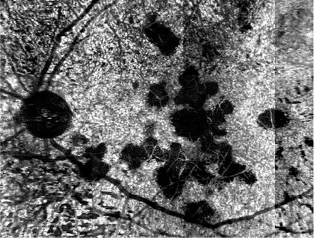
OCT angiography (OCTA) is a non-invasive tool that works on the principle of ‘decorrelation’ and results in the formation of en face images of retinal capillary network and choroidal vasculature without requiring dye. AngioScan OCT Angiography software on the RS-3000 Advance OCT (Nidek) provides ‘panoramic images’ with larger fields of view (Figure 1). Panoramic OCTA (P-OCTA)images allow 3-D analysis, are repeatable on follow-up examinations and to a certain extent are comparable to FA and ICGA in terms of resolution.8
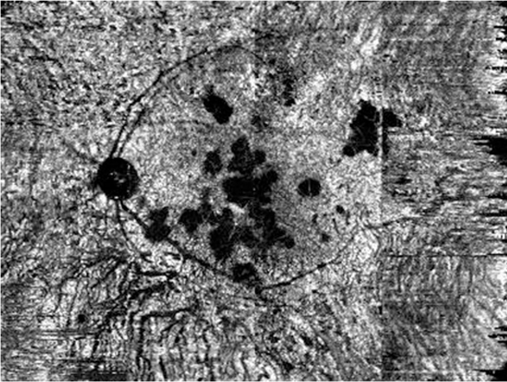
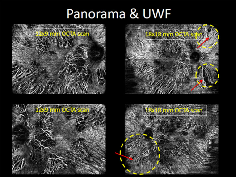
However the size of panoramic cube is 12×9 mm fails to cover the entire lesions as seen in FA and ICGA frames. Some of the lesions are always missed and we need to re-do the OCTA scan by changing the fixation of patient in a different position which is a tedious job both for patient as well as for the examiner. In order to overcome this, we devised a novel innovative way by adding +20D lens in front of OCTA machine undergoing panoramic scan and made the machine more myopic and by anterior displacement of nodal point of patient’s eye, we were able to generate a wider scan measuring 18×18 mm covering all the lesions as provided by invasive FA and ICGA with the highest resolution possible. We term this 18×18 mm scan as ‘Ultra wide field’ OCTA scan(Figure 2). This study aims to compare the non-invasive standard OCTA imaging characteristics of SLC with ‘panoramic’ and ‘ultra wide field’ OCTA imaging and to correlate these findings with ICGA features in SLC.
Material and Methods:
Study design and patient enrolment:
This is a single-center, prospective, observational study approved by the Institutional Review Board. All cases provided informed consent to be enrolled in the study anddata was collected from patients diagnosed as SLC at retina clinic from October 2017 to June 2018.
Eligibility criteria and data collection:
Inclusion criteria were: (1) clinical findings consistent with active stage of SLC pre-treatment at presentationand with healing stage of SLC post-treatment during follow-up on ICGA(2) follow-up of at least 6 months with regular medicationsand (3) complete availability of documentation till the last follow-up. If any one of the above-mentioned inclusion criteria was not met, patients were excluded from the study.
Investigations:
Diagnosis of tuberculosis was made by clinical or relevant laboratory tests, and cases were classified as confirmed, probable, or possible as per the criteria given below: i) Confirmed intraocular TB: microbiological confirmation of tuberculosis from ocular fluids/ tissues. ii) Probable intraocular TB: Chest x-ray consistent with TB infection or clinical evidence of extra- ocular TB or microbiological confirmation from sputum or extraocular sites along with immunological evidence of TB. iii) Possible intraocular TB: positive immunological tests for TB (tuberculin skin test: positive indurationof 10x10mmafter48to72hoursor positive interferon gamma release assay test: positive QuantiFERON of .0.35 IU/mL or positive T-SPOT.TB) or chest x-ray consistent with TB infection.
Treatment Regime:
Patients were treated with tapering course of oral corticosteroids (1–1.5 mg/kg/day) and patients diagnosed with tuberculosis were treated withanti-tubercular therapy9
Study Parameters:
All cases underwent a complete eye examination at presentation and also at every follow-up visit.The examination protocol included best-corrected visual acuity (BCVA) with the help of Snellen’s Chart, dilated fundoscopic examination, color fundus images (Topcon 50 Dx, Medical Systems Inc, Oakland, NJ, USA), fundus autofluorescence (FAF), fundus fluorescein angiography (FFA), indocyanine green angiography (ICGA) (Spectralis, Heidelberg Engineering Inc, Heidelberg, Germany), and spectral domain-optical coherence tomography (SD-OCT)(Spectralis, Heidelberg Engineering Inc., Heidelberg, Germany). Panoramic optical coherence tomography angiography (P-OCTA) (RS-3000 Advance OCT, Nidek) was performed in all eyes centered on the fovea covering 12×9 mm area (40° x 30° field of view). Ultra wide field optical coherence tomography angiography (P-OCTA) (RS-3000 Advance OCT, Nidek) with the help of +20D lens and adjusting machine’s optics was performed in all eyes centered on the fovea covering 18×18 mm area. AngioScan RS-3000 Advance software was used to segment the outer retinal layers and choriocapillaris.
Image Analysis:
For all cases, images acquired at presentation and subsequent follow-up examination, were examined by two independent masked examiners.
Statistical Analysis:
Descriptive analysis was used to compare the appearance of the standard OCTA with panoramic OCTA and ultra wide field OCTA.
Results:
20 eyes of 20 patients were studied. Patients with SLCwho met our inclusion criteria were included in the study. The mean age at presentation was 35.8±11.6 years (range 13–62 years). The gender distribution comprised, 14 eyes of 14 males and 6 eyes of 6 females.All eyes included had lesions in posterior pole.
‘Standard’, ‘Panoramic’ (P-OCTA)& ‘Ultra-wide field’ (UWF-OCTA)Optical Coherence Tomography Angiography Features:
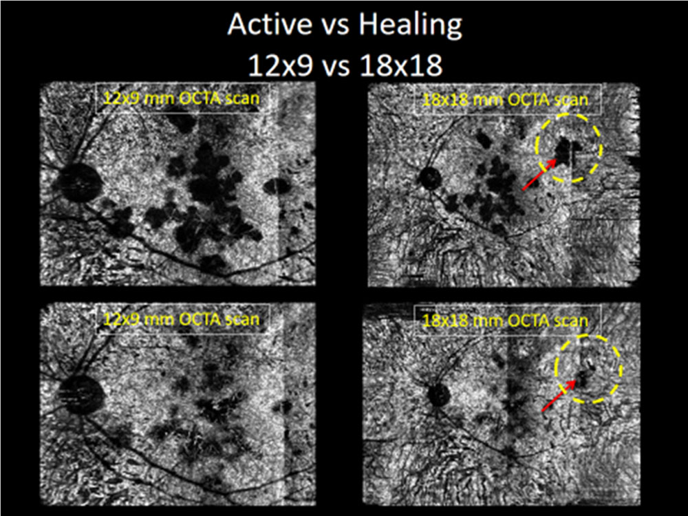
At presentation, all eyes with active lesionson P-OCTA (stage 1 and 2 on FAF) demonstrated areas of large flow void or dark areas with decreased de-correlation signal.These areas were non-contiguous and multi-focal in distribution. However, these featureson OCTA could be attributed to either choriocapillaris hypoperfusion or decreased transmission of signals.
At subsequent follow-up visits, post treatment, in all eyes, the lesions in various stages of healing (stage 3 and 4 on FAF) were studied. These eyes demonstrated areas of reduced large flow void or appearance of increased de-correlation signals with unmasking of choriocapillaris within the flow void areas. Corresponding to active phase, in healing phase, the areas of reduced flow void were againnon-contiguous and multifocal in distribution.The presence of this feature strongly suggests that the areas of large flow void or dark areas with decreased de-correlation signal on OCTA during active phase represent choriocapillaris hypoperfusion.
Comparisons Between Panoramic Optical Coherence Tomography Angiography, Indocyanine Green Angiography, and Fluorescein Angiography
FA in alleyes with active phase demonstrated abnormal sites in early frames as multiple, ill-defined, scattered areas of hypofluorescence (secondary to atrophy of choriocapillaris) with progressive hyperfluorescence at the margins of the lesion (eventual diffuse late staining of the underlying sclera) in late frames. In all eyes with healing phase, FA revealed similar features as of active stage however, the lesions in early frames were less hypofluorescent and in late frames hyperfluorescence at the margins was less intense. The lesions were now better demarcated with well-defined edges and borders.
ICGA during active phase in all eyesrevealed pathological sites as multiple, poorly defined areas of hypofluorescence in the early and late frames (representing choriocapillaris non-perfusion and corresponding to a primary inflammatory choriocapillaropathy) and as demonstrated in clinical fundus picture the lesions were, non-contiguous and multifocal in distribution. Post-treatment, the lesions became less hypofluorescent (possibly due to choriocapillaris re-perfusion), well demarcated and delineated, as the disease progressed from active to healing phase.
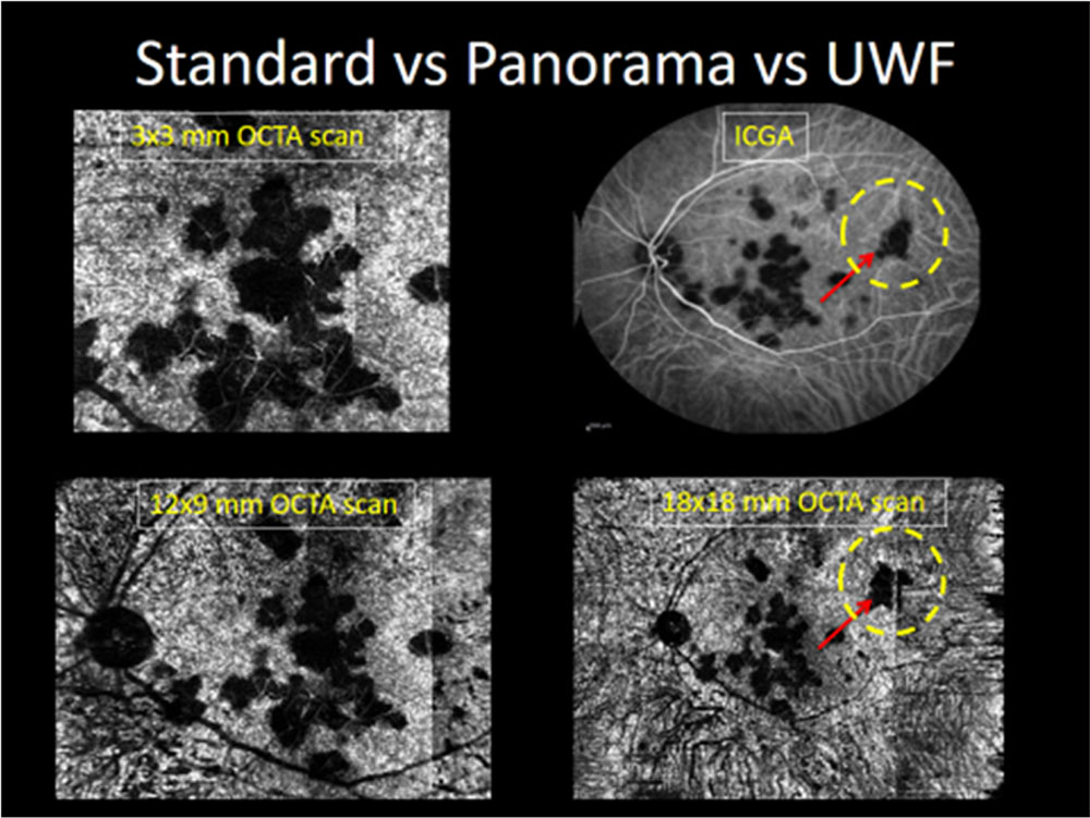
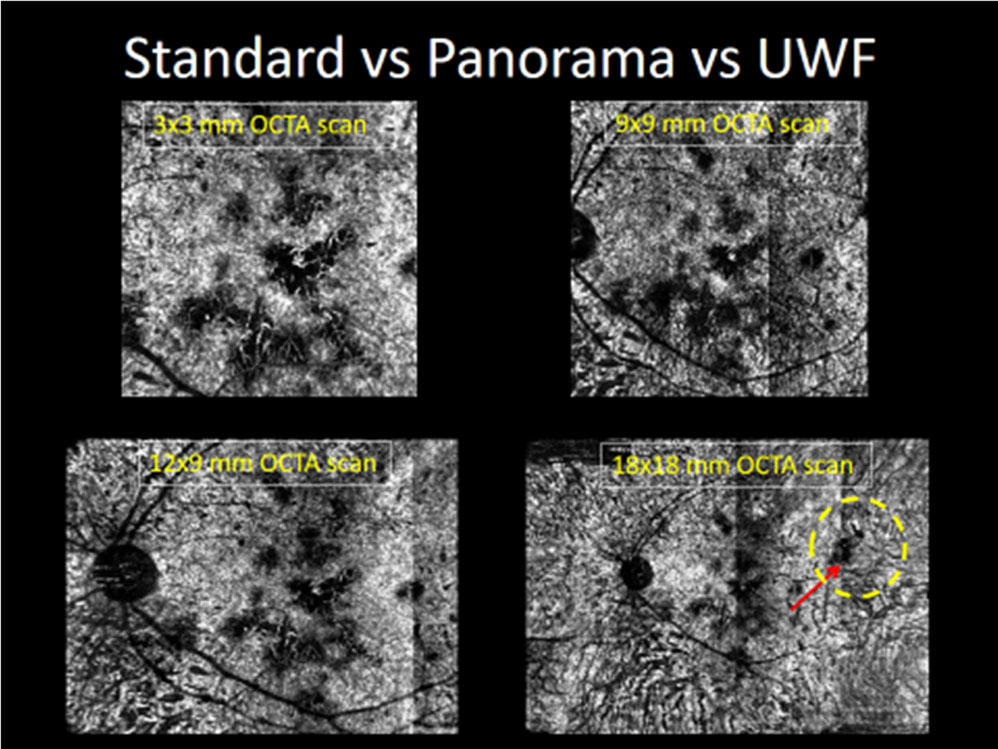
The active lesions seen in all eyes on ICGAexactly correlated with OCTA, in terms of their location and distribution. The large flow void areas or areas of decreased de-correlation signal corresponded with area of hypofluorescence on ICGA. In all eyes with lesions during various stages of healing, there was unmasking of middle and large choriocapillaris architecture possibly due to re-perfusion of choriocapillaris. This feature wasseenonly onOCTA, which was not demonstrated on ICGA.With the advent of ‘Panoramic’ imaging feature, it was possible to obtain large field of view (12 x 9 mm; 40° x 30°)very near to field of view provided byICGA and FA in a particular image, centered at fovea with similar resolution to 3×3 cube of OCTA that enabled a much better analysis, comparison, correlation and repetition of examination (Table 2). But however, some lesions were still missed and it was not a true comparative picture with ICGA and FA to make it 100% comparable and replaceable. But with UWF-OCTA (18x18mm) all lesions were well covered, completely equivalent to ICGA at each and every visit in all cases.
Comparisons between Panoramic Optical Coherence Tomography Angiography, Ultra-wide fied OCTA and routine 3x3mm Optical Coherence Tomography Angiography
‘Panoramic’ OCTA scores over a routine 3x3mm cube of OCTA in terms of field of view and scan acquisition time. ‘Panorama OCTA’ offers a wide field of view of 40×30 degrees (12×9 mm) as compared to a routine 3×3 mm scan, which offers 10×10 degrees field of view only. In terms of scan acquisition time, 3×3 mm routine OCTA scan takes less time (30 seconds to 1 minute) as compared to ‘Panoramic OCTA’ scan (10-15 minutes). But still the lesions were missed in a single Panoramic scan and it was not an 100% replaceable alternative. But with the genesis of wider 18×18 mm scanby novel technique of addition of +20D, UWF-OCTA becomes 100% equivalent to invasive ICGA in each and every respect (size, location, number, distribution, each and every visit).
(Figure 3-6)
Discussion:
Thebasic underlying pathogenic mechanism in patients of SLC, proposed in literature is either ischemia of choriocapillaris (a persistent decrease in choriocapillaris vascularity) or inflammatory choriocapillopathy (destruction of the choriocapillaris associated with choroidal inflammation). This causes disruption of the RPE and photoreceptors, which are fed by the choriocapillaris, resulting in permanent visual loss.5,10 However, enough data is yet not available to explain the alterations or the changes occurring during active, healing and healed stages of SLC.
ICGA is considered as the gold standard test for SLC that can provide information related to state of choriocapillaris. It provides true extent of the condition by identifying and reporting all the lesions. Despite of ICGA’s unique ability to provide information about choriocapillaris, it carries certain major disadvantages. First, it fails to explain the microstructural details of affected choriocapillaris. Second, being an invasive procedure it carries significant inherent risks due to dye injection and is contraindicated in pregnancy, patient’s with raised creatinine levels and is also not preferred in children. Third, being expensive and time-consuming it may not be logistically feasible to perform ICGA during each and every follow-up visit.1,5,11
In our study, the classical finding obtained in all eyes with active lesions of SLC on OCTA was the presence of possible large flow void or decreased de-correlation signals at the level of choriocapillaris. Mandadi et al in their study on SLC cases also reported similar findings of OCTA during active stageand they proposed that this might be due to decreased signal transmission rather than true loss of blood flow.5
However, availability of corresponding B-scans along with OCTA imagesto certain extent show this is not an artifact in the form of reduced signal transmission, but rather there appears to be a true loss of blood flow.
While comparing features seen on OCTA during active stage of disease, with ICGA features during same stage, the hypofluoresecent lesions on ICGA correlated exactly with large flow void or decreased de-correlation signals in terms of location, size and distribution. However the lesions are better demarcated and delineated in OCTA compared to ICGA.
Similarly, during the various healing stages of disease seen in all eyes in our study, the OCTA demonstrated areas of reduced large flow void or appearance of increased de-correlation signals with unmasking of choriocapillaris within the flow void areas. Mandadi et al have proposed that these larger vessels may represent either compensatory response to the atrophy or could be vessels from the Sattler layer that have been pushed toward the RPE–Bruch complex because of choriocapillaris atrophy.5While comparing features seen on OCTA during healing stage of disease, with ICGA features during same stage, the reduced hypofluoresecent lesions on ICGA correlated exactly with reduced large flow void areas in terms of location, size and distribution.
In OCTA during the active stages, the choriocapillaris are fainty visible, probably due to ischemia and hypo-perfusion, but in healing stages due to re-perfusion these choriocapillaris became unmasked and more obvious due to reduction of choroidal ischemia and/or re-perfusion of choriocapillaris post treatment. Hence, it can be mentioned that OCTA non-invasively not only correlates with the ICGA findings but also provides supplement information on morphology of choroidal vasculature in various stages of disease and thus helps to better understand the underlying pathology.
OCTA although is a revolutionary imaging modality, it however carries major limitationswhich needs due attention. It is a nascent technology which still needs lot of understanding, common vocabulary among interpreters, and some technical refinement.12,13 Owing to the presence of projection artifacts of superficial layers onto the deeper layers, the lesions at the level of choroid and choriocapillaris needs to be studied and evaluated carefully. Also, the spectral-domain OCTA has less penetration in acute inflammation of choroid which an examiner can overcome with introduction of swept source OCTA, but it is yet to be commercially available.5,8
Most important disadvantage as reported by previous studies who made an attempt to study the morphological features of SLC during various active and healing stages with OCTA, was the small field of view.5 The field of study obtained in a 3×3 cube of OCTA is not comparable to field offered by FA and ICGA. If the interpreter, increases the field size it reduces the resolution of images significantly, hampering interpretation and correlation to a major extent.The ‘Panorama’ feature overcomes this significant disadvantage to a certain extent but still some of the lesions are missed out and ICGA scores over P-OCTA in this feature (covering the extent of lesion). But with the genesis of novel, 3-D, high resolution, widest UWF-OCTA scans it has become possible in each and every way to replace ICGA in each and every situation. Thus, it allows the examiner to correlate the invasive ICGA findings with non-invasive OCTA findings in the best possible way.8
Our study has few limitations as it needs a small learning curve to master the technique. To conclude, this studydescribes and establishes correlation among features provided by multi-modal imaging techniques during active and healing stages of SLC. Future longitudinal studies with the ongoing development in the field of bio-medical engineering using panorama feature and ultra-wide field OCTA whichwill definitely go a long way in analyzing and further understanding the pathologic alterations seen on OCTA. Nonetheless, UWF OCTA is certainly a promising imaging modality that can be used as an alternative diagnostic test for SLC and all other choroidal lesion.
Abbreviations:
OCTA: optical coherence tomography angiography
ICGA: indocyanine green angiography
FA: fluorescein angiography
SLC: serpiginous like choroiditis
UWF: ultra wide field
References:
1.Crawford CM1, Igboeli O. A review of the inflammatory chorioretinopathies: the white dot syndromes. ISRN Inflamm. 2013 Oct 31;2013:783190.\
2.BansalR, Kulkarni P, Gupta A, et al. High-resolution spectral domain optical coherence tomography and fundus autofluorescence correlation in tubercular serpiginouslikechoroiditis. J Ophthalmic Inflamm Infect. 2011 Dec; 1(4): 157–163.
3.SudharshanS, Ganesh SK, Biswas Current approach in the diagnosis and management of posterior uveitis. Indian J Ophthalmol. 2010 Jan-Feb; 58(1): 29–43.
4.Lim WK, Buggage RR, Nussenblatt RB. Serpiginous choroiditis. Surv Ophthalmol. 2005 May-Jun;50(3):231-44.
5.Mandadi SK, Agarwal A, Aggarwal K, et al; for OCTA Study Group. Novel Findings On Optical Coherence Tomography Angiography In Patients With Tubercular Serpiginous-Like Choroiditis. 2016 Dec 7
6.Oray M, Zakiev Z, Çağatay T, et al. Treatment Results in Serpiginous Choroiditis and Multifocal Serpiginoid Choroiditis Associated with Latent Tuberculosis. Turk J Ophthalmol. 2017 Apr;47(2):89-93.
7.Yee HY, Keane PA, Ho SL, et al. Optical Coherence Tomography Angiography of Choroidal Neovascularization Associated with Tuberculous Serpiginous-like Choroiditis. Ocul Immunol Inflamm. 2016 Dec;24(6):699-701.
8.Nagpal M, Juneja R. Panoramic Imaging With OCTA. Retina Today. 2017 April.
9.Gupta V, Arora S, Gupta A, et al. Management of presumed intraocular tuberculosis: possible role of the polymerase chain reaction. Acta Ophthalmol Scand. 1998 Dec;76(6):679-82
10.Cheng L, Chen X, Weng S, et al. Spectral-Domain Optical Coherence Tomography Angiography Findings in Multifocal Choroiditis With Active Lesions. Am J Ophthalmol. 2016 Sep;169:145-61
11.Ahn SJ, Park SH, Lee BR. Multimodal Imaging Including Optical Coherence Tomography Angiography in Serpiginous Choroiditis. Ocul Immunol Inflamm. 2017 Apr;25(2):287-291
Figures:
Figure 1:
Leave a Comment