

Dr.Kavitha V, Dr.Mallikarjun M Heralgi,Dr.DEEP PATEL,Dr.Sneha Harogoppa
Title
Analysis of macular, foveal and retinal nerve fibre layer thickness in children with unilateral anisometropic amblyopia and their changes following occlusion therapy.
Abstract
Aim: To analyse macular thickness (MT), foveal thickness (FT) and retinal nerve fibre layer thickness (RNFLT)in children with unilateral anisometropic amblyopia and their changes following occlusion therapy.
Material and Method: A prospective, longitudinal and comparative study of 60 children aged between five and 18 years consisted of two groups, group 1: 30 children with unilateral anisometropic amblyopia; group 2: 30 normal children. Best corrected visual acuity(BCVA), a detailed ocular examination, spectral domain optical coherence tomogrraphy (SD OCT) for MT, FTand RNFLT in both eyes were done at visit one (baseline) and every three months for a year following occlusion therapy (initiated one month after first visit) in group 1.
Results: Mean BCVA, MT, FT and RNFLT in amblyopic eyes at first visit were 0.63±0.405, 286.9±6.522, 195.90±8.462 and 100.87±6.240 respectively and at last visit after occlusion therapy were 0.50±0.318, 248.9±11.681, 169.47±10.941 and 99.43±5.722 respectively. At first visit, mean BCVA, MT, FT and RNFLT in non amblyopic eyes (group 1) were 0±0, 240 ± 10.447, 159.27±9.285, 98.63±4.723 and in normal eyes (group 2: average of right and left eyes) were 0±0, 239.8±4.294, 143.6±4.610, 100.5±2.895 respectively. Conclusion: MT and FT which were more in amblyopic eyes as compared to normal fellow eyes and group 2, decreased with improvement in BCVA after occlusion therapy. However there was no difference in RNFLT between amblyopic eyes and normal fellow eyes and group 2 before and after occlusion therapy.
key words: Amblyopia, Macular thickness, Foveal thickness, RNFL thickness, SD OCT, Occlusion therapy.
Introduction
Amblyopia is defined as unilateral or bilateral reduction in best corrected central visual acuity caused by form vision deprivation and/or abnormal binocular interaction, without any visible organic cause to commensurate with visual loss.[1] With advent of OCT, etiopathology of amblyopia is being better understood as it is one of the best tools to study the structural changes in retina.[2]Occlusion therapy has long been the mainstay of amblyopia treatment.[1] Various studies are available documenting changes in MT, FT and RNFLT in amblyopic eyes.[3-8]However, there are only few studies available on analysis of these parameters following occlusion therapy. Hence, the purpose of our study was not only to analyse the structural changes in retina (MT, FT, RNFLT) in unilateral anisometropic amblyopic eyes and compare the same with the normal fellow eyes and normal eyes of normal children but also to understand the same following occlusion therapy.
Material and method
This prospective, interventional, comparative, longitudinal study was carried out at a tertiary care eye hospital in South India between October 2015 and June 2017.The study was approved by the institutional review board and adhered to all the principles mentioned in the Declaration of Helsinki 2000.This study included 60
children: The eyes were divided into 2 groups; group 1-amblyopic eyes (A) and normal fellow eyes (F) of amblyopic patients and group 2- normal eyes of normal children (N).Based on previous literature on outcome variable of visual acuity in Log MAR scale, for 90% statistical power, 5% level of type 1 error, 10 % type 2 error and 95% confidence interval or at 5 % level of significance, the estimated sample size was 60 children, 30 in each group. They were divided equally into two groups; group 1 included children with unilateral anisometropic amblyopic eyes and normal fellow eyes (non-amblyopic eyes), group 2 included normal eyes of normal children. Inclusion
Criteria: children of either sex aged between five and 18 yearswith unilateral anisometropic amblyopia (difference in BCVA of ≥ 0.2 LogMAR between two eyes) were included in group 1; Uncorrected visual acuity of log MAR 0.00 in both eyes were included in group 2; normal neurological, ocular and systemic examination, those willing for follow up. Exclusion
criteria: Previous history of spectacle wear / occlusion therapy / ocular surgery, refractive error correction greater than 6 D spherical power and 3 D cylindrical power, presence of strabismus / nystagmus / hearing impairment / developmental delay, and not compliant for spectacle wear and /or occlusion therapy, children who have come for less than three follow ups. After satisfying the inclusion and exclusion criteria, informed written consent from every child’s parent or parentswas taken after counseling them regarding the nature of the study. In group 2, first two normal children attending the out-patient department every day were recruited in the study. Detailed history regarding any ocular and systemic conditions was noted. UCVA and BCVA for distance using LogMar three meter chart (English letters or Symbols chart) and near vision using snellen chart were recorded by a single person.
All tests to rule out strabismus were done. Intraocular pressure (IOP) was measured using non-contact tonometry wherever possible. Fundus examination was done using indirect ophthalmoscope and 20D condensing lens. Cycloplegic refraction using appropriate drug according to age was carried out in all children. Other visual function tests like Color vision (Ishihara pseudo chromatic plates), contrast sensitivity (Pelli – Robson contrast sensitivity chart), visual fields (Humphrey’s field analysis / confrontation test / Amsler’s chart) and electrophysiology tests were recorded wherever it was required and possible. Detailed ophthalmological examination both anterior and posterior segment was carried out in all children
OCT examinations were performed for measuring MT, FT and RNFLT in each eye using a Topcon 3D Maestro by the same operator through dilated pupils at least 5 mm in diameter. ‘‘Fast RNFL map protocol’’ consisting of three circular scans with diameters of 3.4 mm centered on the optic disc was performed along with the ‘‘Macular Thickness Map’’ protocol consisting of six radial scan lines centered on the fovea, each having a 6 mm transverse length. In order to obtain the best image quality, focusing and optimization settings were controlled and scans were accepted only if the signal strength (SS) was .6 (preferably 9–10). Children who had poor fixation cooperation due to poor vision or low age were excluded from the study
MT was measured using caliper tool 350 µm nasally from the fovea. FT was measured using caliper tool between internal limiting membrane and retinal pigment epithelium. In RNFLT measurement total RNFL thickness was taken in the study.
All findings were recorded for both the groups. Average of right eye(RE) and left eye(LE) values were taken for all the parameters in group 2.One month after first visit (post spectacle wear ), amblyopic children ( group 1) were asked to patch the normal fellow eye for four hours per day and perform near activities such as reading, writing, drawing, mobile games, computer work. Parents were insisted upon maintaining a diary regarding the same to check for compliance. Group 1 children were followed up with BCVA, MT, FT and RNFLT at three, six, nine and 12 months along with patch diary for childrens’ compliance. Data was analyzed using SPSS software (Statistical Package for Social Science) and by using the Paired student‘t’ test.
Statistical methods: MT, FT and RNFLT were considered as outcome variables. Amblyopia was considered as explanatory variable. Descriptive analysis was carried out by mean and standard deviation for quantitative variables, frequency and proportion for categorical variables. Initially the outcome parameters were compared between amblyopic and normal subjects by using independent sample t-test. The mean values of all the outcome variables within amblyopic subjects were compared between the amblyopic and non-amblyopic eye at each follow up interval separately using paired t-test. The change in the outcome parameters over the follow up period within the amblyopic eye was compared by one way repeated measure ANOVA.
Statistical software: Machines IB. IBM SPSS Statistics for Windows, Version 22.0. IBM Corp Armonk, NY; 2013.
Results:
Sixty children aged between five and 18 years were included in the analysis. Out of which 30 children had unilateral amblyopia (Group 1) and 30 children were normal (group 2).The mean age in group 1 and 2 were 9.77±2.674 and 9.70±2.20 years respectively (P = 0.916). In group 1, 56.66 %( 17) and 43.33 %(13) were male and female children respectively. Likewise in group 2, 60% (18) and 40 %( 12) were male and female children respectively. In group 1, 12 (40%) had right eye(RE) amblyopia and 18(60%) had left eye(LE) amblyopia. The baseline values of mean LogMar BCVA, MT, FT and RNFLT in group 1 amblyopic eyes were 0.63±0.405, 286.9±6.522, 195.9±8.462 and 100.8±6.240 and non-amblyopic eyes were 0.00±0.00, 240 ± 10.447, 159.27±9.285 and 98.63±4.723 respectively; in group 2 (average of RE and LE in normal patients) , they were 0.00±0.00 , 239.8±4.294, 143.6±4.610 and 100.5±2.895 respectively.
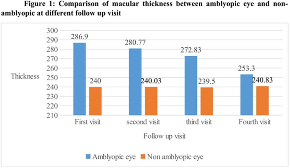
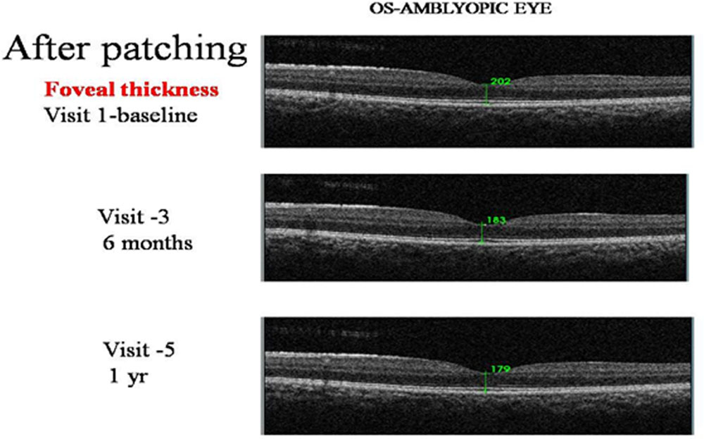
At first visit, the difference in mean BCVA, MT and FT between amblyopic eyes (0.63±0.405, 286.9 ± 6.522 and 195.90±8.462,) and normal eyes of normal group ( 0.00±0.00, 239.8±4.294, 143.6±4.610) was statistically significant (P value <0.001). Similarly, the difference between amblyopic eyes and non-amblyopic eyes (0.00±0.00, 240 ± 10.447, 159.27±9.285) was statistically significant (P value <0.001).However, the difference in mean RNFLT between amblyopic eyes (100.87±6.24) and normal eyes (100.5±2.895) ; and non-amblyopic eyes(98.63±4.723) was statistically not significant (P value : 0.802 and 0.131 respectively). The LogMar BCVA in amblyopic eyes in visit 1 (Baseline) and following occlusion therapy at three, six, nine and 12 months were 0.63±0.405,0.56±0.368, 0.51±0.374, 0.46±0.330, 0.50±0.318 respectively. Table 1 shows comparison of BCVA, MT, FT and RNFLT between amblyopic eyes, non amblyopic eyes and normal eyes at various follow ups. Graph 1 shows comparison of MT between amblyopic and non-amblyopic eyes at various followups. Graph 2 shows comparison of FT between amblyopic and non-amblyopic eyes at various follow ups. Figure 1 shows OCT picture of macula of amblyopic eye at first visit and Figure 2 shows OCT picture of macula of amblyopic eye at last visit following occlusion therapy.
Corelation between improvement in BCVA and decrease in MT and FT : There is a weak negative correlation between change in MT amblyopic eye and change in BCVA amblyopic eye (r value: – 0.026, P value: 0.891). There is a weak negative correlation between change in FT amblyopic eye and change in BCVA amblyopic eye (r value: – 0.020, P value: 0.917)
Discussion
Amblyopia had been thought to be a disease associated with an abnormality of the retina. [9] However, amblyopia- induced cerebral changes were later shown to mainly occur in the visual cortex and the lateral geniculate body. In an experimental study, Von Noorden and colleagues have suggested that the mechanism responsible for amblyopia may be inadequate visual stimulation of the fovea during early childhood, abnormal binocular interaction or incompatibility in the visual information received by the two eyes, or a mixture of these problems.[10] Yen et al. hypothesized that the normal postnatal reduction (apoptosis) of retinal ganglion cells is arrested in amblyopia and predicted that this would cause increased RNFLT. If this does indeed occur, it is likely that the arrest of normal postnatal changes would result not only in increased RNFLT but also would affect the normal maturation of the macula, including movement of Henle’s fibers away from the foveola and a decrease in foveal cone diameter. This would result in increased foveal thickness. Furthermore, because of the reduced apoptosis of retinal ganglion cells, the thickness of the ganglion cell layer in the macula would also be increased.[11]
It may be also contributed to no apoptosis or less apoptosis of retinal ganglion cells in amblyopic eyes due to absence of normal vision stimulation, eventually leading to thicker RNFL of the amblyopic eye than non-amblyopic eye. [12,24]
Few studies have suggested that there is no difference in macular or RNFL thicknesses in children with unilateral amblyopia.[13,14,19,23]
On the contrary few studies, however, have shown that thickening of the RNFL occurs in anisometropic and strabismic amblyopia. [11,12]Huynh et al. and Pang et al. showed a thicker fovea in unilateral amblyopia, [13,14]while Al-Haddad et al. found a thicker macula in anisometropic amblyopia.[15] Other studies, however, have shown thickening of RNFL, macula and fovea in children with amblyopia.[3,15-18]
Optical coherence tomography (OCT) is a rapid, non-invasive, office-based imaging technique allowing objective quantification of retinal structures with high resolution, including determination of peripapillary RNFL thickness and macular thickness.
In our study, of 60 children with anisometropic amblyopia, the MT was more in amblyopic eyes (286.9 ± 6.522) compared to non-amblyopic fellow eyes(240 ± 10.447) and normal eyesof normal children (239.8±4.294) This difference which was statistically significant and was similar to other studies.[4,15,23,24]Foveal thickness in our study was signifiantly more (P < 0.001) in amblyopic eyes (195.9±8.462) compared to normal fellow eyes (159.27±9.285) and normal eyes of normal children(143.6±4.610). This difference was statistically significant. Similar results were found in other studies. [6,24,30]With respect to RNFLT we found no statistically significant difference in amblyopic eyes compared to normal fellow eyes and normal eyes of normal children. This was consistent with other studies.[13,14,19,23]
Furthermore, this study was done to study the effectiveness of occlusion therapy on BCVA, MT, FT and RNFLT at three, six, nine and 12 months. In the process, this study highlights two aspects of amblyopia. First is to treat unilateral amblyopia, the rationale being to optimise visual function and binocular vision, and to try to provide a useful “spare eye” in the event of trauma or pathology in the normal eye. The second aspect is, since our study has included children less than 18 years it brought out the beneficial effect of occlusion therapy in older children similar to study conducted by Paediatric eye disease investigator group. [21]This contradicts the earlier concept that occlusion therapy is not effective in children older than eight years.
In our study, all children with unilaterla anisometropic amblyopia showed improvement in BCVA ≥ 1 line following optical correction and patching during the study period with p value [<0.001] which was statistically significant. Also, performing near activities while patching might have contributed as an additional benefit in treating these children like PEDIG study. [22]
We found statistically significant reduction in MT and FT in each visit compared to baseline [visit 1] but there was no statistically significant difference in RNFLT, post occlusion therapy in amblyopic eyes.Tugcu et al. studied macular thickness in the persistent amblyopic and resolved amblyopic eyes and did not found significant difference between the two groups. [27]Chen et al. compared the macular thickness of the amblyopic eyes with those of fully corrected previous amblyopic eyes and non-amblyopic controls and found there was no significant difference among the three groups.[28] In contrast, Pang et al. reported that the central macular thickness in myopic anisometropic amblyopia significantly reduced after amblyopia treatment. [29] However, the measurements in their study were not adjusted for axial length and refractive error.Analaysis on correlation between BCVA and MT ; FT showed a weak negative correlation which implies that with improvement of BCVA in amblyopic eyes following occlusion therapy there has restoration of MTand FT as revealed in OCT.
Limitations: Sample size was smaller and follow up period was shorter. Bilateral refractive, strabismic and visual deprivation amblyopia were not included in the study. Various types of refractive error and axial lengthwere not analysed. Sustainability of the improved BCVA and changes in anatomical layers of retina following discontinuation of occlusion therapy could not be assessed.
Conclusion:
MT and FT which were more in amblyopic eyes as compared to normal fellow eyes and normal eyes of normal children, decreased with improvement in BCVA after occlusion therapy. However there was no difference in RNFLT between amblyopic eyes and normal fellow eyes and normal eyes of normal children before and after occlusion therapy. Therefore, we can hypothesize that occlusion therapy can result in restoring the process of postnatal reduction of ganglion cells as evidenced by reduction in MT and FT on OCT .This implies that OCT can be prognostic tool in treating children with unilateral anisometropic amblyopia.
References:
- Von Noorden GK, Campos E. Binocular vision and ocular motility, 6th edn. St Louis, MO: Mosby, 2002.
- Liu H, Zhong L, Zhou X, Jin QZ. Macular abnormality observed by OCT in children with amblyopia failing to achieve normal visual acuity after long-term treatment. J PediatrOphthalmol Strabismus. 2010;47:17–23
- Pang Y, GoodfellowGw, Allison C, et al. A prospective study of macular thickness in amblyopic children with unilateral high myopia. Invest Ophthalmol Vis Sci. 2011 Apr; 14; 52 (5): 2444-9.
- Agrawal S, Singh V, Singhal V. Cross-sectional study of macular thickness variations in unilateral amblyopia. J ClinOphthalmol Res. 2014;2:15-7.
- Yoon SW, Park WH, Baek SH, Kong SM. Thicknesses of macular retinal layer and peripapillary retinal nerve fiber layer in patients with hyperopic anisometropic amblyopia. Korean J Ophthalmol. 2005;19:62-7.
- Christiani E AL-Haddad, Georges M E L Mollayess, Carol G Cherfan, et al. Retinal Nerve Fibre Layer and Macular thickness in Amblyopia as measured by spectral- domain Optical Coherence Tomography. Br J Ophthalmol. 2011; 95:1696-1699.
- Abdullah G, Alotaibi, Badriah Al Enazi CO. Unilateral Amblyopia: Optical Coherence Tomography Findings. Saudi Journal of Ophthalmology. October-December 2011; VOl.25 (4): 405-409.
- Atakan M, Culfa S, Calli U, Penbe AD, Atakan TG. Evaluation of Retinal Nerve Fibre Layer and Macular thickness in Ambluopia. J ClinExp Ophthalmol.2015; 6:437.
- McNeil NL. Patterns on visual defects in children. Br J Ophthalmol 1955;39:688-701.
- Von Noorden GK, Dowling JE, Ferguson DC. Experimental amblyopia in monkeys. I. Behavioral studies of stimulus deprivation amblyopia. 1970;84(2):206-214
- May-Yung Yen, Ching-Yu Cheng, and An-Guor Wang. Retinal Nerve Fiber Layer Thickness in Unilateral Amblyopia. IOVS, July 2004, Vol. 45, No. 7
- Lewis TL, Maurer D. Multiple sensitive periods in human visual development: evidence from visually deprived children. Devpsychobial 2005;46(3):163-183
- Altintas O, Yuksel N, Ozkan B, Caglar Y. Thickness of the retinal nerve fiber layer, macular thickness, and macular volume in patients with strabismic amblyopia. J PediatrOphthalmol Strabismus 2005;42:216–221.
- Kee SY, Lee SY, Lee YC. Thicknesses of the fovea and retinal nerve fiber layer in amblyopic and normal eyes in children. Korean J Ophthalmol 2006;20:177–181
- Yen MY,ChengCY,Wang AG. Retinal nerve fibre layer thickness in unilateral amblyopia. Invest Ophthalmol Vis Sci 2004;45(7):2224-30
- Yoon SW, Park WH, Baek SH, Kong SM. Thicknesses ofmacular retinal layer and peripapillary retinal nerve fiberlayer in patients with hyperopic anisometropicamblyopia.Korean J Ophthalmol 2005;19:62–67.
- Huynh SC, Samarawickrama C, Wang XY, Rochtchina E, Wong TY, Gole GA, et al. Macular and nerve fiber layer thickness in amblyopia: the Sydney Childhood Eye Study. Ophthalmology. 2009 Sep;116(9):1604–9.
- Al-Haddad CE, Mollayess GMEL, Cherfan CG, Jaafar DF, Bashshur ZF. Retinal nerve fibre layer and macular thickness in amblyopia as measured by spectral-domain optical coherence tomography. Br J Ophthalmol. 2011 Dec;95(12):1696–9.
- Baddini-Caramelli C, Hatanaka M, Polati M, et al. Thickness of the retinal nerve fiber layer in amblyopic and normal eyes: a scannig laser polarimetry study. J AAPOS 2001;5: 82-4
- Young Rok Lee, JuYounLee.Part-Time Occlusion Therapy for AnisometropicAmblyopia Detected in Children Eight Years of Age and Older.Korean Journal of Ophthalmology 20(3):171-176, 2006.
- Pediatric Eye Disease Investigator Group .Randomised Trial of Treatment of Amblyopia in Children Aged 7 to 17 Years . Arch Ophthalmol.2005; 123:437-447.
- Pediatric Eye Disease Investigator Group. A randomized pilot study of near activities versus non-near activities during patching therapy for amblyopia. J Aapos 2005, 9:129-136.
- Rajavi Z, Moghadasifar H, Feizi M, Haftabadi N, Hadavand MR, Yaseri M, Sheibani K, Norouzi G. Macular thickness and amblyopia. J Ophthalmic Vis Res 2014;9:478-83.
- Wu S-Q, Zhu L-W, Xu Q-B, Xu J-L, Zhang Y. Macular and peripapilary retinal nerve fibre layer thickness in children with hyperopic anisometropic amblyopia. Int J Ophthalmol.2013;6(1):85-9.
- Alotaibi AG, Al Enazi B. Unilateral amblyopia: Optical coherence tomography findings. Saudi J ophthalmol.2011;25(4):405-9.
- Chun-Hsiu Liu, SherineJueOng, Chung-Ying Huang, Wei-Chi Wu, Ling-Yuh Kao, and Meng-Ling Yang. Macular Thickness, Foveal Volume, and Choroidal Thickness in Amblyopic Eyes and Their Relationships to the Treatment Outcome. Journal of Ophthalmology.2018.
- Tugcu, B. Araz-Ersan, E. T. Erdogan et al..Structural and functional comparison of the persistent and resolved amblyopia. DocumentaOphthalmologicavol. 2014;128(2):101–109.
- Chen, J. Chen, F. Zhang, X. Zhu, and F. Lu. Visual outcome in isoametropic amblyopic children with high hyperopia and the effect of therapy on retinal thickness. American Journal of Ophthalmology. 2013;155(3):536–543.
- Pang, K. A. Frantz, S. Block, G. W. Goodfellow, and C. Allison. Effect of amblyopia treatment on macular thickness in eyes with myopic anisometropic amblyopia. Investigative Ophthalmology & Visual Science. 2015;56(4):2677–2683.
- Dong Hee Yoon and Bo Young Chun. Comparison of the Thickness and Volume of the Macula and Fovea in Patients with Anisometropic Amblyopia Prior to and after Occlusion Therapy. Korean J Ophthalmol. 2018; 32(1): 52–58.
Table 1 : Comparison of BCVA, MT, FT and RNFLT between amblyopic eyes, non amblyopic eyes and normal eyes at various follow ups
| Group1- Amblyopic eyes | |||||
| BCVA | MT | FT | RNFLT | ||
| P < 0.001 | (mean±STD) | (mean±STD) | (mean±STD) | ||
| 1st visit
(0 M) |
0.63±0.405 | 286.9 ± 6.022 | 195.90±8.462 | 100.87±6.241 | |
| 2nd visit (3M) | 0.56±0.368 | 280.77±7.677 | 188.800±8.903 | 99.30±4.843 | |
| 3rd visit (6M) | 0.51±0.374 | 272.83±9.476 | 182.633±7.924 | 99.03±5.555 | |
| 4th visit (9M) | 0.46±0.330 | 253.30±11.065 | 176.10±11.309 | 101.23±5.276 | |
| 5th visit (12 M) | 0.50±0.318 | 248.90±11.681 | 169.467±10.941 | 99.43±5.722 | |
| Group 1-Normal fellow eyes | |||||
| 1st visit
(0 M) |
0.00±0.00 | 240±5.977 | 159.27±9.285 | 98.63±4.723 | |
| P < 0.001 | P < 0.001 | P < 0.001 | P 0.131 | ||
| 2nd visit (3M) | 0.00±0.00 | 240.03±5.678 | 164.90±7.840 | 100.60±6.295 | |
| P < 0.001 | P < 0.001 | P < 0.001 | P 0.368 | ||
| 3rd visit (6M) | 0.00±0.00 | 239.50±5.335 | 164.23±8.157 | 99.03±4.642 | |
| P < 0.001 | P < 0.001 | P < 0.001 | P 1.000 | ||
| 4th visit (9M) | 0.00±0.00 | 240.83±5.977 | 164.63±7.981 | 99.03±5.314 | |
| P < 0.001 | P < 0.001 | P < 0.001 | P 0.089 | ||
| 5th visit (12 M) | 0.00±0.00 | 239.47±5.569 | 165.43±7.771 | 99.60±5.500 | |
| P < 0.001 | P < 0.001 | P 0.098 | P 0.917 | ||
| Group 2- Normal eyes of normal children( average of RE and LE values) | |||||
| 1st visit (0 M) | 0.00±0.00 | 239.8±4.294 | 143.6±4.610 | 100.5±2.895 | |
| P < 0.001 | P < 0.001 | P < 0.001 | P -0.802 | ||
BCVA- Best corrected visual acuity; MT- Macular thickness; FT-Foveal thickness; RNFLT -retinal nerve fiber thickness; M-month; RE- Right eye; LE – Left eye
Figure Legends :
Graph 1 : Comparison of macular thickness between amblyopic and non-amblyopic eyes at various follow ups.
Graph 2 :Comparison of foveal thickness between amblyopic and non-amblyopic eyes at various follow ups.
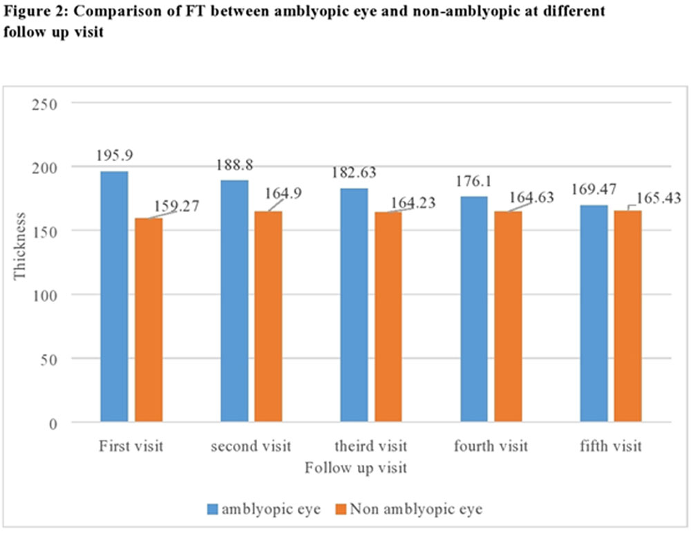
Figure 1: OCT image of macula of amblyopic eye at first visit
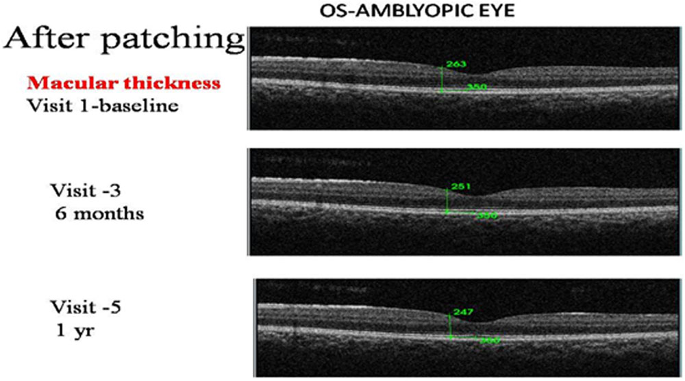
Figure 2: OCT picture of macula of amblyopic eye at last visit following occlusion therapy
Leave a Comment