

Dr.Narayan Bardoloi,Dr.Sandip Sarkar,Dr.Himangshu Das
DROPLESS CATARACT SURGERY- A STUDY
Purpose: To evaluate the clinical outcome following intraoperative transzonularintravitreal injection of Triamcinolone-Moxifloxacin(TM) in patients undergoing phacoemulsification cataract extraction with intraocular lens(IOL) implantation.
Setting: Chandraprabha Eye Hospital, Jorhat,Assam,India.
Design: Prospective,non-randomized,clinical,interventional study.
Methods: A total of 200 eyes were enrolled in the study.Patients who voluntarily gave consent after being informed about dropless cataract surgery along with its pros and cons were included. Those who have glaucoma or are known steroid responders and who lost to follow up were excluded. Dropless cataract surgery is a new technique where 0.1 ml each of moxifloxacin and triamcinolone acetonide is injected transzonularly at the end of IOL implantation in phacoemulsification cataract surgery with the help of 27G curved cannula.This eliminates the need for instillation of eyedrops post operatively. Ophthalmic viscosurgical device is injected into the ciliary sulcus to make space for easy introduction of the cannula.Intraocular pressure rise following injection is counterbalanced with bimanual irrigation and aspiration. Visual acuity ,slit lamp examination, cells,IOP were measured on day 1,7,30,60,90.
Results: Intraoperative complications included bleeding 1 eye(0.5%),leakage in 8 eyes(4%),pain in 3 eyes(1.5%) and IOP rise in 1 eye(0.5%).More than 6/9 UCVA achieved in 96% of patients at the end of 3 months. Mean IOP was normal in all cases in all visits.20 complained of floaters on day 1(D1) postoperatively which decreased to 15 and 13 after 7 and 30 days postoperatively respectively and finally the number came down to 0 after 60(D60) and 90(D90)days postoperatively.100 % patients did not need any eye drop during entire post operative period.
Conclusion:The study clearly demonstrates that this procedure is very effective and safe.
Modern phacoemulsification cataract surgery claims to provide earliest rehabilitation to a patient. It is a walk in walk out procedure. Patient can resume normal activities from the very day of surgery. Inspite of these advantages a patient has to put eye drop into the operated eye from time to time for a period of four to six weeks. That takes away a lot of sheen out of this very beautiful procedure 1, 2. Here arises the problem of compliance, injury to corneal epithelial surface, high cost and frequent callbacks. A compliance monitoring study found that cataract surgery patients did not comply with the prescribed number of postoperative drops3. Some use improper technique during application of post operative drops4 . Examples of improper techniques include the drop missing the eye, instilling an incorrect number of drops in one dose, and contaminating the bottle tip. The same study also reported that less than half of the patients waited less than 5 minutes between instilling different eye-drop medications. Noncompliance with ophthalmic topical drops has been reported to be as high as 40%.5Noncompliance is likely to increase the risk of infection, and may pose a health safety concern; for instance, not adhering to the prescribing schedule for antibiotic eyedrops could increase the risk of bacterial resistance.
It is also known that patients coming for phacoemulsification surgery suffer from dry eye disease of some form6-14. Postoperative eye drop will add insult to the already compromised corneal surface15-17. High cost of postoperative medication adds to the causes of non-compliance in countries like India, Nepal, Pakistan, Bangladesh where half of the population live below the poverty line. How many drops, how many times, which eye are frequent callbacks an ophthalmologist encounters during postoperative period.
Dropless cataract surgery is a new technique practiced by many American cataract surgeons for the last couple of years. A specially prepared compound called Trimoxi containing Triamcinolone acetonide and Moxifloxacin(TM) and Trimoxivanc(TMV) where Vancomycin is added is available in American market(ref). After IOL implantation, the surgeons inject this mixture transzonularly into the vitreous cavity. This eliminates the need for instillation of eyedrop postoperatively and patients can lead a life free from any compulsion. There has been many reports of surgeons resorting to this technique with very good effect and without any major complication. This study is an endeavor to do this technique in India for the first time. Since commercially available mixing compounds of TM is not available in India, we have mixed these two compounds on the operation table from fresh bottles of Moxifloxacineyedrop and fresh ampoule of Triamcinolone acetonide (TA).
MATERIALS AND METHODS
This was a prospective, non randomized, non comparative clinical interventional study. The study period was February 2017- September 2017. The study protocol was approved by institutional ethics committee (IEC) of Chandraprabha Eye Hospital and abide by the tenets of the declaration of Helsinki. All the patients were informed about the pros and cons of the procedure and an informed consent was obtained from all participants. Pre operative exclusion criteria was known cases of glaucoma , steroid responders. Complications during surgery, such as posterior capsular rupture, vitreous loss, zonular dialysis were exclusion criteria as was the inability to deliver the drug .
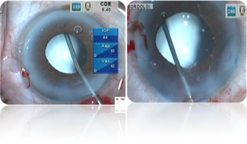
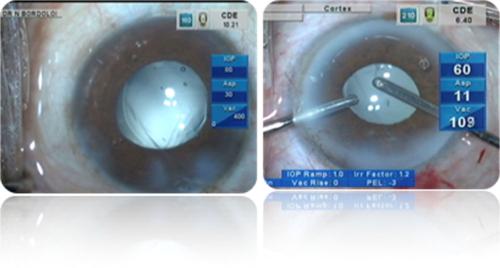
Standard surgical protocols were used in all cases. Local anesthetic (proparacaine 0.5% drops) was instilled in the conjunctival sac 10 minutes and 5 minutes before surgery. A 10% povidone–iodine solution was applied to the skin around the operative site, and a 5% solution was applied to the conjunctival sac 3 min before surgery. Under topical anesthesia, 2.2mm limbal temporal incision given.Phacoemulsification done with chopping technique in active fluidic system (CENTURIAN VISION SYSTEM ALCON LABORATORIES). After IOL insertion into the bag, the AC is filled with cohesive OVD and cilliary sulcus is expanded with viscoelastics prior to injection. A mixture of 0.1ml each of TriamciniloneAcetonide and Moxifloacin (TM) was injected into the anterior vitreous transzonularly with a 27 gauze bent cannula. Plume of visible Triamcinolone suspension with antibiotic was seen within the vitreous. Second or third TM injection was permitted if the surgeon felt the first (or second) injection was inadequate. There was usual rise of IOP with shallowing of the AC and sometimes prolapse of iris. Bimanual irrigation and aspiration is done to remove the OVD and reform the AC. No patch is given and patient is allowed to go home after checkup of vital parameters. All the surgeries were performed by single surgeon (NB). Follow up was done at Day1 (D1), 7, 30, 60, 90. On each visit, visual acquity, intraocular pressure (IOP), cells , flares and signs of endophthalmitis were measured
RESULTS
Out of 196 cases, 128 female (65.3%) and 68 (34.7%) were male. Mean age was 63.73 years (SD 3.848). Cataract density showed NS 1 and 1+ in 17 (8.5%), 2 and 2+ in 57 (29.1), 3 and 3+ in 54(27.6%) and 4 and 4+ in 68 (34.57%) of cases. We encountered intraoperative complications in 12 cases which comprised of bleeding in 1 eye (0.5%), leakage in 8 eyes (4%), pain in 3 eyes (1.5%) and intraoperative IOP rise in 1 eye (0.5%). There was no case of postoperative endophthalmitis.Among 200 cases there were 2 case of posterior capsule rupture and in those 2 cases drug was not injected. Another 2 case lost to follow up. All 196 cases (100%) did not require supplemental medication after surgery. The transzonular injection of TM had advantages in terms of patient compliance.
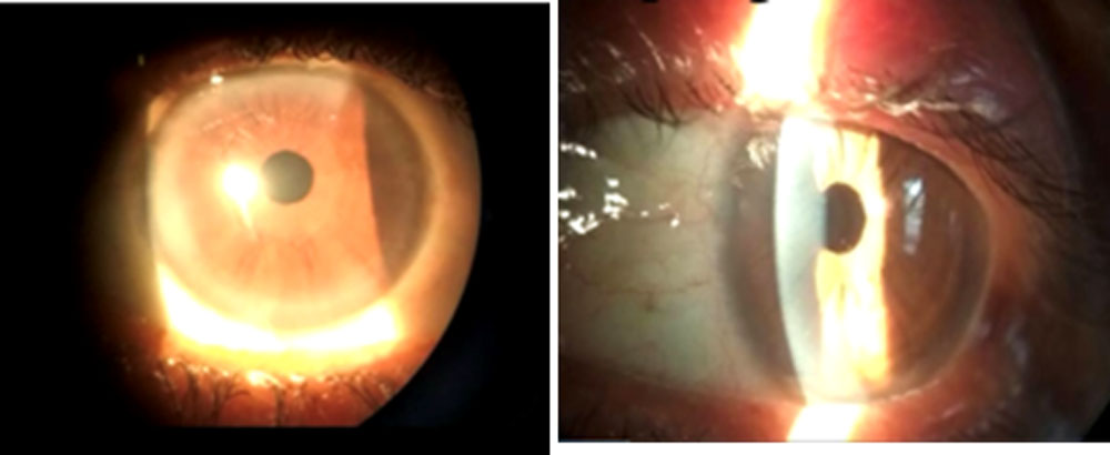
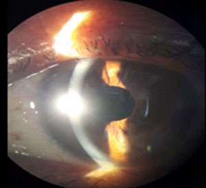
Figure: Post operative day 1 slit lamp examination
OUTCOME
DEMOGRAPHIC PROFILE
| NO OF CASES | 196 |
| Sex [number of patients (%)] | |
| FEMALE | 128(65.3%) |
| MALE | 68(34.7%) |
| AGE( YEARS) | |
| MEAN(SD) | 63.73 + 3.848 |
| MEDIAN | 64.00 |
| CATARACT DENSITY[number of cases (%)] | 196
|
| 1 and 1+ | 17(8.5%) |
| 2 and 2+ | 57(29.1%) |
| 3 and 3+ | 54(27.6%) |
| 4 and 4+ | 68(34.7%) |
| PREOPERATIVE VISION
>6/9 7(3.6%) 6/12 45(23%) 6/18 18(9.2%) 6/24 18(9.2%) <6/36 108(55.1%) Total 196 |
|||||||||||||
|
POST OPERATIVE VISION |
|||||||||||||
| D1 D7 D30 D60 D90
≥ 6/9 175(89.3%) 180(91.8%) 180(91.8%) 186(94.9%) 186(94.9%) 6/12- 6/24 15(7.7%) 11(5.6%) 16(8.2%) 10(5.1%) 10(5.1%) < 6/24 9(4.6%) 5(2.6%) 0 0 0 TOTAL 196 196 196 196 196 CELLS |
|||||||||||||
| NO. OF CASES
OCC + ++ +++ ++++ D1 47 3 10 0 7 D7 3 0 10 0 0 D30 0 0 0 0 0 D60 0 0 0 0 0 D90 0 0 0 0 0 |
|||||||||||||
| FLOATERS
NO. OF CASES D1 20 D7 15 D30 13 D60 0 D90 0 MEAN IOP Number of Days NO. OF CASES mm Hg (SD) D7 196 12.96+ 1.414 D30 196 15.25+ 1.243 D60 196 14.04+ 1.064 D90 196 13.6+ 1.500 |
|||||||||||||
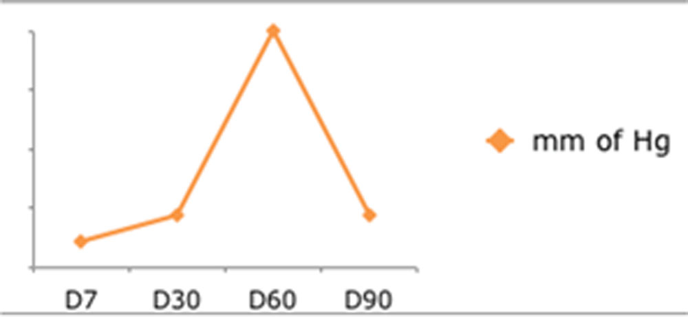
DISCUSSION
Immediate post operative foggy vision, appearance of floaters, postoperative IOP rise were issues that made us apprehensive about the procedure. None of the patients complained of foggy vision, none had rise of IOP and only 20 complained of floaters on day 1 (D1) postoperatively which decreased to 15 and 13 after 7 and 30 days postoperatively respectively and finally the number came down to 0 after 60 (D60) and 90 (D90) days postoperatively. This data provides an assurance and relief about the promising future of this kind of surgery. Another rewarding point is that none of the patients had to resort to instillation of any eye drop during the postoperative period for whatsoever reason.
There has been lot of debates regarding usefulness of perioperative antibiotics in prevention of post-cataract endophthalmitis. The ESCRS study is strong proof that intracameral injection of cefuroxime is responsible for five-fold decrease in post-cataract endophthalmitis18. The study was done across Europe and the results were so astounding that it had to be stopped midway to offer the control group the benefit of the result. Intracameralmoxifloxacin is used as a prophylaxis in India, Canada and many south American countries. A recent large study published by Aravind Eye Hospital confirms its efficacy in prevention of post-cataract endophthalmitis19. Injection of moxifloxacin into the vitreuos cavity should serve the same purpose as done by intracameralmoxifloxacin. Though we did not encounter any endophthamitis in our 200 cases, it should not be taken as conclusive as the strength of this study is very small
Ocular hypertension following systemic, topical, subtenon and intravitreal steroid is a known phenomenon in steroid responders.1-11 It is easy to control with systemic or topical medication as one needs to discontinue it.12,14 In cases of sub tenon and intravitreal, this facility is unfortunately unavailable as long acting steroid is injected for once. Since TA is degradable inside the eye, the initial rise in IOP could be controlled effectively by antiglaucoma medication till the half life of the drug is over In about 3% of cases, and in particular when there is a family history of glaucoma and/or chronic use of steroid (at least 4 years), the ocular hypertensive response has been shown to be irreversible14,16,17 The management of such cases is no different from that for POAG. If the situation demands, urgent removal of intravitreal TA, a simple 25G PPV would suffice to get rid of the complication13,14,15.
Appearance of floaters and not so ‘wow’ vision immediately after surgery may be a disappointment to the patient unless he or she is thoroughly counselled before the surgery. Fortunately we did not have this problem much(ref).
Failure to give the injection on first attempt was an initial hiccup of this procedure. Since the procedure is blind, there is some learning curve. Though we had to repeat the injection on three occasions in one patient, but it was successful in first attempt in 70% cases. Overall it was successful in all cases.
Intraoperative iris prolapse and bleeding were not difficult to manage as the procedure of phacoemulsification is a closed chamber one.
CONCLUSION
The study clearly demonstrates that this procedure is very effective and safe. If further randomized controlled studies can prove its usefulness, the procedure could be a boon to the working rural population of India for whom putting eye drop during working hours is not only difficult, but unsafe as well. If they could be freed from this burden, their post operative lives will be easier.
REFERENCES
1. Bernstein HN, Mills DW, Becker B. Steroid-induced elevation of intraocular pressure.Arch Ophthalmol 1977; 9:1075–1080.
2. Garbe E, Lorier J, Boivin JF, Siussa S. Inhaled and nasal glucocorticoids and the risk of ocular hypertension or open-angle glaucoma. JAMA 1997; 277: 722–727.
3. Cubey RB.Glaucoma following the application of corticosteroids to the skin of the eyelids.Br J Dermatol 1976; 95:207–208.
4. Garrott HM, Walland MJ.Glaucoma from topical corticosteroids to the eyelids.ClinExpOphthalmol 2004; 32:656–657.
5. Kalina RE.Increased intraocular pressure following subconjunctival corticosteroid administration.Arch Ophthalmol 1969; 81:78–90.
6. Behbehani AH, Owayed AF, Hijazi ZM, et al. Cataract and ocular hypertension in children on inhaled corticosteroid therapy. J PedOphthalmol Strabismus 2005; 42:23–27.
7. Smithen LM, Ober MD, Maranan L, Spaide RF. Intravitreal triamcinolone acetonide and intraocular pressure. Am J Ophthalmol 2004; 138:740–743. This article determined the prevalence of IOP elevation following intravitreal triamcinolone injection in patients with and without glaucoma. It allows the clinician to anticipate the elevated IOP approximately 100 days after injection.
8. Singh IP, Ahmad SI, Yeh D, et al. Early rapid rise in intraocular pressure after intravitreal triamcinolone acetonide injection. Am J Ophthalmol 2004; 138: 286–287.
9. Gillies MC, Simpson JM, Billson FA, et al. Safety of an intravitreal injection of triamcinolone: results from a randomized clinical trial. Arch Ophthalmol 2004; 122:336–340.
10. Armaly MF.Statistical attributes of the steroid hypertensive response in the clinically normal eye. Invest Ophthalmol Vis Sci 1965; 4:187–197.
11. Becker B. Intraocular pressure response to topical corticosteroids. Invest Ophthalmol Vis Sci 1965; 4:198–205.
12. Weinreb RN, Polansky JR, Kramer SG, BaxterJD. Acute effects of dexamethasone on intraocular pressure in glaucoma. Invest Ophthalmol Vis Sci1985; 26(2): 170–175. (The chronic corticosteroid response resolves in 1–4 weeks, whereas the rare acute response may resolve within a few days of steroid cessation. – this is the exact sentence of reference)
13. Herschler J. Increased intraocular pressure induced by repository corticosteroids. Am J Ophthalmol1976; 82: 90–93.
14. J. P KERSEY AND D C BROADWAY.Corticosteroid-induced glaucoma: a review of the literature.Eyevolume20, pages407–416 (2006)
15. Jones R III, Rhee DJ. Corticosteroid-induced ocular hypertension and glaucoma: a brief review and update of the literature. CurrOpinOphthalmol. 2006;17(2):163-167.
16. Francois J. Corticosteroid glaucoma. Ann Ophthalmol1977; 9: 1075–1080.
17. Espildora J, Vicuna P, Diaz E. Cortisone-induced glaucoma: a report on 44 affected eyes. J FrOphthalmol1981; 4: 503–508.
Leave a Comment