

Dr.Zelda Dadachanji, Dr.Rohit Shetty,Dr.Pooja Khamar
Introduction
Myopia is one of the main causes of visual impairment throughout the world.Transepithelial Photorefractive keratectomy (TransPRK) ,Small incision lenticule extraction (SMILE) and wavefront guided laser assisted in situ keratomileusis (WFG LASIK) are some of the latest refractive procedures in vogue for correction of low to high myopia.
The transepithelial PRK was launched in 2007 as the Schwind Amaris laser platform (Schwind eye-tech-solutions GmbH & Co. KG) while its smart-pulse technology technique was introduced in 2017.1The uniqueness of this technique is that it removes the corneal epithelium and stroma in a single step with one ablation profile and without flap-related complications.2
Wavefront–guided LASIK is performed using a pupiltracking system and iris registration technique that are intended to obtain a more accurate alignment and correction in corneal refractive surgery. 3
Small Incision Lenticule Extraction (SMILE), introduced in 2011, is a flap-free intrastromal laser assisted refractive surgery technique for the correction of myopia and myopic astigmatism with a reported high efficacy, predictability, stability, and safety for myopic treatments and lower amount of induced corneal aberrations. 4
TransPRK ,SMILE and WFG LASIK use different algorithms to achieve the same refractive change in a patient. Thus, TransPRK, WFG LASIK and SMILE may yield differentialaberrometric outcomes.5
Several studies have reported similar UDVA and CDVA after WFG LASIK and SMILE. The two procedures have also been found to be comparable in their high efficacy and safety.6,7,8 In one study WFG LASIK and SMILE had similar aberrometric outcomes but WFG LASIK had better postoperative visual acuity.8 However no study has made comparisons regarding the visual outcomes between TransPRK, WFG LASIK and SMILE.
The aim of this study was to assess the visual outcomes between TransPRK, WFG LASIK and SMILE (preoperative and 6 month follow-up). It also aimed to study the change in corneal aberrations afterTransPRK, WFG LASIK and SMILE in eyes having low to high myopia and similar scan diameters for aberrometry.
Methods
This was a prospective, interventional and longitudinal study approved by the ethics committee of Narayana Nethralaya Eye Hospital, Bangalore, India. A written informed consent was obtained from all the patients. The study followed the tenets of declaration of Helsinki.
Inclusion criteria were stable refraction (less than -6D equivalent refraction withastigmatism not more than -3D) for a period of one year (change less than 0.25D). In all the eyes, calculated residual stromal thickness was greater than 250 μm.
Exclusion criteria had patients with less than 480 μm central corneal thickness (CCT) or history of keratoconus, diabetes, collagen vascular disease, pregnancy, breastfeeding, any known ocular pathology and any ocular surgery or trauma.
Patients discontinued wearing contact lenses for at least 2 weeks prior to any measurement and surgery
All patients underwent assessment of uncorrected (UDVA) and corrected (CDVA) distance visual acuity preoperatively and postoperatively
Corneal tomography and Corneal wavefront aberration were measured by a rotating Scheimpflug Camera (Pentacam; OCULUS OptikgerateGmbh, Wetzlar, Germany ). Higher order aberrations (HOAs) of the front surface of the cornea were analyzed with Zernike polynomials (6th order and diameter of 4 mm) from the total cornea preoperatively and 6 months postoperatively
A total of 30 eyes( 16 patients) underwent TransPRK on the SCHWIND AMARIS 1050 laser (SCHWIND eye-tech-solutions GmbH, Kleinostheim, Germany)
A total of 30 eyes (14 patients) underwent SMILE with the Visumax femtosecond laser system (Carl Zeiss Meditec, Jena, Germany).
A total of 30 eyes (19 patients) underwent WFG LASIK with STAR S4IR excimer laser and iDesign system for treatment calculation (Abbott Medical Optics, Abbott Park, IL).
Trans PRK was performed on SCHWIND AMARIS 1050 laser (SCHWIND eye-tech-solutions GmbH, Kleinostheim, Germany. Ablations were based on aberration-free algorithms calculated using ORK-CAM software. Before ablation, all patients’ examinations were tested by the statistic cyclotorsion control (SCC) and dynamic cyclotorsion control (DCC) was used through the surgery. For each treatment, the epithelium thickness profile that 50 μm centrally and 60 μm peripherally .The target refraction was emmetropia in all eyes. After surgery, the cornea was irrigated with a cool balanced salt solution and aacuvail soaked soft bandage contact lens was applied for two to three days. The contact lens were removed once the epithelial healing was completed.
WFG-LASIK procedure, flap diameter, optical diameter and flap thickness was set to 9 mm, 6 mm and 100 μm, respectively. After flap creation with the Intralase femtosecond laser (Abbott Medical Optics, Santa Ana, CA), the exposed stromal bed was ablated with the STAR S4IR excimer laser.
In the SMILE procedure, the cap diameter, lenticule diameter and cap thickness was set to 7.7 mm, 6.0 mm and 110 μm, respectively. Laser-cut energy index was preset at 170 nJ. Spot spacing was 2 μm for creation of the lenticule side-cut and 4.5 μm for creation of the lenticule. Track distance was 3 mm. After the incision was made, the lenticule was cut with the laser and extracted manually. Intra-operative cap repositioning was performed in all the SMILE eyes
One drop of moxifloxacin hydro-chloride 0.5% (Vigamox®, Alcon Laboratories, Inc., Texas, USA) was instilled in both eyes after surgery. Routine postoperative regimen included moxifloxacin hydrochloride 0.5% eye drops (Vigamox®, Alcon Laboratories, Inc., Fort Worth, TX, USA) four times a day for 1 week, topical 1% Fluorometholone eye drops (Flarex®, Alcon Laboratories, Inc., Fort Worth, TX, USA) in tapering doses up to 1 month and topical lubricants (Optive, Allergan Inc., Irvine, USA) up to 3 months. The above regimen was the same for all TranPRK , WFG LASIK and SMILE eyes.
Statistical analyses
The normality of distribution was assessed for all the continuous variables. Non-parametric statistical tests were used when the variables weren’t normally distributed. Paired Wilcoxon test was used to compare preoperative with postoperative data within a treatment group. The change in variables due to surgery was calculated and analyzed with independent samples Mann-Whitney test. Factors like keratometry, visual acuity, pachymetry and anterior surface corneal aberrations from Pentacam were obtained and analysed. The median with range of the variables was calculated. All statistical analyses were performed usingMedCalc v18 (MedCalc Inc., Ostend, Belgium). A p-value less than 0.05 was considered statistically significant.
Results:
A total of 90 eyes were included in this study. Each group included 30 eyes. All eyes completed the six-month follow-up. At 6 months, safety index (mean ± standard deviation) of TransPRK, WFG LASIK and SMILE was 1±0.01, 0.99±0.04 and 0.90±0.10, respectively (p<0.0001). Figure 1a, 1b and 1 c show changes in corrected distance visual acuity (CDVA) 6 months after TransPRK, WFG LASIK and SMILE respectively
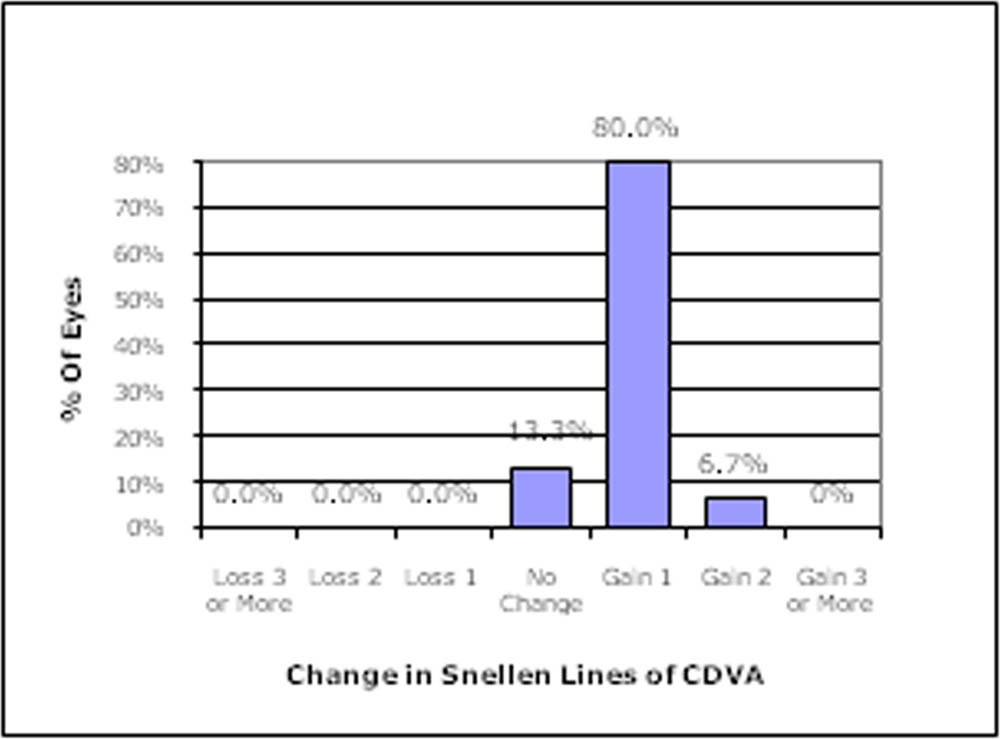
Figure 1a (TransPRK)
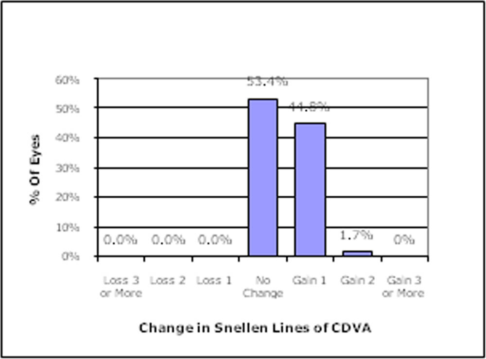
Figure 1 b (WFG LASIK)
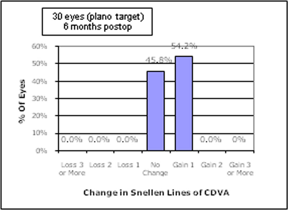
Figure 1 c (SMILE)
Also, efficacy index of TransPRK, WFG LASIK and SMILE was 1.18±0.05, 1.01±0.04 and 1.13±0.14, respectively (p<0.0001).
At 6 months, 100% of eyes achieved a UDVA of 20/20 or better in TransPRK, WFG LASIK and SMILE groups.
Further,73.33%,74.14% and 45.83% of the eyes in WFG LASIK and SMILE group achieved an
UDVA of 20/16 at 6 month follow-up, respectively.
Figure 2a, 2b and 2 c show the Cumulative percentage of eyes achieving uncorrected distance visual acuity (UDVA) 6 months postoperatively after TransPRK, WFG LASIK and SMILE respectively.
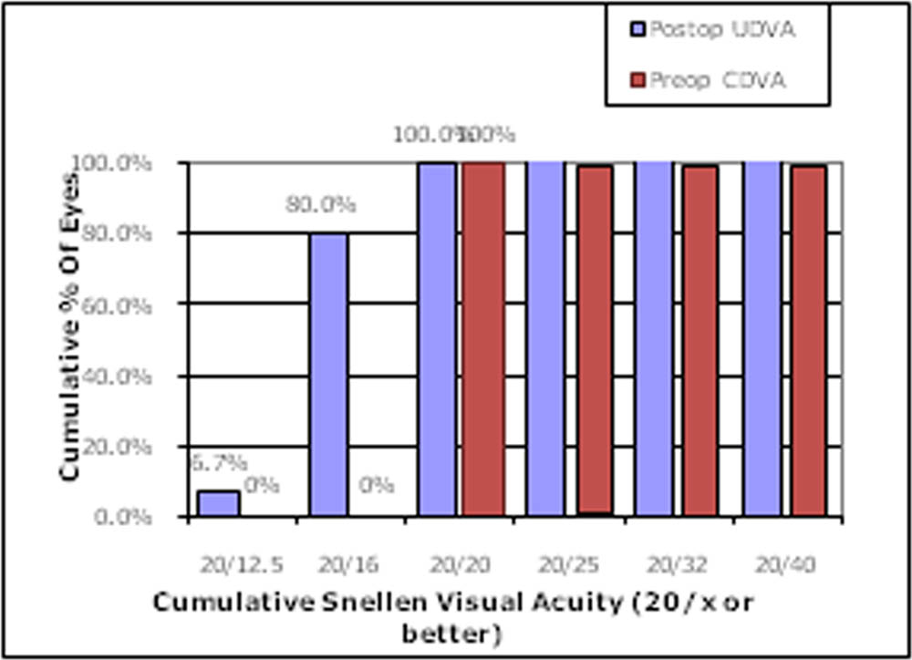
Figure 2a (TransPRK)
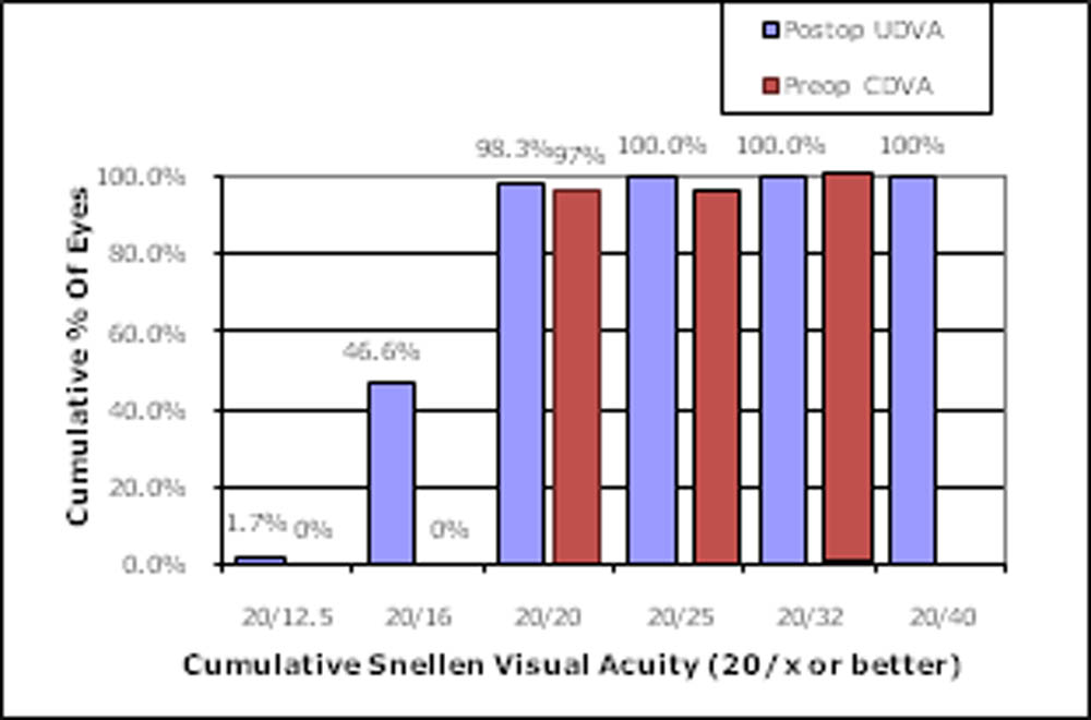
Figure 2b (WFG LASIK)
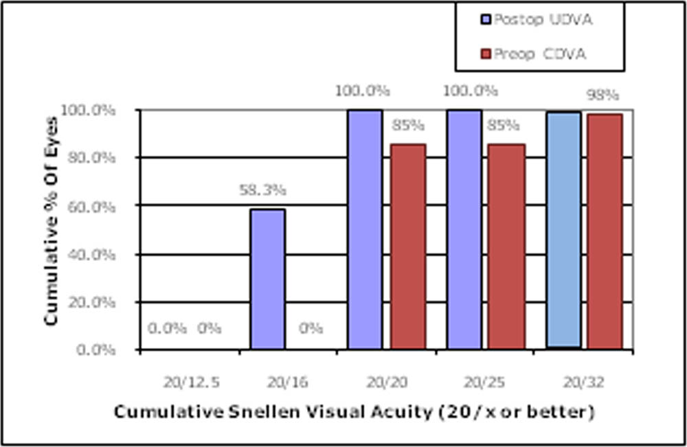
Figure 2c (SMILE)
Table 1.shows a summary of the difference between the preoperative and post operative change in RMS of anterior corneal surface HOA, spherical aberration and coma.
| TRANSPRK | WFG LASIK | SMILE | ||||
| Front HOA Pentacam
(RMS, μm) |
0.11(0.88,0.02) | 0.18(0.1,0.37) | 0.09(0.03,0.18) | 0.15(0.06,0.26) | 0.10(-0.08,0.16) | 0.16(0.08,0.37) |
| Front SA Pentacam
(μm) |
0.05(-0.09, 0.11) | -0.04(-0.14, 0.09) | 0.04(-0.025,0.08) | 0.03(-0.019,0.11) | 0.04(-0.095,0.09) | -0.001(-0.076,0.10) |
| Front coma
Pentacam (RMS, μm) |
0.08(0.02,0.15) | 0.12(0.02, 0.26) | 0.064
(0.013,0.129) |
0.130
(0.034,0.250) |
0.064(0.021,0.134) | 0.130(0.017,0.34) |
Change in mean higher order aberration RMS (preop – post op)for TransPRK, WFG LASIK and SMILE was -0.07, -0.06 and -0.06 respectively. Change in mean spherical aberration (preop – post op)was 0.09, 0.01and 0.041forTransPRK, WFG LASIK and SMILE respectively.Change in coma (preop – post op)was -0.04, -0.07and -0.07 forTransPRK, WFG LASIK and SMILE respectively. All the above changes were statistically similar
Discussion:
This study aimed to study the visual outcomes in terms of visual acuity, safety index and efficacy index of TransPRK, WFG LASIK and SMILE. At 6 months postoperatively, all of the eyes achieved the UCDV of 20/20 or better visual acuity. No patient lost two or more lines of CDVA in either of the procedures. 80% patients showed Gain of 1 line at 6 months postoperatively in TransPRK as compared to 44.8% in WFG LASIK and 54.2% in SMILE. We also studied the primary changes in corneal aberrations after TransPRK, WFG LASIK and SMILE in eye with myopic refractive error. The differences noted in terms of anterior corneal surface aberrometry between TransPRK, WFG LASIK and SMILE were not significant.
Conclusions:TransPRK is a safe and effective surgical option in the treatment of mild and moderate myopia, and showed acceptable safety and efficacy which was comparable to WFG LASIK and SMILE.
References:
- Aslanides IM, Kymionis GD. Trans advanced surface laser ablation (TransPRK) outcomes using SmartPulseTechnology. Cont Lens Anterior Eye 2017; 40:42–46
- Fadlallah A, Fahed D, Khalil K, Dunia I, Menassa J, El Rami H, et al.Transepithelial photorefractive keratectomy: clinical results. J Cataract Refract Surg. 2011;37(10):1852–7.
- Zhang J, Wang Y, Chen X. Comparison of Moderate- to High-Astigmatism Corrections Using WaveFront-Guided Laser In Situ Keratomileusis and Small-Incision Lenticule Extraction. Cornea. 2016; 35(4):523-30.
- Aliódel Barrio JL, Vargas V, Al-Shymali O, Alió JL. Small incision lenticule extraction (SMILE) in the correction of myopic astigmatism: outcomes and limitations – an update. Eye and Vision. 2017;4:26. doi:10.1186/s40662-017-0091-9.
- Khalifa MA, Ghoneim A, ShafikShaheen M, Aly MG, Piñero DP. Comparative Analysisof the Clinical Outcomes of SMILE and Wavefront-Guided LASIK in Low and ModerateMyopia. J Refract Surg. 2017;33(5):298-304.
- Kataoka T, Nishida T, Murata A, et al. Control-matched comparison of refractive and visual outcomes between small incision lenticule extraction and femtosecond laser-assisted LASIK. Clinical Ophthalmology (Auckland, NZ). 2018;12:865-873. doi:10.2147/OPTH.S161883.
- Chen X, Wang Y, Zhang J, Yang SN, Li X, Zhang L. Comparison of ocular higher-order aberrations after SMILE and Wavefront-guided Femtosecond LASIK for myopia. BMC Ophthalmol. 2017;17(1):42
- Shetty R, Matalia H, et al Wavefront guided LASIK has comparable ocular and corneal aberrometric outcomes but better visual acuity outcomes than SMILE in myopic eyes. J Refract Surg. 20182018;34(8):527-532
Leave a Comment