

Dr.Sneha Giridhar, G18165, Dr.Rama Krishnan R, Dr.Shivkumar Chandrashekharan, Dr.Padmavathy Maharajan
Abstract
Aim: To assess ganglion cell complex (GCC) loss and retinal nerve fibre (RNFL) changes in chiasmal compressive lesions.
Methods: Prospective observational study.18 patients with chiasmal compression between 2017 and 2018 were included in the study. Cirrus SD-OCT macular cube 512×128, RNFL scan protocols and 30-2 visual field were performed in all patients. Age matched controls were included for comparison. Correlation between GCC, RNFL and Visual fields were done. Data analysed using SPSS 16.0. P value< 0.05 statistically significant.
Results: Average GCC thickness was 63.14µm in patients and 83.81µm in controls (p<0.05). Average RNFL thickness was 81.78µm in patients and 92.14µm in controls. Effect size was significant for average GCC thickness (d=2.63) when compared to average RNFL (d=0.85).Three patients with no visual field defect showed significant loss in GCC.
Conclusion: GCC loss using SD-OCT is a very sensitive tool to detect optic nerve damage in chiasmal lesions compared to RNFL.
Keywords :- Ganglion cell complex, RNFL, chiasmal compression, visual fields
Body
Introduction
Several methods are available and practiced over the years for assessment of optic nerve function in chiasmal compressive lesions. Traditionally visual field assessment using the Humphrey Field Analyser has been the gold standard for assessment of optic nerve dysfunction in chiasmal lesions. With the development of optical coherence tomography more objective assessment of optic nerve damage is possible. Recently, there has been a few retrospective studies to assess the ganglion cell complex(GCC) loss and RNFL changes using SD OCT in eyes with chiasmal disorders.[1,2,3] More recently high definition OCT algorithms have been established so as to enable automated evaluation of the macula GCC.[4]
The goal of our prospective study was to assess GCC loss and RNFL changes in chiasmal compressive lesions. We also compared average GCC thickness with RNFL thickness and visual field loss to see whether evaluation of GCC thickness is more sensitive than RNFL thickness and visual field loss to detect early chiasmal compression.
Methods
A total of 38 eyes of 19 patients who presented with chiasmal compression confirmed by MRI imaging were recruited for this study. An informed consent was obtained from all the enrolled patients. Evaluation and clinical examination was performed at the neuro ophthalmological clinic of Aravind Eye Hospital, Tirunelveli. A set of age matched was included for GCC analysis and RNFL comparison. The study was approved by the ethics committee of our hospital.
Neuro-ophthalmological examination: This included assessment of best corrected visual acuity, color vision wherever possible with the Ishihara chart, automated visual field assessment using the Humphrey Visual Field Analyser (Carl Zeiss Meditec, Jena, Germany). Macular GCC and peripapillary RNFL was assessed using the spectral domain OCT (Cirrus HD-OCT 5000, Carl Zeiss Meditec, Dublin, CA). For GCC analysis macular cube 512*128 line scan centered on the fovea was used.
RNFL analysis was performed using the optic disc cube (200*200line scan)
Age matched controls also was subjected to a similar examination using the CIRRUS 500 SD OCT. Average and quadrant wise comparison of GCC and RNFL was done between subjects and controls. The average RNFL and GCC thickness were correlated.
Statistical analysis plan
Mean (SD) and frequency (percentage) was used to describe the summary data. Two sample t test / Mann Whitney test was used to compare the mean difference of the ocular parameters between the groups. Categorical variables was compared by Fisher’s exact test / Chi squared test. P value less than 0.05 was considered as statistically significant. All the statistical analysis was performed by STATA 11.1 (Texas).
Results
A total of 38 eyes of 19 patients with chiasmal compression were included in the study.
The mean age of presentation was found to be 49 ±14years. The range of presentation was between 21 and 65 years. Majority of the patients who presented were above 50years of age accounting for 52.6% of study population (Figure1).
The levels of visual acuity are shown in Figure 2. It’s seen that 39.4% of eyes had a near normal visual acuity ranging between 6/6 – 6/9. Sixteen eyes presented with visual acuity ranging between 6/12 and 6/60 forming 42.1% of the eyes studied in the population. 7 eyes presented with very poor visual acuity of <6/60 (18.4%).
The different types of lesions causing chiasmal compression diagnosed on MRI described in Figure 3.Of the 19patients, 9 patents presented with pituitary macroadenoma which represented 47.3% of the study population. This was followed by Meningioma and Craniopharyngioma.
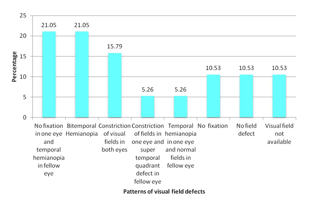
There were different patterns of visual field defects (Figure 4) obtained on HFA 30-2. The fields obtained were in relation to the visual acuity at the time of presentation. Eyes with poor visual acuity were either not able to fixate on the target or showed constriction of fields. The fellow eye was found to have a hemianopic visual field or a supero temporal quadrantic field defect. The above field patterns are due to the decussation of nasal fibres at the chiasma giving rise to temporal field defects. However, there were 2 patients who did not have field defects. A dilated fundus examination with 90D of 38 eyes showed majority of the eyes (63%) with compressive lesions showed temporal pallor of the optic disc.
Optical Coherence Tomography Analysis
Table 1 shows a comparison of average RNFL and quadrant wise measurements between 19 patients and 19 age matched controls. From the table we infer that there is a significant thinning in average RNFL in patients (83.71±16µ) compared to that of controls (93.7±10µ).
We also observe a generalized thinning in all quadrants with maximum thinning seen in the temporal quadrant (49.8±8µ), followed by inferior (105.7±23µ) each showing a significance of p<0.001.
On assessment of average GCC and other GCC parameters across two groups (Table 2), average GCC thickness was 63.34±11µ in patients with compression and 84.18±4µ in controls (p<0.001). Average GCC thinning across all individual sectors were statistically significant. Maximum thinning was observed in the inferonasal quadrant followed by the superonasal quadrant.
The effect size was calculated to evaluate the strength of the difference between patients and controls in various parameters. The effect size was largest for GCC thickness (d=2.4) followed by RNFL thickness (d=0.73).It is also observed that the effect size is greater for nasal GCC thinning as compared to other segments.
We compared RNFL and GCC parameters between 4 patients who showed a bitemporal hemianopia on HFA with their age matched controls (Table 3.1). We see a significant thinning of GCC, especially in the nasal sectors i.e, superior nasal 62.3±9µ and inferior nasal 62.5±6µ compared to their controls(88.3±3µ and 89.1±5µ)respectively. The effect size of -4.5 for the inferonasal sector makes it a sensitive parameter.
2 of the 19 patients with chiasmal compression, did not have visual field defects on HFA (Table 3.2). The average RNFL thickness was within the normal range for their age (93.2±6µ among patients and 88.5±6µ in controls). However, the average GCC was found to be significantly thinner among the patients with compression (70.2±5.3µ) when compared to their controls (82.5±2µ).
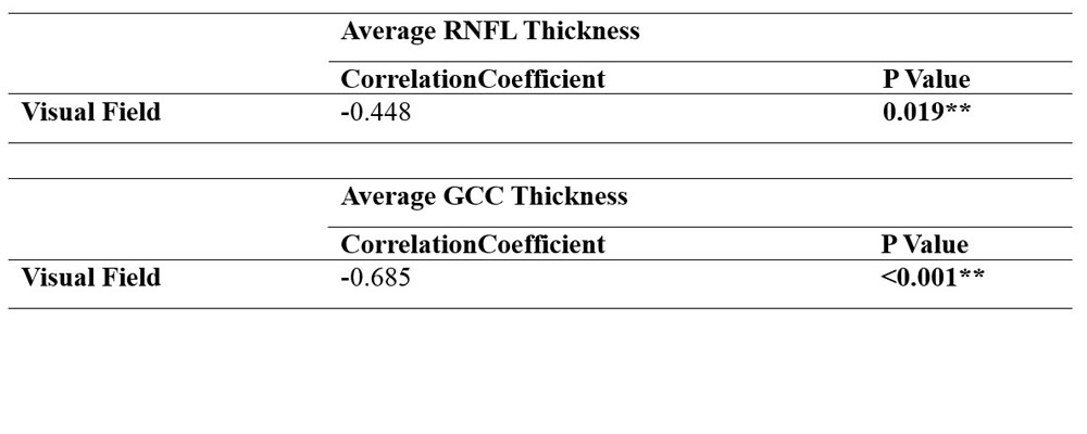
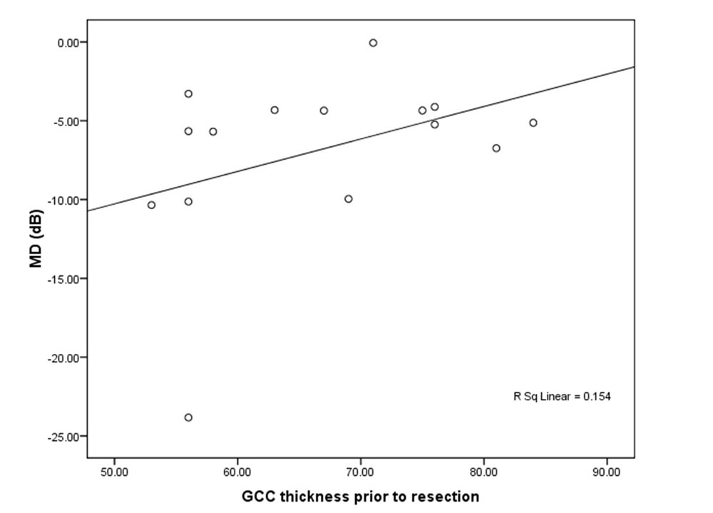
Visual field mean deviation (MD) was correlated to average GCC and RNFL thickness in all patients (Table 4). The graph (Figure 5) shows a significant correlation between mean deviation and GCC (p<0.001) and RNFL (0.019) thickness.The graph also shows that GCC thickness was more strongly correlated with MD (R2=0.46) followed by RNFL thickness (R2=0.20) .
Discussion
This was a prospective observational study of 19 patients. 19 age matched controls were enrolled for RNFL and GCC thickness comparison.
Patients eligible for this study had a history of chiasmal compression documented on MRI imaging or computed tomography.
The mean age of presentation was 49±14yrs and the range varied between 21 to 65 yrs. This is similar to the demographic profile reported by Marisa G Tieger, et al, in a retrospective analysis of 23 patients the mean age of presentation was 52±16yrs.[1] Similar observation was also noted by Shinji Ohkubo et al with a mean age of presentation of 46.3±16 yrs.[6] In the present study 52.6% of the patients were above the age of 50yrs indicating that chiasmal lesions are probably more common in middle age and elderly and its incidence increases as the age advances.
Visual field analysis was not recorded for patients who had a poor visual acuity because they were not able to fix on the target. In our study, 4 of 19 patients presented with bitemporal hemianopia. Four patients showed constriction of visual fields in the affected eye and a homonymous temporal hemianopia in the fellow eye (21.05%). Two patients of the 19 subjects with chiasmal compression did not have field defects. Such varied presentation was also seen in the study by Marisa Tiegra et al where majority of the patients(6) presented with bitemporal hemianopia and there were 6 patients who did not have field defects.[1] We could therefore say that bitemporal hemianopia is a common visual field defect presented among chiasmal compressive lesions. Although, there were patients with normal visual fields in the presence of chiasmal compression.
The goal of this study was to evaluate whether the assessment of GCC thickness using the SD OCT a sensitive tool to identify early damage to the anterior visual pathway following chiasmal compression. For this purpose, the study compared the GCC thickness between patients and controls. Simultaneously the RNFL thickness was also estimated and compared with controls.
Changes in RNFL and its significance
The present study showed a significant thinning of average RNFL (p=0.002). The average RNFL was 83.71±16µ in the subjects compared to 93.71±10µ among controls. The average RNFL was related to visual field mean deviation and a significant correlation was observed (p=0.015). Marisa G Tieger et al obtained similar results in a study.[1] However the observations made by Charlotte et al demonstrated a reduced RNFL thickness in most eyes but not in all eyes and found no correlation between pattern of RNFL loss and visual field defects.[5]
Changes in GCC and its significance
In this study there was significant thinning of GCC parameters in the patient population when compared to controls. The average GCC obtained was 63.34±14µ among subjects and 84±18µ among age match controls and this was statistically significant (p<0.001). In the study by Marisa G Tieger et al, the GCC thickness in patients was 67±9µ when compared to controls (86±5µ).[1] This study also showed that there was greater and significant thinning of the nasal segments especially the inferonasal segment of the macular region (p<0.002). Approximately 50% of the retinal ganglion cells are located within 4.5mm of the fovea and the retinal ganglion cell axon fibers nasal and temporal to the fovea converge around the optic nerve head. Topographical GCC thinning can be revealed using macular ganglion cell complex measurement.[4] In the present study the nasal GCC thickness was found to be 124.9± 25µ in patients and 172.5±8µ in controls. The temporal GCC thickness was found to be 129.8±25µ compared to that of controls (165.6±7µ.) With a greater effect size, d=2.6, the nasal segment thinning was more significant than the thinning of the other segments. These results can be further substantiated by a hypothesis put forward by Montiero et al. in their study. They concluded that with the exception of the nasal area of the optic disc all other sectors receive retinal nerve fibers from both the nasal and temporal hemi retina therefore patients with visual field defects might have loss of RNFL in all quadrants showing lack of its specificity compared to macular thickness reduction which would be exclusively nasal to the fovea with a higher specificity as observed in this study.[3] In the four patients who presented with bi-temporal hemianopia, bi-nasal thinning was observed similar to previous studies.
Studies have also shown GCC thinning to precede visual field loss. Marisa Teiger et al found 6 patients of the 23 patients to have no visual field defect. OCT analysis showed significant GCC and RNFL thinning.[1] Similarly in our study, 2 patients with chiasmal compression were found to have no visual field defects. The average RNFL thickness among these subjects was 93.2±6µ while that of age matched controls 88±5µ. However a significant thinning was obtained in all GCC parameters. The average GCC thickness found to be significantly reduced (p<0.001). The average GCC thickness among subjects was 70.25±5µ when compared to controls (82.5±2µ). Therefore assessment of GCC thickness is a sensitive indicator for early damage to the anterior visual pathway in chiasmal compression
Comparing GCC and RNFL
Therefore both GCC thickness and RNFL thickness parameters are useful in evaluating optic nerve injury. However, a greater sensitivity has been obtained for average GCC (effect size, d=2.4) than for average RNFL (d=0.73).
Average GCC and RNFL were correlated to visual field mean deviation (MD). Both average GCC (p<0.001) and RNFL (p=0.019) had a significant correlation. However, GCC thickness strongly correlated with MD (r2 = 0.46) followed by RNFL thickness ((r2= 0.20).
This is in agreement with a study by Marisa Tieger et al whose GCC thickness (r2 = 0.25) was found to be more significant than RNFL (r2= 0.15) in relation to visual field MD.[1]
Conclusion
The most common etiology causing chiasmal compression is pituitary macroadenoma presenting clinically as bitemporal hemianopia on visual field analysis.
Analysis of RNFL and GCC parameters using SD-OCT showed significant GCC and RNFL loss in eyes with chiasmal compression compared to controls with average GCC showing a greater significance. Moreover, GCC showed a stronger correlation with mean deviation compared to average RNFL.
To conclude, in addition to visual field analysis by HFA, we have in our study showed that GCC thickness can be effectively used as an objective measurement of optic nerve damage.
References
- Tieger MG, Hedges TR, Ho J, Erlich-Malona NK, Vuong LN, Athappilly GK, et al. Ganglion Cell Complex Loss in Chiasmal Compression by Brain Tumors. J Neuroophthalmol. 2017;37:7–12.
- Yang L, Qu Y, Lu W, Liu F. Evaluation of Macular Ganglion Cell Complex and Peripapillary Retinal Nerve Fiber Layer in Primary Craniopharyngioma by Fourier-Domain Optical Coherence Tomography. Med Sci Monit. 2016;22:2309–14
- Monteiro MLR, Costa-Cunha LVF, Cunha LP, Malta RFS. Correlation between macular and retinal nerve fibre layer Fourier-domain OCT measurements and visual field loss in chiasmal compression. Eye (Lond). 2010;24:1382–90.
- Kardon RH. Role of the macular optical coherence tomography scan in neuro-ophthalmology. J Neuroophthalmol. 2011;31:353–61.
- Johansson C, Lindblom B. The role of optical coherence tomography in the detection of pituitary adenoma. Acta Ophthalmol. 2009;87:776-9
- Ohkubo S, Higashide T, Takeda H, Murotani E, Hayashi Y, Sugiyama K. Relationship between macular ganglion cell complex parameters and visual field parameters after tumor resection in chiasmal compression. Jpn J Ophthalmol. 2012;56:68–75
Tables and figures
Figure 1: Age Distribution in the population
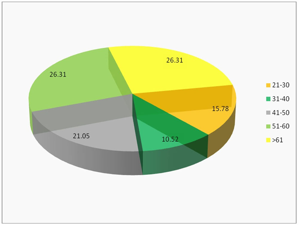
Figure 2: Distribution of BCVA in the study population
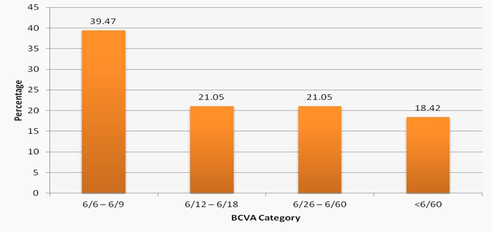
Figure 3: Distribution of the type of tumors in the study
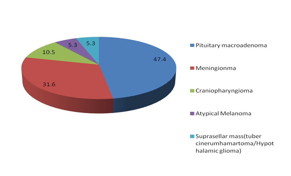
Figure 4: Distribution Of Patterns Of Visual Field Defects among patients
Optical Coherance Tomography Analysis
Table1: Assessment of Average RNFL and other RNFL parameters across two groups
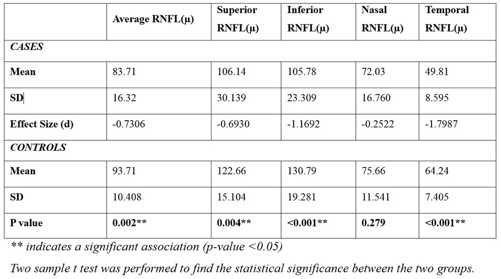
Table 2: Assessment of Average GCC and other GCC parameters across two groups
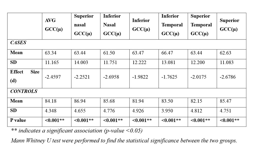
Table 3.1: Assessment of Average GCC and other GCC parameters across two groups- Bitemporal Hemianopia and its controls
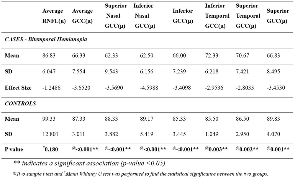
Table 3.2: Assessment of Average RNFL, Average GCC and other GCC parameters between two groups – No VF defect and its control.
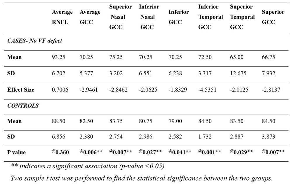
Table 4: Correlation between Visual Field (MD), Average RNFL and Average GCC Thickness
Leave a Comment