

Dr.DEBASIS HAKRABARTI, Dr.ADHIKARI APURBA
INTRODUCTION:
Late-onset bleb leaks after glaucoma filtration surgery are increasingly becoming common with the widespread use of anti-fibrotics like Mitomycin C. The risks of leaking blebs include ocular hypotony and maculopathy, shallow anterior chamber and its sequel; and sight-threatening complications like endophthalmitis. Although many treatment options have been proposed, successful closure of late bleb leaks remains a controversial and challenging problem.
CASE DESCRIPTION:
We report a case series of five eyes of four patients with late-onset bleb leaks who presented to us between January 2017 to December 2017 and who were managed using our technique of trans-conjunctival compression sutures. All patients were reviewed for relevant history and a thorough clinical examination was performed. All were male patients with age-range between 50-68 years. Four had undergone trabeculectomy with MMC 2-11 years before presentation and one eye had undergone Phacotrab with MMC with PCIOL. Two eyes had PACG and one eye had POAG.
All patients complained of watering and discomfort in the eye; two patients complained of a drop in vision. The best corrected visual acuity ranged from 6/12 to 6/36. Two eyes showed mild conjunctival congestion but no signs of blebitis. Blebs were examined for height (elevated or flat), area (localised or diffuse) and wall thickness (thin blebs have transparent conjunctiva with visibility of underlying scleral flap or nylon suture). Four eyes had elevated and localised blebs while one eye had a flattish and diffuse bleb. All blebs had thin walls. Seidel’s test was performed using standard technique and revealed slow leak in four eyes and brisk leak in one eye.
The site of these leaks was from 3-5 mm from the superior limbus. Gentle applanation tonometry was performed taking care not to exert undue pressure and IOP ranged from 0-12 mm Hg. AC was not shallow in any case. Dilated fundus examination revealed normal fundus except glaucomatous discs in all cases and peripheral choroidal detachment with hypotonymaculopathy in one case. All patients were started on topical Moxifloxacin 0.5% four times daily, a protective eye-shield was applied and counselled for surgical intervention.
The surgical technique involved application of multiple radial trans-conjunctival sutures with 10-0 nylon across the full extent of the bleb. The surgery was done under topical anesthesia and using proper aseptic measures in Eye OR.The bites were taken anterior to the anterior extent of the bleb, passed underneath the bleb taking firm bite of the scleral tissue and taken out through the posterior extent of the bleb. The knots were tightened in a 3:2;1 locking fashion, compressing the bleb and reducing bleb height. Suture-knots were buried.
When the bleb height reduced, a few of the initial sutures were found to be loose; they were then cut and new sutures placed. The eye was patched and patients were called next day. Fluorescein-stained strips were applied over the site of bleb leak; leak was found to be absent or scanty. Patients were prescribed topical antibiotic drops for a week and artificial tears for one month. At the end of 2 weeks, bleb leaks had resolved in all cases except one (the eye with the flattish bleb). The latter patient was prescribed Doxycycline 100 mg twice daily for 2 weeks, then once daily for 2 weeks. The bleb leak resolved after 4 weeks.
Follow-up of all these patients ranged from 6 months to 1 year. All of them have well-controlled IOP with no further bleb leak.
DISCUSSION: Management of late-onset bleb leaks include observation, placement of a large-diameter contact lens, autologous blood injection, compression sutures, bleb excision and conjunctival advancement, and argon laser treatment following application of methylene blue.
The technique of compression sutures for overfiltering and leaking blebs was first described byDr. Paul Palmberg. The idea was to create a narrow rectangle or an “ X” made of 8-0 or 9-0 nylon sutures. The anterior suture is passed through 50% depth of the limbal cornea, and the posterior aspect is fixated to the fornix episclera. The suture compresses the problematic bleb. By displacing some of the aqueous away from the hole in the conjunctiva, the leak will have less internal fluid pressure, thus allowing the tissue to heal.
Dr.MarleneMoster had demonstrated a trans-conjunctival technique for compression sutures to treat hypotony in patients with an overfiltering bleb. After topical anesthesia, full-thickness, 10-0 nylon sutures are placed through both conjunctiva and sclera. The sutures are kept as deep and radial as possible, and then tied in the former trabeculectomy site. The sutures can be left to dissolve or are removed. They can be selectively removed if the IOP becomes high at any time point during follow-up.
We have applied the Moster modification of Palmberg compression sutures for the treatment of leaking blebs and found it to be successful in most of our cases. We feel that this technique is easy to adopt and minimally invasive, and it does not preclude further bleb revision surgery if needed.
We have also explored the use of oral doxycycline in a case which did not resolve completely after compression sutures and found it to be an useful adjunct.
REFERENCES:
1.Palmberg, Paul &Zacchei, A.C.. (1996). Compression sutures – A new treatment for leaking or painful filtering blebs. I nvestOphthalmol Vis Sci 1996;37. S444.
2.Management of Conjunctival Bleb Leaks.Abramowitz, Benjamin et al.Advances in Ophthalmology and Optometry , Volume 2 , Issue 1 , 279 – 300
3.Kiranmaye T, Garudadri CS, Senthil S. Role of oral doxycycline and large diameter bandage contact lens in the management of early post-trabeculectomy bleb leak. BMJ Case Reports. 2014;2014:bcr2014208008. doi:10.1136/bcr-2014-208008.
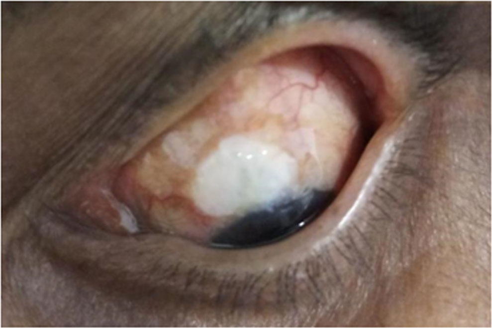
Fig. 1: Bleb leak at presentation
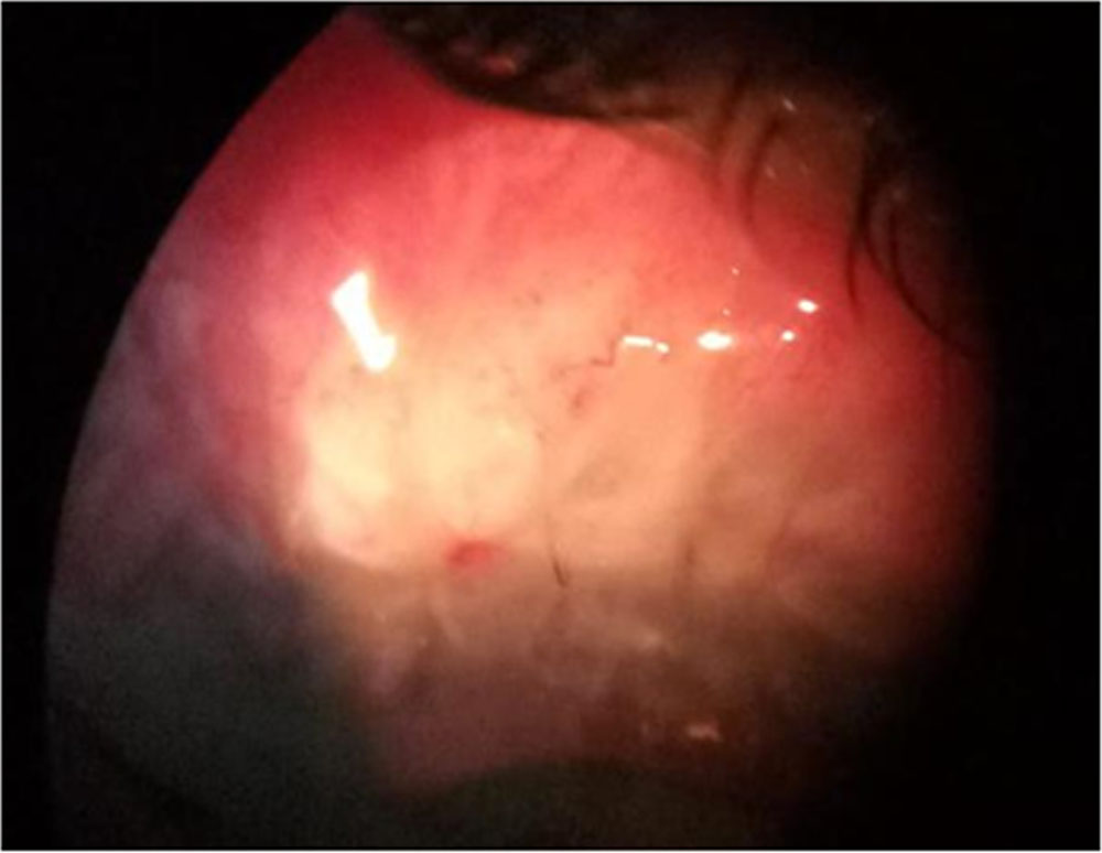
Fig. 2: Post-op day 1 after compression sutures
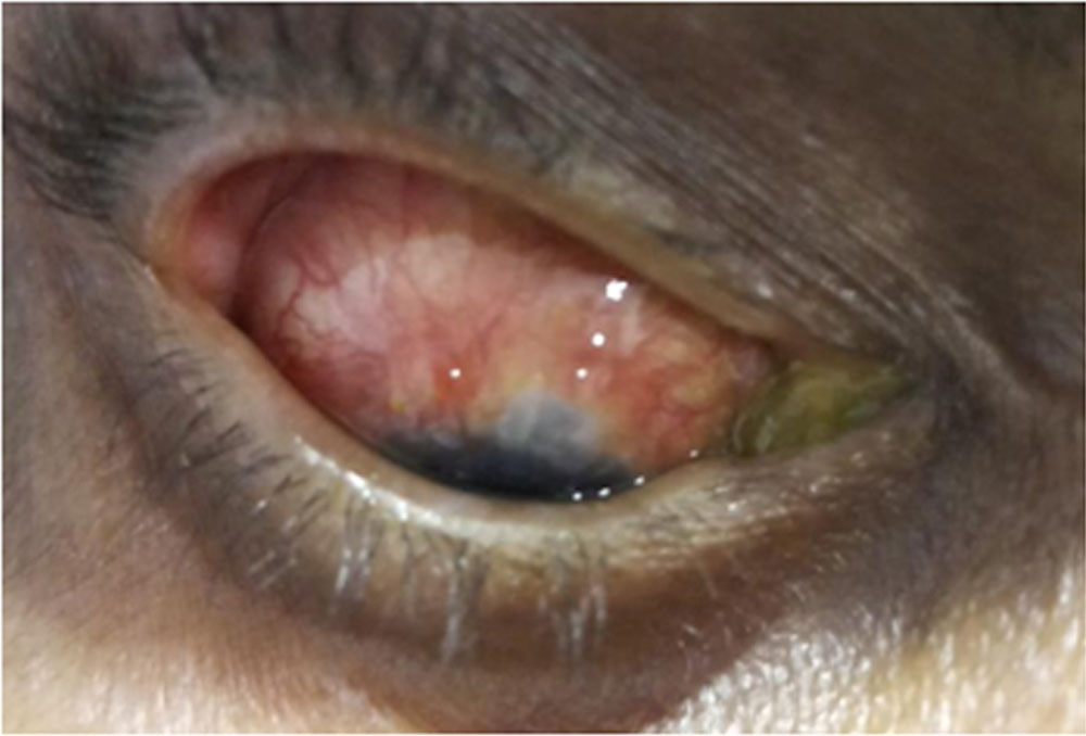
Fi.3: Post-op day 10 after compression sutures.
Leave a Comment