

Dr.Manali Hazarika Dr.Prajna N V
Abstract:
Aim:
To evaluate the release kinetics of voriconazole (VZ) from drug impregnated human amniotic membrane (HAM).
Methods:
6 HAM buttons (1 control, 5 test) were incubated with 1% VZ solution for 3 hours ,6hours , 12 hours , 24 hours and 48 hours. They were then subjected to release kinetics in Simulated Tear Fluid which were studied for 5 weeks . Samples were analysed using High Performance Liquid Chromatography(HPLC).
Results:
There was significant difference in the release kinetics of VZ with different incubation time periods ie, 3 hour versus 6 hour(p=0.0007) , 3 hour versus 12 hour(p= 0.0008), 3 hour versus 24 hour (p=0.0036)and 3 hour versus 48 hours( p= 0.0016). On comparing the release kinetics over a 5 weeks period in each of the incubation time groups, there was significant difference (p=0.000) in the 12 hour, 24 hour and 48 hour group.
Conclusion:
Significant sustained release of VZ from HAM was achieved over 5 weeks, when VZ was incubated with HAM over a time period ranging from 12 to 48 hours. The entrapment efficacy of VZ was significantly higher when the drug was soaked for a minimum of 6h.Thus,HAM can be considered for use as a drug reservoir for VZ, in the treatment of fungal keratitis.
key-words: human amniotic membrane , voriconazole , drug reservoir , fungal keratitis, drug release kinetics.
Key Messages:Human amniotic membrane acts as a drug reservoir.
Introduction:
The human amniotic membrane (HAM) has been termed as a ‘biological bandage’ 1 due to its myriad clinical applications such as in promoting wound healing, preventing scarring, inhibiting vascularisation, arresting corneal stromal melts, facilitating re-epithelisation, expanding limbal epithelial cells ex vivo2, acting as cell delivery matrix in regenerative medicine3 and acting as a drug reservoir.2
Thus, in developing countries , where corneal tissue availability is limited and topical antimicrobial treatment is not commercially available and/or is expensive , we hypothesize that drug- impregnated HAM might serve as an effective method for managing infectious keratitis ;both perforating or non-perforating.
HAM, as an effective drug release system, was first demonstrated by Kim et al4 in an animal model for the slow release of Ofloxacin. More recently , our study evaluated the in vitro, extended drug reservoir function of HAM impregnated with a different antibiotic ie, Moxifloxacin. This study found that HAM can effectively release Moxifloxacin for an extended period of 7 weeks.2 As a sequel to this, our current study investigates the application of HAM as a biological bandage in the treatment of fungal keratitis by providing a sustained drug delivery of Voriconazole.
Voriconazole is a triazole antifungal agent. It is a second-generation synthetic derivative of fluconazole, with enhanced potency and spectrum of activity. In vitro studies have shown promising results with Voriconazole. It was found to have a broad spectrum of action against Aspergillus species, Blastomyces dermatitidis, Candida species, Coccidioides immitis, Cryptococcus neoformans, Curvularia species, Fusarium species, Histoplasma capsulatum, Paecilomyceslilacinus, Penicillium species, Scedosporium species, and others.5 However , in vivo outcomes were poor when compared to Natamycin, particularly among Fusarium species.6
Although Natamycin has long been considered the drug of choice for filamentous fungal keratitis, the results of another study by Prajna et al7, found that Aspergillus flavus isolates had decreased susceptibility to Natamycin. In MUTT I, Aspergillus cases had better clinical outcomes with Voriconazole treatment than natamycin treatment, though this was not significant. Other studies have also shown that Voriconazole treatment is efficacious against Aspergillus ulcers, whereas Natamycin treatment had poor efficacy.8,9
Thus , the role of Voriconazole in the treatment of fungal keratitis remains relevant and our study aims to prove that Voriconazole- impregnated HAM may be beneficial for the extended release of the drug in order to achieve a good therapeutic result.
Subjects and Methods:
The study was approved by the Institutional Review Board of the Aravind Eye Hospital Madurai (IR #: RES2015011BAS).The tissue was handled according to the tenets of the Declaration of Helsinki.
Cell culture plates and dishes were obtained from Corning (Corning, NY). Gratis sample of 1%Voriconazole solution was formulated using commercially available Voriconazole injection,obtained from Aurolab, Madurai. HPLC grade solvents were purchased from Merck Chemicals (Mumbai, India). All other chemicals and reagents were of analytical grade and purchased from the chemical vendors.
Human Amniotic Membrane (HAM) preparation
Human amniotic membrane (HAM) was obtained by elective caesarean section at the Department of Gynaecology, PAMC Hospital, Madurai after getting their informed consent. The chorion was peeled off from the amnion by blunt dissection under sterile conditions. The membranes were washed with sterile phosphate-buffered saline (PBS) containing antibiotics and cryopreserved at 280°C in an amniotic membrane preservation medium [mixture of Dulbecco’s modified Eagle’s medium (DMEM) glutamax medium (Invitrogen) supplemented with penicillin, streptomycin, gentamicin, and glycerol] until use. HAM buttons were prepared using a trephine (12 mm) under sterile conditions and washed with sterile PBS (Gibco; Invitrogen) before incubating them in the drug solution.1
HAM Organ Culture and Drug Treatment
The HAM buttons of uniform weight (mean weight of 40.9 ± 3.9 mg) were used for the present study. HAM Buttons (1Control, 5 Test) were incubated in a freshly prepared (1ml) sterile solution of Voriconazole 1% (w/v) (Aurolab, India) for 3 hours (Group I) , 6 hours (Group II), 12hours (Group III) , 24 hours (Group IV) and 48 hours (Group V) to know the ideal drug soaking time.
In vitro-Drug Release Kinetics
At the end of each incubation time, HAM buttons were given a quick wash with simulated tear fluid (STF) to remove the surface bound drug. Then the drug-soaked HAM buttons were placed into 6-well plate containing 1ml STF (without drug) and incubated at 37oC with relative humidity of 65% and 5% CO2. 100µl of the STF was sampled out at different time intervals and replaced with equal volume of sterile STF in order to maintain the sink condition. The amount of drug released from the drug laden HAM was studied for a period of 5 weeks, to assess the extended release kinetics. The amount of Voriconazole in all the samples was quantified using High performance liquid chromatography (HPLC) after precipitating the proteins with ice-cold methanol (SRL, New Delhi, India).
Estimation of Voriconazole by HPLC
The estimation of Voriconazole was done using a Shimadzu Prominence HPLC system with PDA detector (Shimadzu Corporation, Kyoto, Japan). The analytical separation was achieved with the mobile phase consisting of 0.05M phosphate buffer (pH 3) and acetonitrile in the ratio of 75:25 (vol/vol) pumped at the flow rate of 1 ml/minute into Luna C-18 Column (250 x 4.6mm; 5µm; Phenomenex, California, USA) maintained at 30oC. The quantification of Voriconazole was carried out at lMax of 272 nm and the spectral matching was done with an in-built library matching facility in the PDA detector.
Statistical Analysis
Drug release of HAMs in the 5 groups were compared with each other and with the control. The values are presented as mean ± SEM. Independent samples t-test was performed and a post hoc test called Bonferroni correction was applied to find out which drug release time was statistically significant among the soaking groups. Differences with a p value <0.05 was considered statistically significant. All the statistical analysis was done using STATA ver. 14 (Texas, USA).
Results:
In vitro drug release kinetics based on drug soaking time
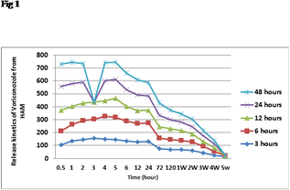
In this study, the extended release kinetics of Voriconazole from 0.5 hours to 5 weeks has been evaluated using HAMs incubated for different soaking periods. The cumulative release of Voriconazole after different exposure times is given in Table 1, and the amount of Voriconazole released at each time point is represented in Figure 1.
On comparing the cumulative drug release values of Voriconazole between the 5 groups, at 5 weeks, our study found that there was a significant increase in the drug entrapment and thereby release; when the soaking time was more than 3 hours.
In case of a 3h (Group I) soaking time with Voriconazole, the cumulative value of drug entrapment was found to be 1433.15µg/ml. When compared with the 6h (Group II) soaking time, that had a cumulative value of drug entrapment of 1549.61µg/ml, the 3h soaking time had significantly lower drug entrapment (p=0.007). Similar result was found when comparison was made between Group I and Group III with a cumulative value of 1400.39 µg/ml (p=0.0008); Group I and Group IV with a cumulative value of 1533.97 µg/ml (p=0.004); Group I and Group V with a cumulative value of 1465.57 µg/ml (p=0.002).
However , there was no statistically significant increase in drug entrapment when groups II, III and IV were compared with each other to determine which soaking time group had better drug entrapment .
Significant sustained release of Voriconazole was achieved from 0.5 hours to 5 weeks, when the drug was soaked over a time period ranging from 12 to 48 hours.
Thus we conclude that to increase the drug entrapment , we have to soak the HAM in Voriconazole for a minimum of 6 hours.
Discussion:
Fungal keratitis is a severe sight-threatening condition that can lead to ocular morbidity and loss of vision especially following corneal perforation due to relentless tissue degradation.12 A study from South India reported that 44% of all central corneal ulcers are caused by fungi.10 The most common etiological agents being the filamentous fungi ie, Fusarium and Aspergillus.11
The therapeutic effect of HAM in managing perforations and deep ulcers due to infective keratitis, is currently limited to addressing the tectonic aspect in a temporary form, ie, it acts by suppressing inflammation and promoting epithelialization till the definitive penetrating keratoplasty takes place.12 However , the management of fungal keratitis , both perforating and non-perforating is challenging because of a limited availability of antifungals and the need for their frequent application. However, topically applied drugs are greatly influenced by blinking, lacrimation, tear turnover rate, and absorption by non-productive adjacent tissues.13 Therefore, with our study, we highlight a third dimension to the utility of HAM ie , as a drug reservoir for the continuous delivery of drugs.
Topical natamycin, a polyene, is the only antifungal agent approved by the US Food and Drug Administration for treatment of fungal keratitis.14 Voriconazole (VZ) is a new triazole anti-fungal agent.It inhibits cytochrome P450 demethylase to alter fungal cell membrane permeability and to arrest growth.This drug is increasingly being considered as the first line treatment due to its broad spectrum of activity and high liposolubility that translates to high penetration through ocular tissues.15 It has low 90% minimum inhibitory concentration (MIC90) values for Candida species, Fusarium, and Aspergillus and has a promisingly low MIC90 (0.5 μg/mL) for Paecilomyceslilanicusand Paecilomycesvariotii. 15The more favorable pharmacology is attributed to its dose-dependent preferential inhibition of the P450 enzyme, sterol 14–demethylase.
Neoh et al 16 investigated the change in Voriconazole concentrations in the aqueous humor of the human eye over time after topical administration of 1% Voriconazole eye drops. They found that Voriconazole concentrations in the aqueous humor is above the MIC90 (0.06 – 8 mg/L) for most fungal species . With an estimated elimination t 1/2 of 1.47 hr, a 2-hourly dosing regimen afforded concentrations that were sufficient to eradicate Aspergillus and Candida keratitis, but not Fusarium keratitis. This correlates with other studies that have found poorer clinical resolution with topical 1% Voriconazole in Fusarium keratitis.6,17,18Senthilkumari et al11 also advised a 2 hourly regimen as repeat hourly administration of Voriconazole resulted in a higher aqueous concentration above 6 µg/ml, which was comparable with a 2 h interval. Furthermore, the 1% formulation was stable at maintaining its antifungal efficacy at least 30 days from the day of reconstitution.
Even though these reports are promising, a 2 hourly regimen amounts to frequent instillation and this maybe a barrier in the successful treatment of fungal keratitis in a developing nation due to low compliance. A drug -impregnated HAM can overcome this barrier by acting as a drug reservoir for the sustained release of the drug over a long period of time. Our study concluded that when HAM is soaked in 1% Voriconazole solution in vitro for a minimum of 6 hours, it can successfully release the antifungal agent over a period of 5 weeks.
References:
1.Dua HS, Rahman I, Miri A, Said DG.Variations in amniotic membrane: relevance for clinical applications.Br J Ophthalmol. 2010;94:963-4.
2.Yelchuri ML, Madhavi B, Gohil N, Sajeev HS, Venkatesh Prajna N, Srinivasan S.In Vitro Evaluation of the Drug Reservoir Function of Human Amniotic Membrane Using Moxifloxacin as a Model Drug.Cornea. 2017;36:594-599.
3.Niknejad H, Deihim T, Solati-Hashjin M, Peirovi H.The effects of preservation procedures on amniotic membrane’s ability to serve as a substrate for cultivation of endothelial cells.Cryobiology. 2011;63:145-51.
4.Kim HS, Sah WJ, Kim YJ, Kim JC, Hahn TW.Amniotic membrane, tear film, corneal, and aqueous levels of ofloxacin in rabbit eyes after amniotic membrane transplantation.Cornea. 2001;20:628-34.
5.Vemulakonda GA, Hariprasad SM, Mieler WF, Prince RA, Shah GK, Van Gelder RN.Aqueous and vitreous concentrations following topical administration of 1% voriconazole in humans.Arch Ophthalmol. 2008;126:18-22.
6.Prajna NV, Krishnan T, Mascarenhas J, Rajaraman R, Prajna L, Srinivasan M et al.The mycotic ulcer treatment trial: a randomized trial comparing natamycin vs voriconazole.JAMA Ophthalmol. 2013;131:422-9.
7.Lalitha P, Sun CQ, Prajna NV, Karpagam R, Geetha M, O’Brien KS et al.In vitro susceptibility of filamentous fungal isolates from a corneal ulcer clinical trial.Am J Ophthalmol. 2014;157:318-26.
8.Johnson EM1, Szekely A, Warnock DW.In-vitro activity of voriconazole, itraconazole and amphotericin B against filamentous fungi.J Antimicrob Chemother. 1998;42:741-5.
9.Pradhan L, Sharma S, Nalamada S, Sahu SK, Das S, Garg P.Natamycin in the treatment of keratomycosis: correlation of treatment outcome and in vitro susceptibility of fungal isolates.Indian J Ophthalmol. 2011;59:512-4.
10.Sharma S, Srinivasan M, George C. The current status ofFusarium species in mycotic keratitis in South India. J MedMicrobiol.1993;11:140–147.
11.Senthilkumari S, Lalitha P, Prajna NV, Haripriya A, Nirmal J, Gupta P, et al.Single and multidose ocular kinetics and stability analysis of extemporaneous formulation of topical voriconazole in humans.Curr Eye Res. 2010;35:953-60.
12.Solomon A, Meller D, Prabhasawat P, John T, Espana EM, Steuhl KP et al.Amniotic membrane grafts for nontraumatic corneal perforations, descemetoceles, and deep ulcers.Ophthalmology. 2002;109:694-703.
13.Lee VHL, Robinson JR. Topical ocular drug delivery: recent developmentsand future challenges. J OculPharmacol. 1986;2:67–108.
14.Prajna NV, Krishnan T, Rajaraman R, Patel S, Srinivasan M, Das M et al.Effect of Oral Voriconazole on Fungal Keratitis in the Mycotic Ulcer Treatment Trial II (MUTT II): A Randomized Clinical Trial.JAMA Ophthalmol. 2016;134:1365-1372.
15.Lau D, Fedinands M, Leung L, Fullinfaw R, Kong D, Davies G et al.Penetration of voriconazole, 1%, eyedrops into human aqueous humor: a prospective open-label study.Arch Ophthalmol. 2008;126:343-6.
16.Neoh CF, Leung L, Chan E, Al-Badriyeh D, Fullinfaw RO, Jhanji V et al.Open-Label Study of Absorption and Clearance of 1% Voriconazole Eye Drops.Antimicrob Agents Chemother. 2016;60:6896-6898.
17.Prajna NV, Mascarenhas J, Krishnan T, et al. Comparison of Natamycin andVoriconazole for the treatment of fungal keratitis. Arch Ophthalmol2010;128:672–8.
18.Sharma S, Das S, Virdi A, Fernandes M, Sahu SK, Kumar Koday N et al.Re-appraisal of topical 1% voriconazole and 5% natamycin in the treatment of fungal keratitis in a randomised trial.Br J Ophthalmol. 2015;99:1190-5.
Table 1 : Cumulative Release of Voriconazole from HAM in each group
|
TIME (h) |
Group I | Group II | Group III | Group IV | Group V | ||||||||||
| Mean Con (µg/ml) | SEM | Cumulative amount (µg/ml) | Mean Conc (µg/ml) | SEM | Cumulative amount (µg/ml) | Mean Conc (µg/ml) | SEM | Cumulative amount (µg/ml) | Mean Conc (µg/ml) | SEM | Cumulative amount (µg/ml) | Mean Conc (µg/ml) | SEM | Cumulative amount (µg/ml) | |
| 0.5 | 106.58 | 9.40 | 106.58 | 107.08 | 18.76 | 107.08 | 159.6994 | 7.40 | 159.70 | 183.30 | 10.23 | 183.30 | 172.11 | 15.48 | 172.10 |
| 1 | 132.92 | 13.38 | 239.50 | 131.11 | 29.8 | 238.19 | 140.5089 | 8.45 | 300.21 | 173.29 | 14.04 | 356.60 | 166.89 | 7.07 | 338.99 |
| 2 | 146.74 | 13.71 | 386.25 | 145.33 | 19.17 | 383.52 | 138.09 | 12.87 | 438.30 | 161.11 | 11.29 | 517.71 | 143.55 | 4.68 | 482.54 |
| 4 | 151.20 | 19.25 | 537.44 | 173.10 | 34.06 | 556.62 | 124.4007 | 9.18 | 562.70 | 154.05 | 11.87 | 671.76 | 139.43 | 18.92 | 621.97 |
| 5 | 146.52 | 23.65 | 683.97 | 172.71 | 36.55 | 729.33 | 146.5271 | 5.09 | 709.23 | 144.82 | 13.17 | 816.57 | 132.36 | 5.74 | 754.33 |
| 6 | 135.04 | 22.78 | 819.002 | 153.78 | 33.36 | 883.11 | 112.4907 | 14.58 | 821.72 | 131.93 | 13.42 | 948.51 | 125.44 | 12.11 | 879.77 |
| 12 | 129.09 | 22.93 | 948.10 | 141.06 | 31.01 | 1024.17 | 100.925 | 10.30 | 922.64 | 120.55 | 13.32 | 1069.05 | 117.86 | 4.36 | 997.62 |
| 24 | 129.37 | 20.38 | 1077.46 | 144.28 | 27.03 | 1168.45 | 98.23682 | 9.97 | 1020.88 | 110.55 | 13.82 | 1179.60 | 105.33 | 6.19 | 1102.95 |
| 72 | 76.51 | 10.38 | 1153.97 | 80.21 | 15.82 | 1248.66 | 88.03671 | 10.65 | 1108.92 | 88.47 | 7.06 | 1268.07 | 93.70 | 11.28 | 1196.65 |
| 120 | 70.22 | 11.93 | 1224.19 | 74.98 | 14.47 | 1323.64 | 83.4107 | 11.25 | 1192.33 | 71.23 | 6.00 | 1339.30 | 75.38 | 6.59 | 1272.03 |
| 168 | 66.84 | 12.47 | 1291.03 | 70.20 | 14.62 | 1393.84 | 79.77076 | 6.08 | 1272.10 | 63.78 | 3.996 | 1403.09 | 63.78 | 2.04 | 1335.81 |
| 336 | 60.29 | 9.65 | 1351.32 | 66.25 | 13.39 | 1460.08 | 63.38594 | 0.35 | 1335.48 | 55.28 | 3.003 | 1458.37 | 58.93 | 2.35 | 1394.74 |
| 504 | 44.10 | 6.30 | 1395.42 | 51.14 | 9.44 | 1511.22 | 35.90647 | 8.06 | 1371.39 | 44.62 | 3.67 | 1502.99 | 41.43 | 4.01 | 1436.18 |
| 672 | 25.22 | 4.77 | 1420.63 | 30.06 | 5.99 | 1541.29 | 28.47173 | 5.87 | 1399.86 | 29.30 | 1.16 | 1532.29 | 26.64 | 5.66 | 1462.81 |
| 840 | 12.51 | 3.58 | 1433.15 | 8.32 | 0.45 | 1549.61 | 0.526897 | 0.07 | 1400.39 | 1.68 | 0.26 | 1533.97 | 2.75 | 1.76 | 1465.57 |
Acknowledgement: Mrs. Iswarya , Department of Statistics , Aravind Eye Hospital , Madurai
Leave a Comment