

Dr.ANNU JOON, Dr.Kirti Singh,Dr.Mainak , hattacharyya,Dr.Divya Kishore
PORTABLE FUNDUS CAMERA ON PHONE
Abstract
Aim:
To validate a generic, portable fundus on phone (FOP).
Material & Methods:
FOP (Remidio Innovative solutions Pvt Ltd) was used to capture disc images of undilated, consecutive cases referred for glaucoma evaluation. Expert examiners reviewed images using the Disc damage likelihood scale (DDLS)grading into suspect, mild, moderate and severe glaucoma. The result was then compared it with the gold standard 90D slit lamp biomicroscopy for disc evaluation. The similarities & differences were noted, a discrepancy of >1 scale DDLS was taken as significant.
Result:
Consecutive 103 eyes of 30 males & 23 females, aged 12 -72years were assessed. Grading by both methods was similar in 75% eyes, underestimation by FOP 10%. FOP overestimated suspects/ mild glaucoma by 3% and underestimated advanced damage by 5%. Limitations encountered in FOP imaging were minimal and related to media haze, disc pallor.
Conclusion
: Digital imaging on phone is a promising tool for disc damage assessment in undilated glaucoma patients and compared favourably with standard 90 D slit lamp biomicroscopy.
Key words:
Glaucoma screening, portable Fundus-on-phone, disc photography, DDLS
Introduction
Glaucoma is the second leading cause of blindness globally and the first cause of irreversible blindness.1Worldwide estimate of 37 million people being blind due to glaucoma in 2002,it is expected to increase with increasing life span and enhanced diagnostics.2 The economic burden due to glaucoma increases as the disease progresses. A 4-fold increase in direct costs has been documented as glaucoma severity increases from early to end-stage disease.3Glaucoma-related blindness itself leads to a 30-fold increase in indirect costs to society due to lost productivity and additional burdens on families.4Indian scenario is alarming with a 2.6% prevalence of glaucoma, of which 90% being undetected.5
In this situation early diagnosis and intervention by an ophthalmologist is imperative. In a developing country like India, with ophthalmologist-patient ratio a dismal 1:100,000, and inequitable spread, with 70% nation’s citizens residing in rural areas while 70% of its eye care professionals living in urban areas,6 this remains a Utopian concept. The huge volume of glaucoma cases or those at risk of the disease at 40 million, cannot be screened by the very few trained glaucoma specialists.7Tonometry, gonioscopy and perimetry being either contact procedures or laborious, subjective tool, disc screening remains a viable option to diagnose the rampantly pervasive disease. Disc evaluation and identifying at risk cases at risk cases, by trained paramedical personnel at field level would be greatly helpful in administering tele-ophthalmology.
Teleophthalmology
holds great potential to improve the quality, access and affordability in eye care especially for patients in rural areas by reducing the need for travel and providing virtual access to a super-specialist right at their doorstep. Screening modalities using non-mydriatic single field (monoscopic) photography provide an efficient tool. To expand these screening programs into rural areas, through highly distributed primary care facilities or through mobile imaging units, it would be beneficial to have access to low-cost, portable, easy-to-operate, and high image quality fundus cameras. This is of particular importance in low and middle income countries.
Low-cost, handheld, nonmydriatic eye fundus cameras have recently been used in developed nations, namely Smartscope Pro (Optomed, Oulu, Finland), Pictor (Volk Optical, Mentor, OH, USA), VersaCamTM DS-10 (Nidek, Gamagori, Japan), Horus DEC 200 (MiiS, Hsinchu, Taiwan), and enesis-D (Kowa, Nagoya, Japan). Smartphone-based solutions are the way ahead and some products like Peek Vision (Nesta, London, UK),23PanOptic + iExaminer (Welch Allyn, Skaneateles Falls, NY), and D-Eye (D-EYE, Padova, Italy).8 are in the marked. Fundus-On-Phone (Remidio, Bengaluru, India) is one such smartphone-based, portable device which has been indigenously developed.
This study is the first of its kind to validate this devise for diagnosing suspicious discs.
Materials and methods
Patient Recruitment
Consecutive patients diagnosed as glaucoma and referred from the ophthalmology out-patient department for glaucoma evaluation at the Glaucoma Services, Guru Nanak Eye Centre, attached to Maulana Azad Medical College, New Delhi in the month of May 2018 were invited to participate in this study. An informed written consent was taken from all study participants. The study adhered to the tenets of the Declaration of Helsinki.
Comparative Modalities
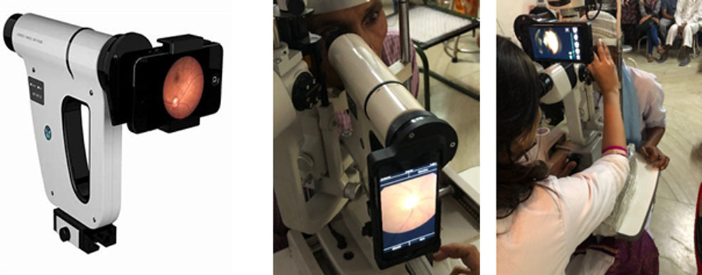
Nonmydriatic disc evaluation was compared by two methods in this study- a slit lamp biomicroscopic examination using a 90D lens (standard protocol) and non mydriatic fundus photography for disc evaluation using a portable Fundus on Phone (FOP) by Remidio Innovations Pvt Ltd, Bengaluru, India(Figure 1).
Figure1- FOP, disc evaluation by FOP and 90D
Examination Protocol
All examinations were conducted in ambient light environment and non mydriatic pupils. First, the FOP (Remidio Innovations Pvt Ltd) was used to capture disc images, by a trained technicians from Remidio Innovations. Subsequently a glaucoma expert reviewed the captured image using the rubrics provided by Disc damage likelihood scale (DDLS) and categorised it as no/ suspect glaucoma, mild/ moderate/ severe glaucoma. The same expert then did and compared 90D disc assessment of the same patient, to remove the inter-observer variability. The similarities and differences in two disc evaluations were noted. A discrepancy of > 1 scale of DDLS was taken as significant. Difficulties in image acquisition such as media opacities like advanced cataract and corneal haze, and miotic pupils on pilocarpine therapy were documented. The process was done within 2-3 minutes to prevent patient fatigue.
Results
Fifty three patients were enrolled in this study comprising 30 males and 23 females, with a total of 103 eyes (3 patients being one eyed with atrophic bulbiand no visibility of fundus). Patients ranged from 12- 72 years of age,49 years old on average. The diagnosis ranged from disc suspects to open angle glaucoma, angle closure glaucoma, normal tension glaucoma, secondary glaucomas (post traumatic, post uveitic, steroid induced) and few cases of non- glaucomatous disc (such as disc coloboma).
A clear disc photograph could be was captured in 83 eyes (80.5%).
Nine eyes had a hazy media due to moderate cataract, healed endothelitis and vitrectomised eyes with aphakia.In this group 7 eyes could be evaluated better with 90D examination, 90D had a comparatively hazy view in 1 eye, whereas it was not possible in 1 eye where FOP captured a hazy scan.
In eleven cases with media haze arising from pan uveitis, advanced cataract, miotic pupils, advanced pterygium, corneal oedema, and silicone oil in situ eyes, FOP photo acquisition was not possible. In this group 3 of which could not be assessed by 90D examination either, but a blurred view of the disc on 90D evaluation permitted DDLS grading in the others.
The FOP capture was better in 3 eyes with hazy cornea, posterior subcapsular cataracts and small pupil and lent itself to grading whereas 90D assessment was not possible.
DDLS scoring of the disc by both methods revealed similar grading in 77 eyes (75%). FOP underestimated the disc score in 10 eyes (10%), the discrepancy being one grade in 50% eyes and more than one grade in the remainder. Overestimation of disc cupping by FOP was less frequent, and occurred by one grade in 2 eyes (2%). In the 8 eyes where FOP captured a hazy scan and 90D disc grading was possible, 6 eyes scored the same by both modalities on DDLS whereas 2 eyes were underestimated by 1 grade by FOP. Comparison could not be attained in 14 eyes in whom either disc photograph could not be captured or disc visualisation was not possible on 90D examination(Table 1).
Table 1- Comparison of DDLS by FOP vs 90D evaluation
| DDLS | FOP grading vs 90D (no. of eyes/ %) |
| Same grade | 77 (74.8) |
| underestimation | 10 (9.7) |
| overestimation | 2 (1.9) |
| NA | 14 (13.4) |
| Total | 103 |
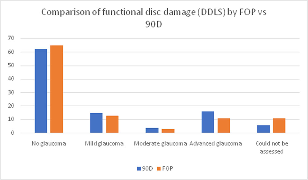
The two modalities were also compared for functional disc damage and divided into no glaucoma, mild glaucoma, moderate glaucoma and advanced glaucoma.
FOP slightly overestimated eyes with no functional damage by glaucoma (63% vs 60% by 90D) and underestimated those with glaucomatous damage;12% vs 14% on 90Din mild glaucoma, 3% vs 4% on 90D in moderate glaucoma, and 11% vs 16% on 90D in advanced glaucoma. FOP data was not available for 11% eyes and 90D grading was unavailable for 6% eyes(Table2).
Table2- Comparison of functional disc damage (DDLS) by FOP vs 90D
Limitations encountered in FOP imaging were hazy scans to no image acquisition due to hazy media and doubtful assessment of cupping in eyes with severe pallor.
Of 103 discs examined, 3 with severe pallor lent themselves well to FOP image acquisition. Similar grade DDLS estimation was seen in one eye, doubtful in one eye and for the third FOP image could not be distinguished from optic atrophy, but stereoscopic 90D examination could.
Discussion
Technological advances in android phones with sophisticated image capture software has revolutionized today’s world. This technology boom aided bystart-ups has led to the development of one such innovative design by the Remidio Innovative Solutions Pvt Ltd and is called Remidio fundus on phone (FOP). FOP provides an advanced, affordable, and portable technology for retinal viewing.
Glaucoma screening by photographic disc evaluation is still new as compared to widespread retinopathy screening programmes for diabetes. Optic nerve photography has been found to compare reasonably with the more complex and expensive devices like scanning laser ophthalmoscopes, scanning laser polarimetry to optic coherence tomography (OCT).9-11 The traditional fundus cameras however are table mounted, bulky, expensive, and need a trained technician. Lack of portability makes these devices less well suited as screening tools.
Non‐mydriatic fundus cameras permit image capture without pupil dilation and facilitate quick and easy photography of the posterior segment.12
The advent of non‐mydriatic and portable fundus cameras is making ophthalmic imaging more accessible to the community. It has been successfully used for diabetic retinopathy and glaucoma screening.13 The indigenous FOP has been validated as a diabetic retinopathy screening tool by various authors. Rajalakshmi R et al compared Remidio FOP to a traditional Zeiss FF450 desktop imaging system as a screening tool for diabetic retinopathy and found FOP’s clinical sensitivity to be 93% and specificity to be 98%.14 They also reported a high sensitivity for detection of diabetic retinopathy (DR) and sight-threatening DR (STDR) (above 95% for DR, 99% for STDR) using Artificial Intelligence (EyeArt software).15 FOP has also been incorporated into large scale diabetic retinopathy screening. The low-cost indigenous cameras handled by paramedical work force/health volunteers used telemedicine for remote reporting by trained doctors/reading centres. Sensitivity and specificity with smart phone as compared to the gold standard in 100 randomly assigned patients was found to be 82% and 98%, respectively.16
Monophotographic optic disc evaluation has been found to be equally sensitive as compared to stereoscopic disc evaluation (by sterophotography). Parkin B et al17compared stereoscopic and monoscopic evaluation of optic disc topography using a digital optic disc stereo camera in 150 patients and found little benefit of stereoscopic imaging over monoscopic imaging. This was echoed by Chan et al18 who conducted a prospective observational study to determine if monoscopic vs stereoscopic viewing impacted evaluation of optic disc photographs for glaucoma diagnosis in their Glaucomatous optic neuropathy evaluation (GONE) project byanalysing 20 pairs of high-quality monoscopic and stereoscopic photographs of similar size and magnification. They concluded that monoscopic optic disc photographs did not represent a significant disadvantage compared to stereoscopic photographs.
Hand held non mydriatic fundus camera as a glaucoma screening modality, is a relatively new modality. Hark et al.’s prospective randomized trial in 906 subjects at high risk for glaucoma with handheld non‐mydriatic disc photography (Volk Optical, Mentor, OH, USA)reported 28.5%suspicious discs, 17.5% unreadable images, with 11% being diagnosed with diabetic retinopathy.13Miller SE et al.19 compared cup-to-disc ratio (CDR) measurements from images taken with a portable, 45-degree nonmydriatic fundus camera (Pictor) to images from a traditional tabletop mydriatic fundus camera (Topcon) in a prospective study done on 422 eyes, and diagnosed glaucoma in 47% cases.
This is the first validation study of Remidio FOP for glaucoma screening. It can be used for documentation of disc at baseline and on follow up and study progression like a routine fundus camera. Combined with basic tools such as non contact tonometry for intraocular pressure measurement, it may provide added advantage in early detection of glaucoma and referral of patients by non-ophthalmologists. FOP has the potential to significantly impact preventable blindness programs, through tele-ophthalmology-based screening, globally.
Limitations of the study
The present study was a pilot project with a small sample size. It needs to be continued to include more number of patients to enhance the validation process. The data of each patient was analysed by one expert examiner and masking of patient was not performed while comparing 90D assessment to FOP images. Randomisation in the present study design with each patient’s data being analysed by more than one examiner and assessing inter-observer variation will be more affirmative.
Conclusion
Digital imaging on phone is a promising tool for disc damage assessment in undilated glaucoma patients and compared favourably with standard 90 D slit lamp biomicroscopy.
References
- Mariotti SP, Pascolini D. Global estimates of visual impairment. Br J Ophthalmol. 2012;96:614–618.
- Resnikoff S, Pascolini D, Etya’ale D, et al. Global data on visual impairment in the year 2002. Bull World Health Organ. 2004 Nov;82(11):844-851.
- Mowatt G, Burr JM, Cook JA, et al. Screening tests for detecting open-angle glaucoma: Systematic review and meta-analysis. Invest Ophthalmol Vis Sci. 2008;49(12):5373–85.
- Wang YX, Xu L, Zhang RX, Jonas JB. Frequency-doubling threshold perimetry in predicting glaucoma in a population-based study: The Beijing Eye Study. Arch Ophthalmol. 2007;125(10):1402–6.
- Kingman S. Glaucoma is second leading cause of blindness globally. Bull World Health Organ. 2004 Nov;82(11):887-8.
- Gullapalli NR. Ophthalmology in India. Arch Ophthalmol. 2000:118: 1431-1432.
- Tejwani S, Murthy SI, Gadudadri CS, et al. Impact of a month-long training program on the clinical skills of ophthalmology residents and practitioners. Indian J Ophthalmol. 2010;58:340-343.
- Russo A, Morescalchi F, Costagliola C, Delcassi L, Semeraro F. Comparison of smartphone ophthalmoscopy with slit-lamp biomicroscopy for grading diabetic retinopathy. Am J Ophthalmol. 2015;159:360–364.
- Lin SC, Singh K, Jampel HD et al. Optic nerve head and retinal nerve fiber layer analysis: a report by the American Academy of Ophthalmology. Ophthalmology 2007; 114: 1937–49.
- Kotowski J, Wollstein G, Ishikawa H, Schuman JS. Imaging of the optic nerve and retinal nerve fiber layer: an essential part of glaucoma diagnosis and monitoring. 2014;59:458–67.
- Vessani RM, Moritz R, Batis L, Zagui RB, Bernardoni S, Susanna R. Comparison of quantitative imaging devices and subjective optic nerve head assessment by general ophthalmologists to differentiate normal from glaucomatous eyes. J Glaucoma.2009;18:253–61.
- Panwar N, Huang P, Lee J, et al. Fundus photography in the 21st century- A review of recent technological advances and their implications for worldwide healthcare. Telemed J E Health.2016;22:198–208.
- Hark LA, Katz LJ, Myers JS, et al. Philadelphia telemedicine glaucoma detection and follow‐up study: methods and screening results. Am J Ophthalmol.2017;181:114–24.
- Rajalakshmi R, Arulmalar S, Usha M, et al. Validation of Smartphone Based Retinal Photography for Diabetic Retinopathy Screening. Merigan WH, ed. PLoS ONE. 2015;10(9):e0138285.
- Rajalakshmi R, Subashini R, Anjana RM, Mohan V. Automated diabetic retinopathy detection in smartphone-based fundus photography using artificial intelligence. 2018 doi:10.1038/s41433- 018-0064-9
- Gadkari SS. The Pune diabetic retinopathy awareness and screening model. J Clin Ophthalmol Res.2015;3:23-6.
- Parkin B, Shuttleworth G, Costen M, Davison C. A comparison of stereoscopic and monoscopic evaluation of optic disc topography using a digital optic disc stereo camera.Br J Ophthalmol. 2001 Nov;85(11):1347-51.
- Chan HH, Ong DN, Kong YX, et al. Glaucomatous optic neuropathy evaluation (GONE) project: the effect of monoscopic versus stereoscopic viewing conditions on optic nerve evaluation. Am J Ophthalmol. 2014 May;157(5):936-44.
- Miller SE, Thapa S, Robin AL, et al. To compare cup-to-disc ratio (CDR) measurements from images taken with a portable, 45-degree nonmydriatic fundus camera to images from a traditional tabletop mydriatic fundus camera. Am J Ophthalmol. 2017;182:99-106.
Conflict of interest– None
Source of funding– the Fundus on phone was made available to the Glaucoma Services, Department of Ophthalmology, Guru Nanak Eye Centre, New Delhi for the study period by Remidio Innovations Pvt Ltd, Bengaluru.
Financial interests– None
Leave a Comment