

Dr.Manushree Gautam,Dr.PARUL MALVIYA,Dr.Shweta walia,Dr.Preeti Rawat
Clinical profile , visual field changes and progression in patients with NTG
Presenting Author: Dr Manushree Gautam
Co Authors: Dr Preeti Rawat , Dr Shweta Walia , Dr Parul Malviya
Introduction
Early recognition of glaucomatous optic neuropathy in the setting of physiologic IOP was described by Von Graefe in the mid 19th century[1].
Given the unclear etiology of the condition, many clinical definitions have been put forth guided by proposed mechanisms and efforts to describe the disease[2]
There is considerable debate in the literature whether Normal Tension Glaucoma (NTG) represents a distinct entity or is simply POAG with IOP within the normal range.(1)
The definition of NTG thus should be based on clinical history, co-morbidities, characteristic optic disc cupping and specific visual field defects.
We studied the Clinical profile , visual field changes and progression in patients with NTG
Methodology
It was a prospective and retrospective observational study conducted in MYH, Indore from Oct 2014 to Feb 2017
30 eyes of 15 patients with NTG were studied
Detailed history was taken
Disc evaluated with slit lamp biomicroscopy with 90 D.
Visual fields analyzed by HFA 30-2.
Glaucomatous fields was defined as per Anderson Criteria
PSD, MD and defects extending within 15° of fixation (PCFD) were noted and CCT was measured .
All patients were followed for minimum 2 years with minimum 4 visual fields at 6 monthly interval.
Progression was defined by Modified Anderson Hodapp criteria and/or by GPA.
First two tests were not taken and test was repeated for any field with progression.
Inclusion Criterion
NTG was diagnosed as glaucomatous optic neuropathy on Disc examination,characteristic visual field defects ,open anterior chamber angles on gonioscopy ,pretreatment IOP never exceeding 21 mmHg, by GAT( IOP was corrected as per CCT values).
Age 15 years or more
Exclusion Criterion
Eyes with other visually significant ocular pathology
Patients on medications that could affect visual sensitivity and IOP
History of ocular surgery
High myopes >-6 D
Results
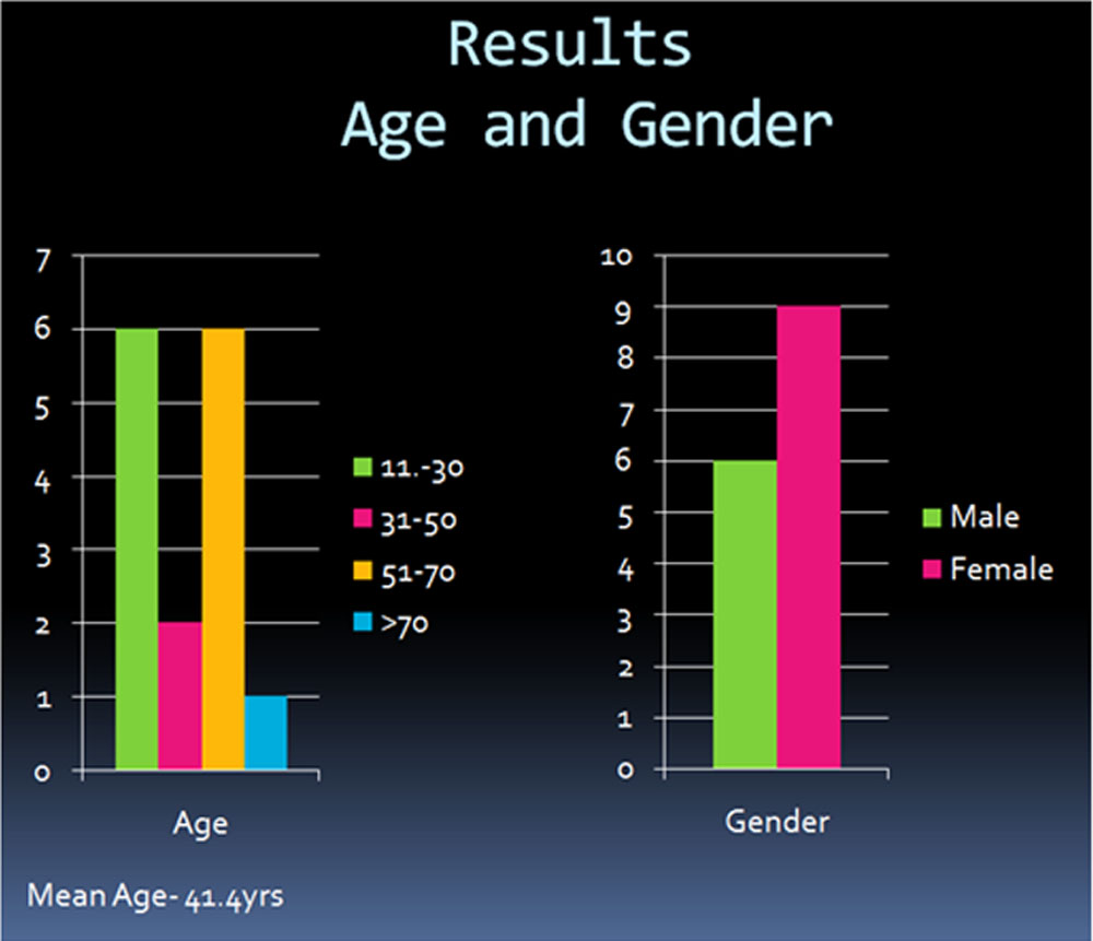
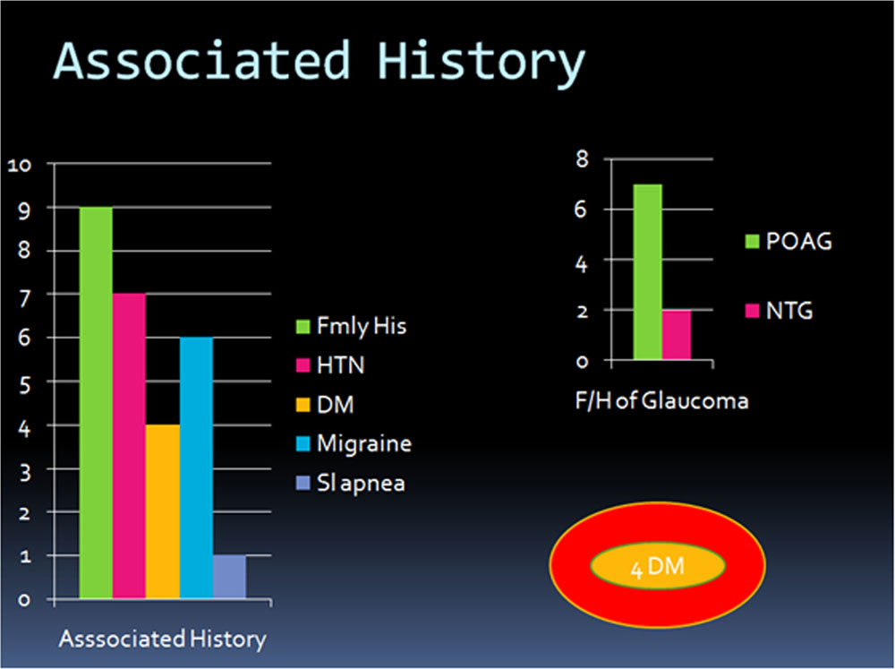
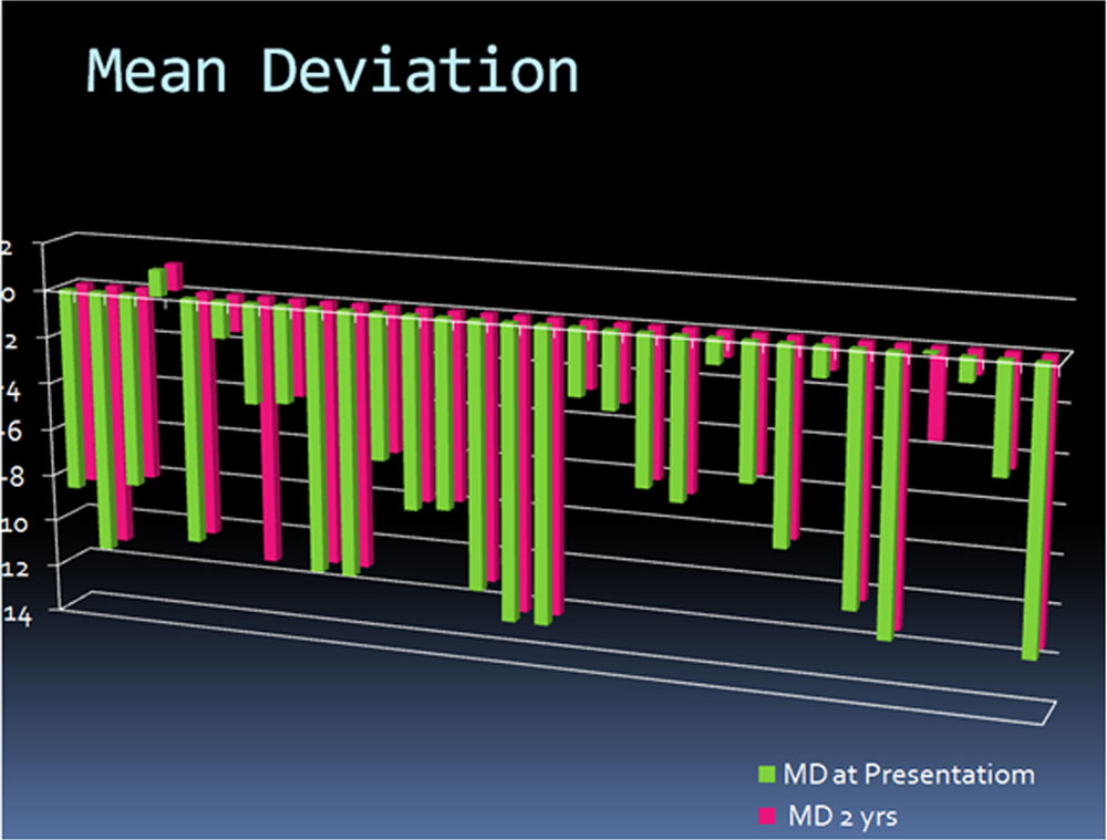
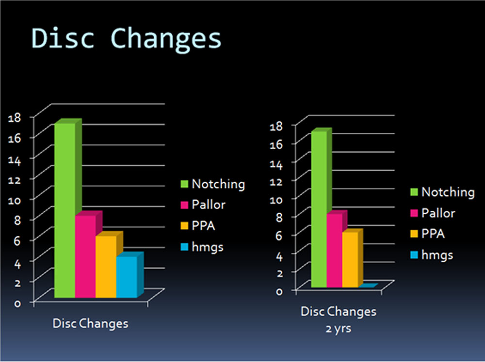
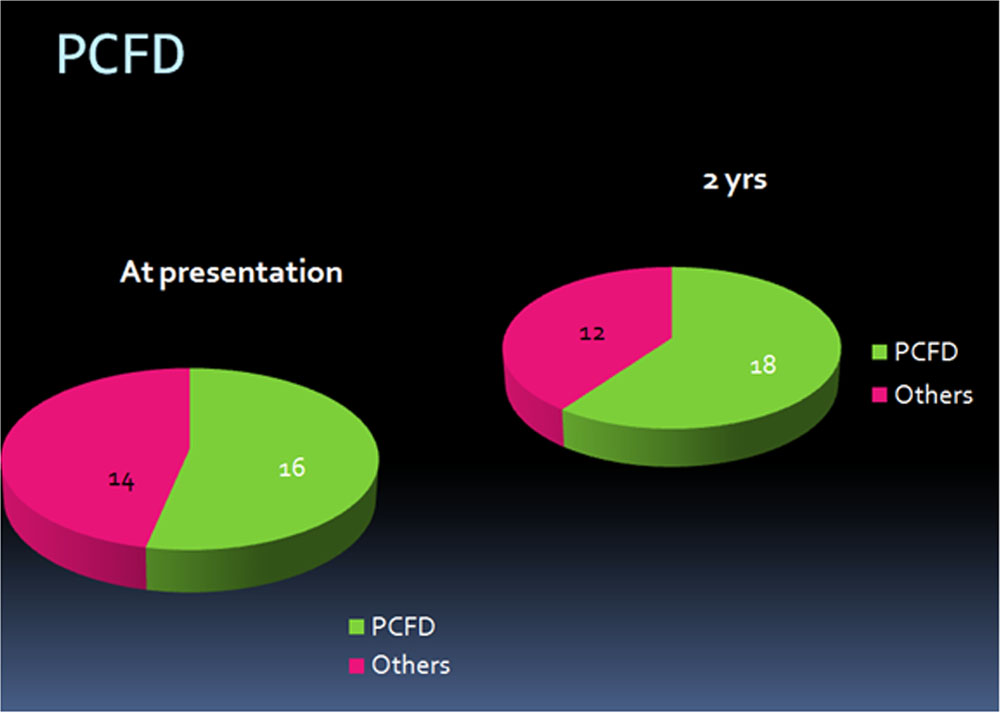
The mean age of patients was 41.4 years with female predilection. Family history(40%POAG,13% NTG) ,HTN(46%), DM(26%), Migraine(40%), sleep apnea (6%)and stroke(6%) was found. NRR showed focal thinning especially inferotemporally. Disc haemorrhages were observed. PSD was high which increased with progression. MD change was small . PCFD present in 53% eyes at presentation, increased to 60% over two years
Progression:
4 Eyes of 3 patients (13.33%) had progression on visual fields :
20 year old male with h/o DM and HTN had progression in LE.
60 year old female with h/o Migraine had progression in left eye which was normal on presentation
64 year old female with h/o DM and HTN had progression in both eyes with peripapillary hemorrage at presentation
Discussion
The mean age of patients in this study was 41.4 years. Previously considered as a disease of elderly persons.
The mean reported age in clinical studies generally is in the 60s; myopic patients with this disease are significantly younger.(2)
However Leung et al calculated age of onset for patients with progressive NTG and recommended screening for NTG from 40 yrs of age.(2)
Younger populations may also be affected.
We observed a female predilection in our study. Rotterdam Study demonstrated increased risk of primary open angle glaucoma (POAG) in women with early menopause.(3)
The changes in the level of female sex hormones may influence intraocular pressure (IOP) as well as vascular resistance that might effect the optic nerve head circulation. (4)
In most case optic disc rim showed focal thinning especially inferotemporally.
Optic disc hemorrhages and parapapillary disc atrophy was also observed as seen in previous studies.5,6 and were associated with progression.
Studies show visual field defects more focal, deeper, and closer to fixation.7 which can be emphasised by higher values of PSD in our study which increased with progression.
The MD change was typically small and insufficient to measurably affect the MD index.
We found PCFD in 53% eyes at presentation which increased to 60% over two years, NTG has been found as a risk factor for progression into central fields in one of the previous studies. 8
Conclusion
NTG patients show charachteristic clinical profile
Clinical profile can help in early diagnosis and estimating prognosis of NTG .
Progression in Patients with NTG was found to be rare and associated with HTN, Disc hmg and PCFD
References
- Sommer A, Tielsch JM, Katz J, et al. Relationship between intraocular pressure and primary open angle glaucoma among white and black Americans. The Baltimore Eye Survey. Arch Ophthalmol 1991;109:1090-1095
- Current Concepts in Normal Tension Glaucoma James Tsai, MD New York City A. Karim, MD Chittagong, Bangladesh Review of Ophthalmology June 2006
- Hulsman CA, Westendorp IC, Ramrattan RS. Is open-angle glaucoma associated with early menopause? The Rotterdam Study. Am J Epidemiol. 2001 Jul 15;154(2):138–144.
- Higginbotham EJ. Does sex matter in glaucoma? Arch Ophthalmol. 2004
Mar;122(3):374–375. - Caprioli J, Spaeth GL. Comparison of the optic nerve head in high- and lowtension
glaucoma. Arch Ophthalmol 1985;103:1145. - Tezel G, Kass MA, Kolker AE, et al. Comparative optic disc analysis in normal
pressure glaucoma, primary open-angle glaucoma, and ocular hypertension.
Ophthalmology 1996;103:2105. - Chauhan BC, Drance SM, Douglas GR, et al. Visual field damage in NTG and
high-tension glaucoma. Am J Ophthalmol 1989;108:636. - Visual Field Progression differences between Normal Tension and Exfoliative
High Tension Glaucoma IOVS March 2010 Vol51 Issue 3
Leave a Comment