

Dr.Neha Khanna, N17070, Dr.Sri Ganesh, Dr.Mamatha Nagamalli, Dr.Mahesh Kumar
Introduction:
Vitreous consists of 98% water and macromolecules with collagens and hyaluronan as most important component organized in an exquisitely clear gel.[1,2] With advancing age, there is liquefaction and lacunae formation in the vitreous body. The walls of these lacunae interfere with transmission of photon to the retina, which in turn contributes to the perception of floaters. Floaters can be considerably symptomatic when they increase in size and number. When situated in the visual axis they are most noticeable and are known to reduce contrast sensitivity and deteriorate quality of life.[3,4]. Structural changes within the vitreous body can also result from other causes like myopic[5] and diabetic vitreopathy inflammation, vitreoretinal dystrophies, but most commonly it occurs due to aging.[6]
Weiss ring is a prominent type of floater resulting from Posterior vitreous detachment (PVD), the remnant of vitreopapillary attachment and peripapillary glial tissue at the optic disc.[6,7,8]A Weiss ring can be visualized by biomicroscopy as an annular structure which is adjacent and anterior to the optic disc. This ring can cast a prominent circular or semicircular shadow onto the retina, particularly when it is only a few millimeters anterior to the retina. PVD allows the vitreous body to move with the movement of head or eye and are perceived as floaters.
Management of floaters consists mainly of Patient education and observation. Floaterectomy by vitrectomy and Floaterrhexis, via Nd:YAG laser vitreolysis are other treatment options. YAG vitreolysis is an untested treatment for floaters. In this approach Laser energy is used to fragment the vitreous opacities via a non-invasive approach. Intense pulses of laser energy are focussed into opaque regions of the transparent vitreous which raise the temperature of these confined spots. At this temperature, plasma is produced, and ’optical breakdown’ occurs, with the successful photodisruption mechanism alleviation of floater symptoms occurs.
Historically, using standard YAG laser systems, the clinician could not see the middle and posterior vitreous while with the Ultra Q ReflexTM system (Ellex Medical Lasers Ltd, Adelaide, Australia, the combination of on- and off-axis allows for a safer and more efficacious treatment. On-axis feature helps to visualize a floater against the red-glow background (floaters in the middle and posterior vitreous) while Off-axis determine how far the floater is behind the lens and also allows a negative contrast to help visualize them better.[9] The aim and objective of this study isto see the effect of Nd YAG Vitreolysis for Symptomatic Floaters and assessing the Patient Quality Life Index (PQLI)andto study and assess the efficacy and complications of Nd YAG Vitreolysis using Ultra Q Reflex™ in treatment of floaters.
Review of Literature
The vitreous is a translucent extracellular matrix filling the region of the eye between the retina and lens. It is approximately 4.0 mL in volume. The vitreous is responsible for maintaining the transparency of the eye and the uniform transmission of photons towards the retina for photoreception.[1,10,11]As the vitreous ages, the collagen aggregates into tight parallel bundles bound by cross-links. This leaves pockets of liquid within the vitreous which have a paucity of these structural macromolecules. This molecular rearrangement is termed vitreous liquefaction, or syneresis causing symptomatic floaters.[12]Using a utility value analysis approach to establish the effects of vitreous floaters on the quality of life, these studies found this negative effect to be so severe in younger patients (< 55years of age) that they are willing to accept a 7% risk of blindness to get rid of floaters. [13]
Laser energy can be used to fragment the vitreous opacities via a non-invasive approach. Typically, Neodymium-doped Yttrium Aluminium Garnet (YAG) laser is the one which is utilised for this purpose. [14]
Webb et al.,[15]reported that of 603 smartphone users, an overwhelming 76% (n=458) noticed floaters; of these patients, 199 had noticeable vision impairment. Furthermore, myopes and hyperopes were 3.5 and 4.4 times (respectively) more likely to report moderate-to-severe floaters. This study highlights quite clearly that floaters are very common in the general population, irrespective of age, race, gender and eye colour.
Wagle[13]found that symptomatic floaters had a negative impact on health-related quality of life. Study by Zou et al, also showed that the presence of floaters considerably diminished patients’ life perception.[16]
YAG lasers were traditionally only equipped for capsulotomies and iridotomies; however, with improvements in the technical design, Laser Floater Removal (LFR) now offers more efficient and safer energy profiles with reliable and repeatable outcomes that provide low complication and high success rates. [17]
Geller, S in his study treated 112 eyes with Nd YAG laser with success rate of 85% while Kirchhoff J in his Formal Floater Study under the supervision of the INOVA Fairfax Hospital Institutional Review Bard found 92% success rate in 200 eyes treated.[18]
Singh IP in a retrospective, observational study included 326 eyes of n=218 patients (mean age, 66 years [range, 38 to 89 years]) who underwent YAG vitreolysis with the Ultra Q Reflex™ (Ellex Medical Lasers, Adelaide, Australia), a Neodymium-Doped (Nd) YAG laser in an office outpatient setting. [Power range 2.0 mj-7.0mj] showed that 93% of patients were satisfied with the procedure.[9]
Inclusion Criteria
- Patients with symptomatic Weiss rings resulted in inclusion of eyes with at least 6 months of symptoms
- Weiss rings at least 3 mm from the retina and 5 mm from the posterior lens capsule
Exclusion Criteria
- Subjects with any of the following conditions will not be enrolled in the study:
- Younger patients (< 45 years
- Vitreous haze
- Acute posterior vitreous detachment (PVD), with inherent debris
- Very mobile opacities
If the floater(s) was located within 2 mm of the retina or crystalline lens
- Media haze at any level
- Family history of retinal detachment, prematurity, feeding problems, arthritis, and midline defects
- Vitreous base infiltrates and pars planitis
- Macular edema and premacular membranes with macular pucker
- Perfusion disorder
Materials and Methods
This is a Prospective, unicentric, interventional observational study with a goal to enroll patients symptomatic for floaters coming to the vitreoretinal department in Nethradhama Superspeciality Eye Hospital, Jayanagar, Bangalore from August 2017 to July 2018. Institutional Ethics Committee approval was obtained and special informed consent from patients was taken in their own language before recruitment in study explaining about the probable complications and need for multiple sittings.
Patients recruited underwent previtreolysis clinical evaluation, fundus examination, B scan to measure the distance of the floater from the retina and posterior capsule of the lens, HD analyser to see the optical quality and scattering index and I trace to note the improvement in aberrations caused by floaters treatment.
Following parameters were noted during the treatment with Ultra Q Reflex™ Nd Yag Vitreolysis:
- Type of floater [Single (Weiss) / Multiple (Amorphous clouds)]
- Power
- Duration
- Number of shots
- Interval
- No of Sessions
Post Vitreolysis in 1 week post op period patient underwent contrast sensitivity, HD analyser and I trace and a Questionnaire to assess the patient’s response to the treatment.The patients were questioned with regard to duration of their symptoms prior to presentation, laterality, severity and number of their floaters, activity most inconvenienced by the presence of floaters and subjective improvement in symptoms following laser vitreolysis. The patients were asked to quantify their post-operative improvement as a percentage as well as choose a descriptive analogy.
The options given were:
(a) Failure: floaters are the same or worse
(b)Partial success: some improvement but still floaters of moderate inconvenience
(c) Significant success: significant improvement with only slight inconvenience
(d) Complete success: complete resolution of floaters
If measured on a self-rated scale of 1-10, then failure (1-2), partial success (3-5), significant success (5-8), complete success (9-10)
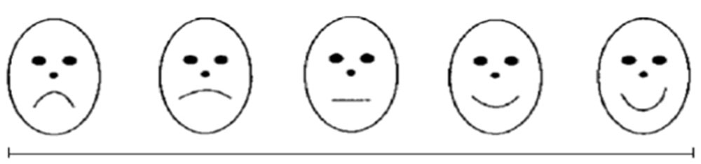
0 1 2 3 4 5 6 7 8 9 10
Ethical Issues
Written informed consent will be obtained from the patients before participation in the trial. Patients will be explained that they will be monitored for any signs of deterioration during the study period, they would be treated accordingly. Each patient will be subjected to minimal routine investigations and hence explained that it was unlikely to impose any extra risk to the patient. Each patient will be free to withdraw any time during the study and he would not be denied treatment at any time. All the needed investigations will be provided by the hospital as per its policy, so no extra financial burden will be put on patient’s families. This is a nonfunded study. The thesis committee of the institute has approved this study and the consent form and patient leaflet form has been attached along.
Results
Floaterswithin the safe zone 3mm away from the retina and posterior lens capsule confirmed on Bscanwere treated to avoid the risk of inducing a retinal damage or in phakic eyes to minimize pitting and lens damage. On-axis setting was used to provide coaxial illumination to focus the laser beam on the floater and assess its relation to the retina accordingly. Average number of sessions were 1.7, where treatment was repeated, there was an interval of 15days between two laser sessions.
Average no of shots used were 331. More no of shots and sittings were used for amorphous clouds as compared to the Weiss ring. Average power was 8.53mJ.The energy used initially was set at 3 mJ and titrated to an appropriate level at which the surgeon observed disruption of the floater. Post procedure patients were advised topical anti-inflammatory for 1week and reviewed 1day post procedure, after 1week and 1month and were assessed for any rise in intraocular pressure, anterior segment and dilated fundus evaluation. All recruited patients were given scaled questionnaire 1month post procedure. Fifty eyes were included in the study out of which 93.7% of patients were satisfied and showed improvement in daily visual functioning. On average patients rated 7.8/10 in PQLI scale which was considered significantly successful.Better results were seen with solitary “Weiss rings” as compared to amorphous “clouds”. Eleven eyes were phakic and the remaining 39 eyes were pseudophakic with 1.8yrs being the average time from cataract surgery to vitreolysis.
We observed 3 cases of Intraocular pressure spike which was treated with topical medications, Inadvertent posterior capsule rupture was documented in one patient and retinal hole nasal to disc in one eye,which was lasered immediately.HD Analyzer did not show any significant change in OSI (optical scattering index) values by comparing pre and post procedure. We did not observe any significant change inoptical scattering index(OSI) on pre and post HD analyser. The iTrace Wavefront Aberrometer and Corneal Topographer which provides a break-down of contribution between corneal and internal aberrationsincluding the lens, the posterior cornea, macula and vitreous.It objectively measures quality of vision which matches patient’s subjective response. It was found that the high order aberration quality was significantly reduced along with improvement in contrast sensitivity post procedure.
Limitations
In initial phase of learning there were certain problems we faced, like the view of vitreolysis lens is very less which made it difficult to relocate the floater which keeps moving with minimal eye movements. Complete pupillary dilatation is very important as the floater hides behind the iris with the movement. There are chances of injuring the surrounding tissue by inexperienced hands. Due to difficulties faced initially we had biased patient selection criteria with preference to pseudophakic and Weiss rings in comparison to phakic and patients with dense amorphous clouds.
Discussion
Vitreous floaters are most commonly caused by posterior vitreous detachment (PVD)[19,20]vitreous syneresis.[20] The majority of patients tolerate their symptoms without any inconvenience. However, there is a significant minority for whom the floaters are very troublesome.In general, Ophthalmic YAG lasers are designed for use in the anterior segment to perform capsulotomy following IOL implantation, and to perform iridotomy prior to implanting an anterior chamber lens, or in cases of angle closure glaucoma. In posterior segment this Laser energy can be used to fragment the vitreous opacities via a non-invasive approach.
Unlike vitrectomy, which carries a high risk of bleeding and infection, vitreolysis is a non-invasive, pain-free procedure that can eliminate the visual disturbance caused by floaters. Unlike vitrectomy, which being an invasive procedure with significant chances of side effects like chances of infection, retinal detachment, macular edema, glaucoma and development of cataract, vitreolysis has been proved to be highly effective with low complication rate and offers a high degree of patient satisfaction. It delays or obviate the need for surgery. Typically, neodymium-doped yttrium aluminium garnet (YAG) laser is utilised for this purpose [14]
Nd:YAG Laser Vitreolysis (vitreolysis) is a highly effective, outpatient-based procedure, which involves the use of a co axial illumination which assists in viewing both anterior and posterior segment. It uses nano-pulsed YAG laser to vaporize vitreous strands and opacities. Use of short, intense pulses of laser energy to cause ’optical breakdown’ can be used to disrupt the vitreous floater.
Ultra Q Reflex™ has been optimized for use in both the posterior and anterior segment. Featuring a proprietary slit lamp illumination tower design that converges the operator’s vision, the target illumination and the treatment beam onto the same optical path, and focuses them to the same optical plane, Ultra Q Reflex™ minimizes the potential for focusing errors and the risk of damage to the natural lens or the retina. This makes it ideal for targeting vitreous strands and opacities.[9]
Lasered beam is focussed accurately with the help of various lenses designed by Peyman and by Karickhoff. We used Singhs Midvitreous lens to focus the laser beam onto the front surface of the floater. The laser emits a short 3nanosecond (0.000000003 seconds) burst of energy. It is important to note that the laser energy does not simply break the floater into smaller pieces. Instead, the high power density of this laser energy converts the collagen and hyaluronin molecules to a gas, which is then resorbed into the eye.The impact symptomatic floaters have on quality of life was reported to be as profound as diabetic retinopathy, colon cancer and asymptomatic human immunodeficiency virus (HIV) infection.[12,13]Hence, it is important to establish the safe and effective means of treating floater symptoms appropriate patient selection is vital for success in YAG vitreolysis treatment.[21,22]
Conclusion
To conclude Yag Vitreolysis can be a novel approach for treating symptomatic floaters.We often underestimate thePatients’ symptoms from vitreous floaters for which if we consider Nd:YAG vitreolysis, it can be safe and effective treatment. If we look towards risk/benefit ratio this procedure can be considered as a primary modality of treatment for symptomatic floaters deteriorating patient’s quality of life.
Bibliography
- Bishop PN. Structural macromolecules and supramolecular organisation of the vitreous gel. Prog Retin Eye Res. 2000;19(3):323-44.
- Reardon AJ, Le Goff M, Briggs MD et al. Identification in vitreous and molecular cloning of opticin, a novel member of the family of leucine-rich repeat proteins of the extracellular matrix. J Biol Chem. 2000;275(3):2123-9.
- Sebag J, Balazs EA. Morphology and ultrastructure of human vitreous fibers. Invest Ophthalmol Vis 1989;30(8):1867-71.
- Sebag J. Age-related changes in human vitreous structure. Graefes Arch Clin Exp Ophthalmol.1987;225(2):89-93.
- Gale J, Aiello LP, Sebag J. Diabetic vitreopathy. In: Sebag J, editor. Vitreous – in Health and Disease. New York: Springer-Verlag; 2014. p. 57-79.
- Tozer K, Johnson M, Sebag J. Vitreous aging and Posterior Vitreous Detachment. In: J. Sebag, editor. Vitreous – in Health and Disease. New York: Springer-Verlag; 2014. p. 131-50.
- Halfter W, Sebag J, Cunningham ET. Vitreo-retinal interface and inner limiting membrane. In: J. Sebag, editor. Vitreous – in Health and Disease (New York: Springer-Verlag; 2014. p. 165-91.
- Sebag J. Anatomy and pathology of the vitreo-retinal interface. Eye. 1992;6(6):541-52.
- Singh IP. Treating vitreous floaters: patient satisfaction and complications of modern YAG vitreolysis. Paper presented at: American Society of Cataract and Refractive Surgery annual meeting; May 7, 2016; New Orleans, LA.
- Sebag J. Vitreous: the resplendent enigma. British Journal of Ophthalmology 2009;93(8):989–91.
- Yanoff M, Duker JS. Ophthalmology. 3rd Edition. Mosby Elsevier, 2008.
- Sebag J. Floaters and the quality of life. American Journal of Ophthalmology 2011;152(1):3–4.
- Wagle AM, Lim WY, Yap TP et al Utility values associated with vitreous Am J Ophthalmol. 2011;152(1):60-5 e1.
- Kwasniewska S. Some applications of the neodymium:YAG laser operating in the thermal and photodisruptive modes. Vitreolysis. In: Fankhauser F, Kwasniewska S editor(s). Lasers in Ophthalmology: Basic, Diagnostic, and Surgical Aspects: a Review. Kugler Publications, 2003:403–13.
- Webb, Blake F, Schroeder MC et al Prevalence of vitreous oaters in a community sample of smartphone users. Int J Ophthalmol. 2013; 6(3): 402–405.
- H, Liu. H, Xu. X et al The impact of persistent visually disabling vitreous oaters on health status utility values. Quality Life Res. 2013; 22 (6), 1507-1514.
- Data on file. Ellex Medical Pty Ltd.
- Geller S: Nd YAG laser treatment effective for oaters. OSN, Dec. 1, pg. 37, 2001.
- Murakami K, Jalkh AE, Avila MP, Trempe CL, Schepens CL. Vitreous floaters. Ophthalmology 1983; 90: 1271–1276.
- Morse PH. Symptomatic floaters as a clue to vitreoretinal disease. Ann Ophthalmol 1975; 7: 865–868.
- Tsai WF, Chen YC, Su CY. Treatment of vitreous floaters with neodymium YAG laser. Br J Ophthalmol 1993;77:485-8.
- DelaneyYM,OyinloyeA,BenhaminL.Nd:YAGvitreolysisandparsplana vitrectomy: surgical treatment for vitreous floaters. Eye 2002;16:21-6.
Leave a Comment