

Dr. GOMPA MOHANA PREETHI, Dr. Prajna N V
Abstract:
Context:
Creation of digital, quantified measures of microbial corneal ulcer characteristics using slit lamp photographs helps understand healing trajectories and may serve as a useful prognosticating tool to predict microbial ulcer outcomes.
Aims:
To digitally quantify corneal ulcers and predict healing trajectories between different microbes
Settings and Design:
50 corneal ulcers were recruited from a tertiary eye care centre in South India
Methods and Material:
Serial slit lamp photographs of corneas with presumed bacterial or fungal ulcers were used from which measurements were made. An Indian Visual Function (IVF) questionnaire and an 8 item interval visit questionnaire were administered to assess the impact of ulcer on general function, psychosocial behaviour and visual function. Valid digital imaging software was used to develop predictive algorithms and the differences between clinical and algorithmic measurement were noted
Statistical analysis used:
Kruskal Wallis H test, Fishers exact measures, Pearson’s correlation, Dice coefficients
Results:
The height of the epithelial defect between different growths as measured by clinicians was statistically significant by a Kruskal –Wallis H test. No statistically significant difference due to the microbe or the location of ulcer was observed over the quality of life measured through the questionnaires
Conclusions:
With algorithms that predict heterogeneity of patients, ophthalmologists can personalize treatments of corneal ulcer at an early onset.
Key-words: Corneal ulcer, healing, slit lamp imaging
Key Messages:
Currently, monitoring of ulcers is done qualitatively subject to physician variability. [7, 8, 9, 10] Patients’ potential outcomes from microbial corneal ulcers could be distinguished if researchers addressed critical barriers in how we collect, measure, and analyze corneal ulcers. To achieve the long-term goal of predicting ulcer outcomes and to personalize treatments, the overall objective of this study is to collect and create digital, quantified measures of ulcer characteristics and to analyze healing trajectories and differences in microbial ulcer outcomes.
Introduction: Corneal ulcers are a leading cause of blindness in developing countries with an annual 113 per 100, 000 person years in Madurai, South east India. [1] Ulcers vary depending on microbial, morphologic, patient, and environmental factors. [2, 3, 4, 5, 6] Despite variation, powerful broad-spectrum antimicrobials are given to many patients because ulcers require rapid intervention. Ophthalmologists can change to targeted medications, but only days later and only if culture results are conclusive.
Currently, monitoring of ulcers is done qualitatively subject to physician variability. [7, 8, 9, 10] Patients’ potential outcomes from microbial corneal ulcers could be distinguished if researchers addressed critical barriers in how we collect, measure, and analyze corneal ulcers. To achieve the long-term goal of predicting ulcer outcomes and to personalize treatments, the overall objective of this study is to collect and create digital, quantified measures of ulcer characteristics and to analyze healing trajectories and differences in microbial ulcer outcomes.
Subjects and Methods:
Participants:
The primary inclusion criterion for participants to be eligible for this study was the presence of a corneal ulcer which was untreated and clinically-significant, defined as ≥1 mm in greatest linear dimension as measured by the gold-standard examination by an ophthalmologist .A sample of fifty patients recruited from the cornea clinic at a tertiary eye care centre during the period of June 2017 to May 2018 were observed and images were gathered. Participants were stratified by gender and age and there was no change to their standard of care or treatments given. They were excluded if: (1) pregnant, (2) have had prior incisional corneal surgery, (3) had no light perception vision, or (4) had a corneal perforation or impending perforation.
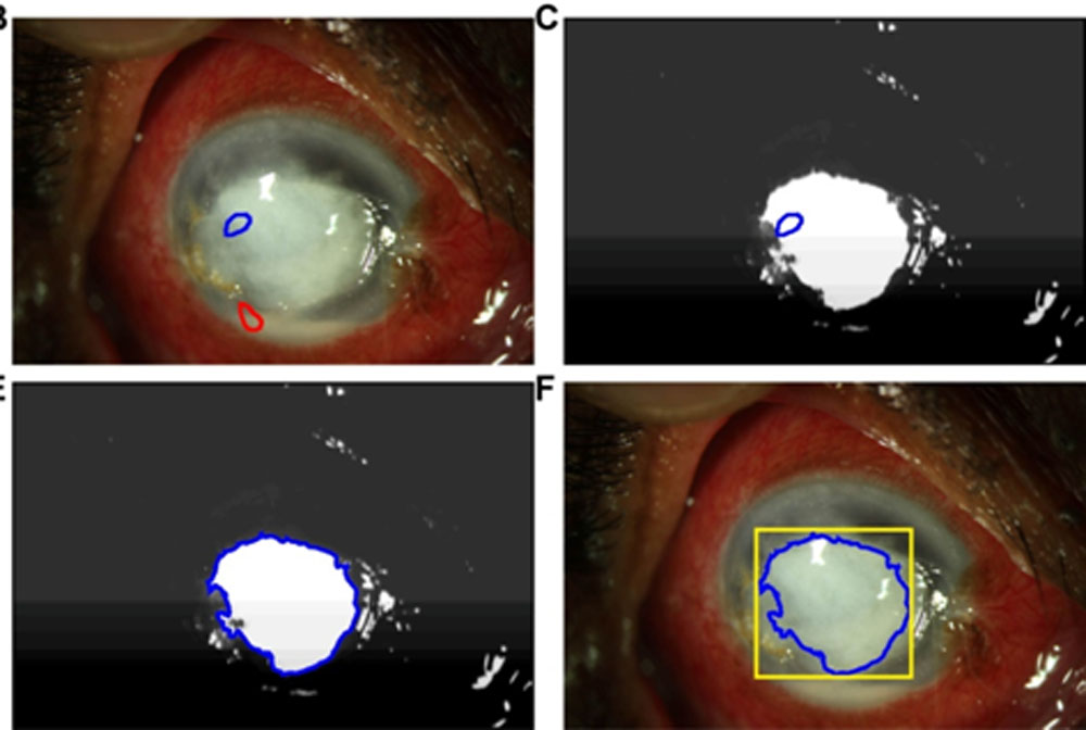
Data collection content: All participants underwent examination by a cornea specialist, corneal scraping, patient-reported outcome questionnaires, and slit lamp photography and AS OCT for white to white diameter measurement. Demographic information, risk factors, and patient symptoms were gathered. The height and width of epithelial defect (ED) and the stromal infiltrate (SI) were measured by the cornea specialists. Also, the ulcer location, depth of the infiltrate, degree of corneal thinning, presence of a hypopyon, and consolidation at the ulcer edge were noted. Health-related quality of life at the initial recruitment and at the completion of the study was evaluated by Indian Visual Function questionnaire [11] (VFQ-33), validated in English and Tamil). A reliable, accurate algorithm for image-based measurements using Quantitative Corneal Monitoring (QCM) semi-automated analysis was done. [10] (Figure 1).
Results:
Demography:
The average age of the cohort was 47 years with a majority being males (54.8%)
Clinical characteristics:
At the initial presentation, the gold standard measurement by ophthalmologists of the mean height and width of ED and SI was 1.8 x 1.6 mm and 2.6 x 2.3 mm respectively .After epithelization, the mean ulcer measurement for the same were 0.2 x0.2 mm (mostly neurotrophic) and 1.7×1.4 mm. ( Table 1) The mean algorithmic measurements of ED and SI from the photos were 2.1×2.1 mm and 2.6 x2.6 mm respectively.(Table 1) Ulcers were predominantly non central (56%) and hypopyon was present in 32.2% cases. Depth of the ulcer reduced from 37 % at presentation to 27.7 % after epithelization /scarring.
Microbiology:
Fungi (52%) were the predominant cause of corneal ulcers followed by smear and culture negative cases (28%). Among the 20% bacterial cases, gram positive cocci (GPC- 60%) were the majority followed by gram negative bacteria (GNB- 40%).
AS-OCT:
The mean inner White to White diameter as measured by AS-OCT was 9.5 mm
Statistics:
The percentage of absolute measurements that differed by ≥1.0 mm was 15% for ED height and width, 10% for SI height, and 6 % for SI width. A Kruskal –Wallis H test showed that there was a statistically significant difference in the gold standard measurement of height of the ED between fungi, GPC, GNB and culture negative cases. An overall quality of life affected due to corneal ulcer by VFQ showed a greater impact on general and visual function in comparison to psychosocial function, yet a Kruskal –Wallis H test showed no statistical significance between them either due to the morphology or location of ulcer.
Discussion:
Effective treatments for corneal diseases require early and definitive disease diagnosis, well-defined staging, and clinically relevant end points. Despite the high prevalence and wide variety of microbial ulcer types, we have insufficient knowledge to formally stage microbial corneal ulcers or to monitor response to treatment. Currently, ophthalmologists monitor ulcers qualitatively by measuring the size of the overlying epithelial defect and the size of the stromal infiltrate. [9] Accordingly, they manage medications after noting the response to treatment. [19] Clinicians’ morphology measurements, while accurate in aggregate, have lower reliability. [10]
Computer aided quantified automated measures have transformed the way that patients are managed in macular degeneration, glaucoma, and corneal ectasia, [20,21,22,23] yet a quantified algorithm doesn’t exist for corneal ulcer, which if present would become an earlier clinical endpoint measure for impact of interventions. Corneal ulceration provides an ideal disease to develop morphologic analysis tools. The rapid disease course enables data collection, measurement, and analysis in a relatively short timeframe. Patients’ potential outcomes could be distinguished if researchers addressed critical barriers in how we collect, measure, and analyze corneal ulcers.
The project team consists of expert cornea specialists and applied ophthalmic engineers. To achieve the long-term goal of predicting corneal ulcer outcome and to personalize treatment, we aim to collect and create digital, quantified measures of ulcer characteristics and to analyze healing trajectories and differences in microbial ulcer outcomes. Our central hypothesis is that combining reliable, accurate corneal morphology measurements with host and pathogen characteristics will effectively distinguish patient risk levels for visual disability and corneal transplantation.
Image analysis:
External, diffuse light, slit lamp photograph of the ulcerated eye under white light and cobalt blue light illumination (with fluorescein) were taken by a cornea specialist prior to corneal scraping. Photographs were taken with a Canon EOS 7D camera mounted on a Haag-Streit International BX 900 model slit lamp biomicroscope that captures high-quality digital images of corneal ulcers. A semi automated Quantitative Corneal Measurement (QCM) software package [10] was used for algorithmic measurement of ulcers from the images. Performance of the algorithmic measurement of primary ulcer features was evaluated in comparison to measurement by a corneal specialist using the percentage of ulcers with an absolute difference between algorithm and specialist measurements ≥1.0mm, correlation coefficients, and Dice coefficients to compare ulcer area from manual tracing of images by a specialist to area obtained from the algorithm.
Statistical Analysis:
Descriptive statistics of the measured height and width of ED and SI were calculated, including mean, standard deviation (SD), range, and median for both gold standard clinician and algorithmic measurements. Scatter plots were used to assess the agreement in measurements between examiner and semi automated method, and the degree of linear association was assessed with Pearson’s correlations (). Absolute differences in ED and SI measurements between examiners and semi-automated methods were investigated and displayed with histograms. A threshold of≥ 0.5 mm absolute difference in measurement length was deemed a clinically significant difference. The absolute differences in ED and SI measurements between examiners and methods were tested for deviations from 0.5 mm with Wilcoxon signed rank tests. In the present study which constituted a majority of fungal corneal ulcers, a Kruskal –Wallis H test showed a statistical significance in the gold standard measurement of height of epithelial defect between different microbes. Neither the microbe nor the location of the ulcer had any statistical impact on the general, visual and psychosocial functions of the patient.
Limitations:
A less sample size and inconsistencies in noting the dimensions of corneal ulcer by clinicians during the follow up was a limitation to the study. Computerized image analysis requires good, consistent quality of the photographs. Photographs attained by different photographers would also impact the measurement variability
Future directions:
Visualized ulcer trajectories with healing over time will be created and statistical analysis will be done by linking this data to the host pathogen. Novel inquiries to predict quantitative healing of corneal ulcers is possible with a very large sample of patients who can follow up on a long term basis. We aim to provide iterative algorithms that predict heterogeneity of patients in the long run thus helping the ophthalmologists to personalize ulcer treatments at disease onset. The rationale for the research is that with building blocks to automate and quantify corneal ulcer features, we can distinguish among healing trajectories and link data to outcomes resulting in innovative approaches to staging, monitoring, and managing corneal ulcer patients.
References:
- Gonzales CA, Srinivasan M, Whitcher JP, et al. Incidence of corneal ulceration in Madurai district, South India. Ophthalmic Epidemiol. 1996;3:159-166.
- Lin C. C., Prajna L., Srinivasan M., Prajna N. V., McLeod S. D., Acharya N. R., et al. . (2012). Seasonal trends of microbial keratitis in south India. Cornea31, 1123–1127.
- Bharati MJ, Ramakrishnan R, Meenakshi R, Padmavathy S, Shivakumar C, Srinivasan M. Microbial Keratitis in South India: Influence of Risk Factors, Climate, and Geographical Variation. Ophthalmic Epidemiol2007;14:61-69.
- Leck AK, Thomas PA, Hagan M, Kaliamurthy J, Ackuaku E, John M, et al. Aetiology of suppurative corneal ulcers in Ghana and south India and epidemiology of fungal keratitis. Br J Ophthalmol2002; 86:1211-1215.
- Titiyal JS, Negi S, Anand A, Tandon R, Sharma N, Vajpayee B. Risk factors for perforation in microbial corneal ulcers in north India. Br J Ophthalmol2006; 90:686-689.
- Krishna S, Shafiyabi S, Sebastian L, Ramesha R, Pavitra D. Microbial keratitis in Bellary district, Karnataka, India: Influence of geographic, climatic, agricultural and occupational risk factors. Int J Pharm Biomed Res2013; 4:189-193.
- Aris Konstantopoulos , Jennifer Kuo , David Anderson, Parwez Hossain. Assessment of the Use of Anterior Segment Optical Coherence Tomography in Microbial Keratitis. American Journal of Ophthalmology 2008 ; 146 , Pages 534-542.
- Toutain-Kidd CM, Porco TC, Kidd EM, et al. Evaluation of fungal keratitis using a newly developed computer program, Optscore, for grading digital corneal photographs. Ophthalmic Epidemiol. 2014;21:24-32.
- Parikh PC, Valikodath NG, Estopinal CB, et al. Precision of Epithelial Defect Measurements. 2017;36:419-424.
- Patel TP1, Prajna NV2, Farsiu S3, Valikodath NG1, Niziol LM1, Dudeja L2, Kim KH1,4, Woodward MA1,5. Novel Image-Based Analysis for Reduction of Clinician-Dependent Variability in Measurement of the Corneal Ulcer Size. 2018 ;37:331-339
- Murthy GVS, Gupta SK, Thulasiraj RD, et al.The development of the Indian vision function questionnaire: questionnaire content. Br J Ophthalmol 2005; 89:498–503.
- Whitcher JP, Srinivasan M. Corneal ulceration in the developing world-a silent epidemic. Br J Ophthalmol1997;81:622–3.
- Lalitha P1, Prajna NV2, Manoharan G1, Srinivasan M2, Mascarenhas J2, Das M2, D’Silva SS2, Porco TC3, Keenan JD4. Trends in bacterial and fungal keratitis in South India, 2002-2012. Br J Ophthalmol 2015 ;99:192-4
- Mónika Homa,1,2László Galgóczy,2,3 Palanisamy Manikandan,4,5,6 Venkatapathy Narendran,4 Rita Sinka,7Árpád Csernetics,2 Csaba Vágvölgyi,2 László Kredics,2 and Tamás Papp1,2. South Indian Isolates of the Fusarium solani Species Complex From Clinical and Environmental Samples: Identification, Antifungal Susceptibilities, and Virulence. Front Microbiol. 2018; 9: 1052.
- Berger S., El Chazli Y., Babu A. F., Coste A. T. (2017). Azole resistance in Aspergillus fumigatus: a consequence of antifungal use in agriculture? Microbiol. 8:1024. 10.3389/fmicb.2017.01024
- Pinna A, Usai D, Sechi LA, Molicotti P, Zanetti S, Carta A. Detection of virulence factors in Pseudomonas aeruginosa strains isolated from contact lens-associated corneal ulcers. 2008; 27:320–6.
- Ly CN, Pham JN, Badenoch PR, Bell SM, Hawkins G, Rafferty DL, McClellan KA. Bacteria commonly isolated from keratitis specimens retain antibiotic susceptibility to fluoroquinolones and gentamicin plus cephalothin. Clin Experiment Ophthalmol. 2006; 34:44–50.
- McLeod SD1, Kolahdouz-Isfahani A, Rostamian K, Flowers CW, Lee PP, McDonnell PJ. The role of smears, cultures, and antibiotic sensitivity testing in the management of suspected infectious keratitis. 1996 ;103:23-8.
- Krachmer JH, Mannis MJ, Holland EJ. Cornea Fundamentals, Diagnosis and Management. St. Louis, MO: Mosby; 2010.
- Wintergerst MWM, Schultz T, Birtel J, et al. Algorithms for the Automated Analysis of Age-Related Macular Degeneration Biomarkers on Optical Coherence Tomography: A Systematic Review. Transl Vis Sci Technol. 2017;6:10.
- Hark LA, Katz LJ, Myers JS, et al. Philadelphia Telemedicine Glaucoma Detection and Follow-up Study: Methods and Screening Results. Am J Ophthalmol.
- Lichter PR. Variability of expert observers in evaluating the optic disc. Trans Am Ophthalmol Soc. 1976;74:532-572.
- Baumeister M, Terzi E, Ekici Y, et al. Comparison of manual and automated methods to determine horizontal corneal diameter. J Cataract Refract Surg. 2004;30:374-380.
Tables
Table 1:
| Ulcer Dimension | N | Mean | SD | Min | Max | Median | |
| Gold Standard Measurement | |||||||
| ED Height | 50 | 1.8 | 1.5 | 0.0 | 5.0 | 2.0 | |
| ED Width | 50 | 1.6 | 1.5 | 0.0 | 6.5 | 1.0 | |
| SI Height | 50 | 2.6 | 1.3 | 0.5 | 6.0 | 2.5 | |
| SI Width | 50 | 2.3 | 1.3 | 0.5 | 6.0 | 2.0 | |
| Algorithm Measurement from Photo | |||||||
| ED Height | 50 | 2.1 | 1.5 | 0.0 | 7.1 | 1.9 | |
| ED Width | 50 | 2.1 | 1.4 | 0.0 | 6.0 | 1.8 | |
| SI Height | 50 | 2.6 | 1.2 | 0.7 | 6.4 | 2.6 | |
| SI Width | 50 | 2.6 | 1.2 | 0.7 | 5.7 | 2.4 | |
Table 2 :
| Gram Stain | N | Mean | SD | Min | Max | Median | P-value* |
| Gold Standard ED Height | |||||||
| Fungus | 25 | 1.3 | 1.3 | 0.0 | 5.0 | 1.0 | 0.0159 |
| GNB | 4 | 3.8 | 1.0 | 3.0 | 5.0 | 3.5 | |
| GPC | 6 | 1.6 | 1.1 | 0.0 | 3.0 | 2.0 | |
| Negative | 14 | 2.1 | 1.6 | 0.0 | 5.0 | 1.5 | |
Table 3 :
| Gram Stain | N | Mean | SD | Min | Max | Median | P-value* |
| General Function Score | |||||||
| Fungus | 21 | 44.7 | 14.1 | 22.0 | 68.0 | 46.0 | 0.1993 |
| GNB | 4 | 33.5 | 12.2 | 22.0 | 48.0 | 32.0 | |
| GPC | 4 | 35.5 | 6.6 | 27.0 | 42.0 | 36.5 | |
| Negative | 12 | 36.3 | 13.2 | 21.0 | 60.0 | 33.0 | |
| Psychosocial Impact Score | |||||||
| Fungus | 22 | 11.1 | 3.8 | 5.0 | 18.0 | 11.0 | 0.1445 |
| GNB | 4 | 8.0 | 2.9 | 5.0 | 12.0 | 7.5 | |
| GPC | 4 | 10.5 | 2.4 | 7.0 | 12.0 | 11.5 | |
| Negative | 12 | 8.4 | 2.9 | 5.0 | 13.0 | 7.5 | |
| Visual Symptom Score | |||||||
| Fungus | 22 | 17.7 | 3.3 | 12.0 | 26.0 | 18.0 | 0.1336 |
| GNB | 4 | 13.8 | 3.1 | 11.0 | 18.0 | 13.0 | |
| GPC | 4 | 16.3 | 1.7 | 14.0 | 18.0 | 16.5 | |
| Negative | 12 | 15.2 | 4.8 | 7.0 | 23.0 | 14.5 | |
Figure legends
Figure 1: Semi-automated segmentation (delineation) of stromal infiltrate from slit lamp photo – the user seeds regions to denote the foreground (stromal infiltrate or epithelial defect, depicted in blue) and the background (clear cornea, depicted in red).
Acknowledgement:
Dr. Amrita Kapoor
Dr. Nitant Shah
Dr. Aparna Paranjpe
Dr. Tushar Waghule
Dr. Dhanashree Ratnaparkhi
Dr. Sonalika Dubey
Dr. Arindam Bhattacharyya
Dr. Raghavendra Borgaonkar
Ms. Renu Wadhawa
Leave a Comment