

Dr.Aditi Nair,Dr. SMITESH K. SHAH,Dr. SONAL SHAH
Introduction:
Increased intraocular pressure (IOP) is still considered as the most important risk factor for the
development and progression of glaucoma.All our treatment strategies target intraocular pressure reduction.Hence, accurate intraocular pressure measurement becomes important.Corneal biomechanics, central corneal thickness and corneal curvature affectapplanation tonometry readings. In a busy ophthalmology clinic it is imperative we know whether performing applanation tonometry would affect CCT measurement on RTVue OCT. This way we can set a standard operating protocol for evaluation of glaucoma patients.
Aim:
To assess the impact of Goldmannapplanation tonometry (GAT) on central corneal thickness (CCT) measurement.
Materials and Methods:
It was a prospective study where subjects aged between 20-75 years were included.All subjects underwent CCT measurement using RTVue OCT (Optovue Inc.) before and after GAT. RTVue XR -100 OCT with CAM-L (low – magnification corneal lens adaptor) anterior segment OCT pachymetry mapping protocol was used for measurement of corneal thickness. Subjects were asked to look at the internal fixation target and the scan was obtained when a bright, well centered infrared image of the central cornea was visualized and displayed on the real time OCT video image on the screen and the circular overlay. The operator positioned the corneal vertex at the center of the scan image to obtain the maximum, bright vertex flare reflection within 0.5 mm of the center of the scan and it was ensured that the scan is perpendicular to the corneal surface. The OCT software automatically delineates anterior and posterior corneal boundaries in the cross-sectional images along the 8 meridians.
Only scans with the good demarcation of anterior and posterior corneal boundaries and good measurement reliability rating with signal strength index (SSI) >60, were included in the analysis. All readings of CCT on RTVuewere taken by same trained technician. A drop of topical anestheticProparacaine was installed. Subsequently ocular surface was stained using the fluorescent strip. Then Applanation tonometry was performed by single ophthalmologist on all subjects. First CCT reading was recorded before applanation tonometry and second reading of CCT was recorded 5 minutes after applanation tonometry, without instillation of lubricating eye drop.
Inclusion criteria: Patients who presented to our clinic, aged between 20-75 years
Exclusion criteria: Subjects with any corneal disease, intraocular inflammation, corneal scar, poor fixation and those using antiglaucoma medication for > 1 month were excluded.
Results: Sixty eyes of 30 subjects with mean±sd age of 51.3±12.4 years were included. The mean±sd intraocular pressure was 14.7±3.1 mmHg. The pre and post mean±sd CCT was 498.6±21.9 and 495.2±22.7 microns respectively. Paired t test revealed statistically significant differences in CCT pre and post GAT (p<0.001).
Table1:Mean age and CCT
| MEAN | |
| AGE | 51.33 YEARS |
| CCT PRE GAT | 498.65 MICRONS |
| CCT POST GAT | 495.18 MICRONS |
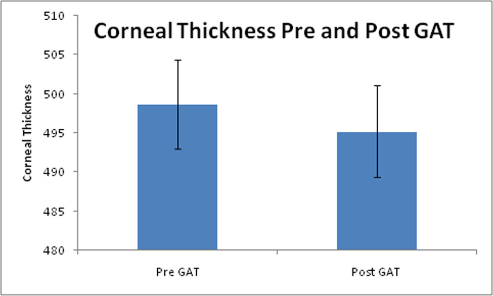
Fig1:Box plot showing difference in CCT before and after applanation tonometry (p<0.05)
Discussion:
GAT is stillthe golden standard in glaucoma practice for IOP measurement and CCT helps us in the appropriate interpretation of the GAT measurements. The Ocular Hypertension Treatment Study was thefirst study to document prospectively that the central corneal thickness appeared to be a strong risk factor for the development of primary open angle glaucoma (1).
Each clinician will have their own protocol depending on patient flow and ease with which investigations can be done with minimum patient waiting time.
Some believe that CCT measurement after GAT might be influenced by punctate erosions of the epithelium surface or its desquamation (2,3).
In a study done by Pourjavan. S, et al, mean CCT measurement before and after GAT was 565.22 ±32. 99 µ and 566.04 ±33.50 µ (p=0.34, r=0.98) respectively. The study suggested that the sequence of the two examinations didn’t matter (4).
This differs from our study, where we have noted significant differences in CCT before and after GAT. The reason for this could be that, in a study conducted by Pourjavan. S et al, pachymetry was measured using ultrasonic pachymeter with probe, whereas in our study, we have recorded CCT inRTVue OCT. It can be hypothesized that CCT measurement would be affected if epithelial surface is disturbed as optical coherence tomography relies on imaging of the anterior and posterior surface.
Also the use of topical anesthetic further causes punctate erosions again affecting CCT readings.
Conclusion:We noted significant changes in CCT post GAT. Thus, it is advisable to measure CCT using a non contact techniquebefore.
References:
1.BRANDT J.D., BEISER J.A., KASS M.A., GORDON M.O. − Central corneal thickness in the Ocular Hypertension Treatment Study. Ophthalmology 2001;108:1779-88.
2.LIM R., DHILLON B., KURIAN K.M., ASPINALL P.A., FERNIE K., IRONSIDE J.W. − Retentionof the corneal epithelial cells following Goldmann tonometry: Implications for CJD risks. BrJ Ophthalmol. 2003;87:583-6.
3.YEUNG K.K., KAGEYAMA Y.K., CARNEVALI T.− A comparison of Fluoracaine and Fluorox oncorneal epithelial cell desquamation after Goldmann applanation tonometry. J Am Optom Assoc. 2000;71:49-54.
4.Pourjavan S, Deghislghe C, Van Maelderen , Zeyen T (2005) Pachymetry before or after applanation tonometry. Does it matter? Bull SocBelgeOphthalmol 296:51–55
Leave a Comment