

Dr.Revathy Subramany, S14321
Objective:
To characterize the incidence of papilledema and obesity in females
Method:A retrospective analysison 59females, age12-35yrs withbilateral papilledema and decrease in vision seen at ourcentrefrom2014to 2017. History collection, Height and weight, Body Mass Index (BMI), Neurological tests,visual acuity, Slit lamp examination, (SLE), IOP, fundoscopy and optic coherence tomography (OCT) were done.
Results: Of59 patients, 10in puberty age. 11% had hormonal treatment. 68% had sudden increase of weight. 90% had BMI >37. SLE, IOP, CT &MRI appeared normal. 80% Uncorrected visual acuity between 20/30 and 20/40. OCT and fundoscopy confirmed papilledema. Lumbar puncture showed increase in pressure in all cases. 60% of cases had Lipid profile changes.
Conclusion:Our study revealed that the Recent weight gain and the degree of obesity, could be the reason for papilledema. The onset of puberty and hormonal variations also could provoke the occurrence of papilledema.
Introduction
Papilledema, is a condition of optic disc swelling that is secondary to elevated intracranial pressure1 Although Papilledema is common in obese patients, majority of patients with papilledema with complaint of decreased vision were seen in our Centre were in the reproductive age group which led us to do a study to identify various factors concerned with this. The condition may be self-limiting or enter a chronic phase with significant morbidity because of headache and visual loss. Population based surveys indicate a overall rate of visual impairment between 10 and 20%2-3.The incidence is approximately 1/100 000/year rising to 13/100 000/ year in women between 20 and 44 years who are 10% above ideal body weight and 19/100 000/ year in those 20% above ideal body weight.3-4
Method
A retrospective case analysis performed on all females patients with papilledema at our centre from 2014 to 2017. Age group were between 12 and 35 yrs. All patients had been undergone baseline examination of height, weight, blood pressure and Body Max index (BMI). Neuro Ophthalmic examinations includes Corrected and uncorrected visual acuity,pupillary evaluation for a relative afferent defect, color vision, extraocular motility, slit lamp exam, dilated fundus exam and visual fields Intraocular pressure, fundus photography, Optic Coherence Tomography (OCT).Neuroimaging to rule out an intracranial mass , lumbar puncture , visually Evoked potential (VEP), MRI were also performed .Detailed history collections which includes duration of symptoms, family history, past medical & surgical history, systemic disease specifically Diabetes, hypertention, thyroid dysfunction, ovarian diseases, cancers etc, use of medications particularly steroids, oral contraceptives, immune suppressant agents, protein supplements, antibiotics, estrogen receptor modulators, vitamin A preparations and associated factors of recent weight gain, puberty, pregnancy, sleep apnea were also recorded.
All patients had the complaints of disturbance in vision. BMI was divided into categories corresponding to World Health Organization BMI cutoff points 5-6; Normal (18.5-24.9), overweight (25-29.9), class1/11 obesity (30-39.9) and class 111 obesity (>_40).
Visual field examination was performed with Humphrey visual field machine (Carl Zeiss, Germany) were systematically reviewed and graded on a 0 to 3 scale as 0- normal, 1- enlargement of blind spot, 2- visual field defect Nasal or temporal, grade 3 – constricted visual field7.
Results:
59patients of papilledema were included in our study with the age group between 12 and 35yrs. 17% were in the puberty age(between 13 and 18yrs)pic1. Among the puberty age group, 8patients were obese and 1 had overweight but no obesity1 had normal BMI. 63% of patients had abnormal lipid profile who had BMI >37
90% of the total patients,were in the class 11 or class 111 obesity who had BMI >37. Only 4 patients had normal BMI;1in puberty age group and 3 in adult age. 68% of the patients had sudden increase in weight of 15 to 20kg within 6 to 12months and off these, 2 patients were sisters.
All patients had visual disturbances and were categorized as blurred vision 80% and 12% had double vision. 90% patients had complaint of headache .Vomiting reported by 5% and Dizziness in 25% of patients.pic2
11% patients were on progesterone treatment;3%were on tamoxifen treatment for breast cancer. One patient was taking ricotenemedication for acne vulgaris. 15% had serum cortisol level of >25 mcg/dL. 20% had high Thyroid Stimulating Hormone >10 mU/l.and were on Eltroxin treatment. 80% of patients had visualacuity (VA) 20/40 to 20/30, and not improving with pinhole ; 20% of the patients had corrected visual acuity (CVA) of 20/20.
Field abnormalities were detected with blind spot enlargement in 92%pic3 cases patients where as 7% had constricted visual fieldpic4and Nasal and temporal field defects in 11% cases pic5
Lumbar spinal tap in lying position was done initially for all cases to measure the CSF opening pressure and for CSF; all had CSF value of above 300mmH2O. Cytology results were within the normal values in all cases..Contrast CT and MRI were also appeared to be normal in all cases.
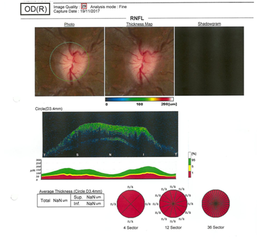
OCT showed the presence of papilledema in all casespic6.
Discussion
Our study showed that occurrence of papilledema strongly associated with obesity which is correlated with few previous studies whichalso addressed the role of the degree of obesity in vision loss in idiopathic intracranial hypertension. Although other known risk factors and systemic disease associations exist, weight remains the primary risk factor for IIH. The rates of obesity among patients with IIH have been examined in several studies worldwide. A retrospective study from Detroit Medical Center determined that more than 90% of IIH patients are obese 8Similar literature exists worldwide, with another study from Israel reporting that 97.2% of IIH patients were obese 9. A study done in the UK demonstrated that the incidence of IIH in obese women was 11.9 per 100,000 10.
The mechanism behind weight gain in idiopathic intracranial pressure and its association with raised ICP remains a debated issue. Adipose tissue has been increasingly viewed as a neuroendocrine tissue releasing chemical signals, in particular adipokines such as leptin, which could act directly on the choroid plexus or arachnoid granulation tissue, or indirectly via peripheral mechanism with consequent secondary central effects that modify CSF secretion and absorption11.A number of studies have evaluated cytokine and adipokines profiles in the serum and CSF of patients with IIH, but have been limited by small numbers and suboptimal control groups. Results have not been consistent, which may also be related to differences in the sensitivity of the assays used. Leptin, an adipokine which regulates satiety at the hypothalamus, was elevated in the serum in one study, but this effect was absent when BMI was controlled for.
CSF leptin levels appear elevated in IIH compared with controls matched for BMI, age and gender. However, it remains to be established whether dysregulation of adipokines and cytokines are pathogenic in dysregulating ICP, or merely reflect a consequence of the disease12,13.
Suggestions that centrally distributed adiposity transmits pressure, thereby generating raised intracranial pressure (ICP), are questionable, as few obese patients have elevated ICP. Additionally, studies of waisthip ratios in patients with IIH suggest that adiposity is predominantly in the lower body in IIH patients, by contrast with central adiposity of typical obesity14.
In contrast to absolute weight gain alone, papilledema was associated with 68% of our cases who had recent weight gainwhich was observed in within one year period. Similar association reported by another case control study of 50 patients, the amount of recent weight gainwas significantly higher among the five patients who experienced visual deterioration15
One of our patient was on ricotene treatment for Acne. Vitamin A toxicity has long been known to cause IIH. This association was first reported amongst explorers consuming polar bear liver, which has very high levels of vitamin A.16,17 Medications containing vitamin A are commonly prescribed to adolescents for acne. Retinol (a vitamin A derivative) has also been implicated in IIH. Arctic explorers developed elevated ICP following excessive ingestion of vitamin A rich polar bear liver16. Levels of retinol and retinol-binding protein have been evaluated in the serum and CSF of patients with IIH, with results of both elevated and decreased levels18,19,.
20 percent of our patients had high TSH values and were on eltroxine treatment whichcould be hypothesized that rapid correction of hypothyroidism with thyroxine, a major regulator of sodium transport, may result in altered CSF dynamics. Similar study reported that a child with hypothyroidism who developed IIH prior to thyroxine treatment has been reported 20In both these study groups were females and obese; so it cannot be confirmed that hypothyroidism and IIH were associated. Cases of pediatric IIH following thyroxine replacement therapy in juvenile hypothyroidism have been described 21-23. There is also a report of a child with concurrent new diagnoses of Graves’ disease and IIH who had resolution of IIH in the setting of thyroid suppressive therapy 24
IIH is predominantly seen in obese females of childbearing age; therefore, it is possible that female steroid hormones have a pathogenic role. However, a distinctive hormonal profile has not yet been described in IIH. Case reports have suggested a link between the development of pseudo tumorcerebri and oral contraceptives, as well as pregnancy, although the latter may also be associated with weight gain. These cases suggest a potential role for female sex hormones in IIH 25-27
In 2012, a theory unifying various effects on the mineralocorticoid receptor was proposed by Salpietro et al to explain a possible mechanism for increased CSF production and hence ICP in secondary IIH. The theory describes that stimulation of the mineralocorticoid receptors in the choroid plexus epithelium increases the activity of Na+/K+ ATPase, an active transporter of sodium for potassium ions. Thus, the movement of sodium ions into the cerebral ventricles increases, creating an osmotic gradient to drive CSF secretion and increase ICP28
Conclusion
Our study revealed that the Recent weight gain and the degree of obesity is strongly related to the development of papilledema but it is not a lone factor; The onset of puberty and hormonal variations also could provoke the occurrence of papilledema. Other factors are abnormal lipid profile, thyroid hormone, serum cortisol, medications, hormone therapy.
Fig.1
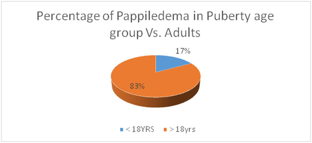
Fig.2
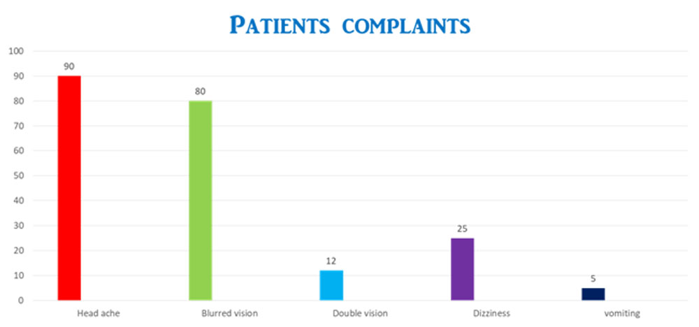
Fig. 3
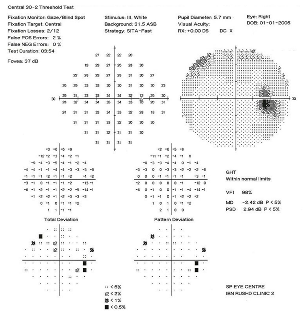
Fig. 4
Blind spot enlargement with constricted visual field
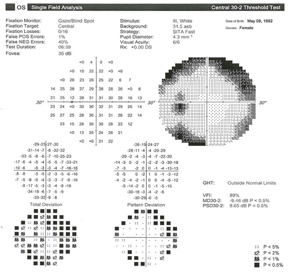
Fig.5
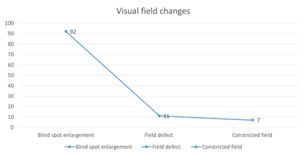
Fig.6
OCT of patient with bilateral Papilledema
References
- Garrity J. Papilledema. Merck Manual: Professional Version. April 2016; https://www.merckmanuals.com/professional/eye-disorders/optic-nerve-disorders/papilledema.
- Corbett JJ, Savino PJ, Thompson HS (1982) Visual loss in pseudotumorcerebri. Follow-up of 57 patients from five to 41 years and a profile of 14 patients with permanent severe visual loss. Arch Neurol,39, 461–474.
- Radhakrishnan K, Ahlskog JE, Cross SA, Kurland LT, O’Fallon WM (1993) Idiopathic intra-cranial hypertension (pseudotumorcerebri). Descriptive epidemiology in Rochester, Minn,1976 to 1990.Arch Neurol ,50 , 78–80.3,4
- Radhakrishnan K, Thacker AK, Bohlaga NH, Maloo JC, Gerryo SE (1993) Epidemiology of idiopathic intracranial hypertension: a prospective and case–control study.JNeurol Sci116,18–28
- Bruce BB, Kedar S, Van Stavern GP, Corbett JJ, Newman NJ, Biousse V. Atypical idiopathic intracranial hypertension: normal BMI and older Neurology. 2010; 74:1827–1832.[PubMed: 20513819]
- Physical status: the use and interpretation of anthropometry. Report of a WHO expert committee.World Health Organization Technical Report Series. 1995; 854:1–452. [PubMed: 8594834]\
- Frisen L. Swelling of the optic nerve head: a staging scheme. J NeurolNeurosurg Psychiatry. 1982;45:13–18. [PubMed: 7062066]
- Andrews, Laura A, Liu, Grant T, and Ko, Melissa W. “Idiopathic Intracranial Hypertension and Obesity.” Horm Res Paediatr 2014; 81: 217–225.
- Kesler A, Gadoth N. “Epidemiology of idiopathic intracranial hypertension in Israel.” J Neuroophthalmol 2001; 21: 12–14.
- Raoof N, Sharrack B, Pepper IM, et al. “The incidence and prevalence of idiopathic intracranial hypertension in Sheffield, UK.” Eur J Neurol 2011; 18: 1266–1268.
- Keira A Markey,1 Maria Uldall,2 Hannah Botfield,1 Liam D Cato,Idiopathic intracranial hypertension, hormones, and 11β-hydroxysteroid dehydrogenases J Pain Res. 2016; 9: 223–232.
- Ball AK, Sinclair AJ, Curnow SJ, et al. Elevated cerebrospinal fluid (CSF) leptin in idiopathic intracranial hypertension (IIH): evidence for hypothalamic leptin resistance? ClinEndocrinol (Oxf) 2009;70:863–9. doi:10.1111/j.1365-2265.2008.03401.x [PubMed]
- Lampl Y, Eshel Y, Kessler A, et al. Serum leptin level in women with idiopathic intracranial hypertension. J NeurolNeurosurg Psychiatry 2002;72:642–3. doi:10.1136/jnnp.72.5.642 [PMC free article] [PubMed.
- Kesler A, Kliper E, Shenkerman G, et al. Idiopathic intracranial hypertension is associated with lower body adiposity. Ophthalmology 2010;117:169–74. doi:10.1016/j.ophtha.2009.06.030 [PubMed]
- Wall M, George D. Idiopathic intracranial hypertension. A prospective study of 50 patients. Brain. 1991;114(Pt 1A):155–180. [PubMed].
- Rodahl K, Moore T. The vitamin A content and toxicity of polar bear and seal liver. Biochem J 1943;37:2:166-168.
- Lee AG. Pseudotumorcerebri after treatment with tetracycline and isotretinoin for acne. Cutis 1995;55:3:165-168.)
- Warner JE, Bernstein PS, Yemelyanov A, et al. Vitamin A in the cerebrospinal fluid of patients with and without idiopathic intracranial hypertension. Ann Neurol 2002;52:647–50. doi:10.1002/ana.10377 [PubMed]
- Tabassi A, Salmasi AH, Jalali M. Serum and CSF vitamin A concentrations in idiopathic intracranial hypertension. Neurology 2005;64:1893–6. doi:10.1212/01.WNL.0000163556.31080.98 [PubMed
- Adams C, Dean HJ, Israels SJ, Patton A, Fewer DH: Primary hypothyroidism with intracranial hypertension and pituitary hyperplasia. PediatrNeurol 1994;10:166–168.
- Campos SP, Olitsky S: Idiopathic intracranial hypertension after L-thyroxine therapy for acquired primary hypothyroidism. ClinPediatr (Phila) 1995;34:334–337.
- Raghavan S, DiMartino-Nardi J, Saenger P, Linder B: Pseudotumorcerebri in an infant after L-thyroxine therapy for transient neonatal hypothyroidism. J Pediatr 1997;130:478–480.
- Van Dop C, Conte FA, Koch TK, Clark SJ, Wilson-Davis SL, Grumbach MM: Pseudotumorcerebri associated with initiation of levothyroxine therapy for juvenile hypothyroidism. N Engl J Med 1983;308:1076–1080.
- Merkenschlager A, Ehrt O, Muller-Felber W, Schmidt H, Bernhard MK: Reversible benign intracranial hypertension in a child with hyperthyroidism. J PediatrEndocrinolMetab 2008;21:1099–1101.
- Finsterer J, Kues EW, Brunner S. Pseudotumourcerebri in a young obese woman on oral contraceptives. Eur J ContraceptReprod Health Care. 2006;11(3):237–240. [PubMed]
- Chan JW. Idiopathic intracranial hypertension associated with depot medroxyprogesterone. Eye (Lond) 2006;20(12):1396–1397. [PubMed]
- Kesler A, Kupferminc M. Idiopathic intracranial hypertension and pregnancy. ClinObstet Gynecol. 2013;56(2):389–396. [PubMed]
- Salpietro V, Polizzi A, Berte LF, et al. Idiopathic intracranial hypertension: a unifying neuroendocrine hypothesis through the adrenal-brain axis. Neuro Endocrinol Lett. 2012;33(6):569–573. [PubMed]
Leave a Comment