

Dr.DIVYA BETALA,Dr.Meenakshi Swaminathan,Dr.Srikanth R
ABSTRACT
AIM:
To compare the efficacy of cycloplegia and extended fogging with open field auto-refractometer as methods of accommodation control in teenage children in India
METHODS:
This prospective study was carried out at the pediatric outpatient department of Sankara Nethralaya, Chennai between April 2017 and February 2018. Children between 11-17 years with refractive error and without any associated ocular and systemic co-morbidities were included in the study. All subjects underwent a comprehensive eye examination, dry retinoscopy, closed field autorefraction (CF) and open field auto refraction (OF) with extended fogging for 20 minutes with +2.00 DS lenses. Following this, cycloplegic refraction was carried out with retinoscopy and closed field auto refraction. This study tested the null hypothesis that there is no significant difference between the cycloplegic autorefraction and dry open field autorefraction with extended fogging.
RESULTS:
The mean (SD) age of the 99 subjects was 13 (1.8) with 53 males. The correlation between right and left eye refraction data was >0.9 (Pearson’s correlation, p <0.001) and thus data from right eye was used for analysis. The spherical and cylindrical component ranged between +8.50 DS to -7.00 DS and -0.50 DC to -5.50 DC respectively. A 2×2 repeated measures ANOVA (RM-ANOVA) revealed statistically significant difference between cycloplegic and non-cycloplegic refraction techniques for spherical component (RM ANOVA, F (1, 96) = 36.5; p<0.001). There was no statistical and clinically significant difference between cycloplegic retinoscopy and open field autorefraction with extended fogging (RM ANOVA, F (1, 96) = 2.843; 95% CI for difference in mean: -0.2 to -0.5 DS; p>0.05). The cylindrical component also did not show statistically significant difference between refraction techniques under cycloplegic and non-cycloplegic conditions (RM ANOVA, F (1, 85) = 1.46. p>0.05).
Conclusion:
Open field autorefraction with extended fogging is clinically comparable to cycloplegic closed field autorefraction among teenage children.
INTRODUCTION
Refractive errors have since long known to be the major cause of visual impairment in India. Several studies have addressed the magnitude of this problem. In a study done in rural India, refractive error was the main cause of visual impairment in children aged between 7 and 15 years.1 Systematic reviews of previous studies have also been performed.2
Teenage children are particularly involved and are ideal candidates for research as they are co operative for reliable measurement taking. Therefore, large number of research projects have been done involving school going children, especially middle to high school. 3Myopia in particular seems to be a growing problem, in view of increasing prevalence and progressive nature of the disease. Hence, it has been extensively addressed in studies.4
The above mentioned studies, however, cannot be compared as they have all used different methods of accommodation control. Most screening studies have performed an automated refraction, sans cycloplegia, while others have used cycloplegic retinoscopy as the preferred method, it being the gold standard in refractive error measurement due to its accommodation control properties.6-9 Latent errors are common, especially in cases of high hyperopia in the absence of adequate accommodation control.10
Cycloplegic drops, however, have their own disadvantages in terms of time taken, cost, inconvenience and difficulty in instillation especially in younger children. Hence, cycloplegic refraction is not a very popular choice in research settings where large populations have to be screened.11
An alternative to cycloplegic drops has been found in the form of optical fogging, where plus lenses placed before the eyes help in relaxing accommodation when placed for a sufficient amount of time. The amount of fogging varied between studies: +1.50D fogging lenses were used in one study12 whilst increasing amounts of plus lens power applied in a stepwise procedure were used in another.13
Hopkins et al nominated a 20-minute time period in their study because it was considered a sufficiently conservative amount of time to relax accommodation,14 whilst having the advantage of being shorter than the time required for onset of cycloplegic agents. In our study, we compared spherical refractive error results measured at baseline and using two different accommodation control methods: extended optical fogging and cycloplegia in teenagers. Autorefraction and retinoscopy were used to measure the most plus spherical refractive power, with the aim of determining whether extended optical fogging was comparable to cycloplegia for either or both measurement techniques.
METHODS:
This prospective study was carried out at the pediatric outpatient department of Sankara Nethralaya, Chennai between April 2017 and February 2018. Institutional Review Board clearance was obtained after drafting the project proposal. Based on Hopkins et al’s study, 14 sample size for comparison between two or more groups at 95% confidence interval and 80% power, the calculated sample size was 34 in each refractive group, amounting to a total of 102 subjects, for comparison between the three refractive techniques of retinoscopy, open field and closed field autorefraction.Open field autorefraction was done using the Grand Seiko Auto Ref/Keratometer WAM-5500 and closed field autorefraction using the Topcon KR 800 machine.
Myopia was defined as spherical refractive error of at least -0.50 D and hyperopia as +1.50 D or more. Emmetropia was defined as refractive errors in the range of -0.25 D to +1.25 D without an astigmatic component exceeding 0.50 D. Refractive errors with astigmatic component greater than 0.50 D were classified as simple, compound myopic, and compound hyperopic and mixed astigmatism based on the spherical component. For analysis purposes, compound myopic, hyperopic and mixed astigmatism were considered as myopia and hyperopia based on the spherical component.
All children had best-corrected visual acuities of 6/7.5 or better. Subjects with refractive errors and without any associated ocular and systemic co morbidities were included. Subjects with allergic conjunctivitis but no complications of the same, were included. All participants and their guardians were given a full explanation of the experimental procedures. Written informed consent was obtained from both the participant and his or her guardian before involvement, with the option to withdraw from the study at any time.
After a preliminary eye examination, including visual acuity testing, subjects were fogged using +2 dioptre lenses in a trial frame with loose lenses. They were asked to be seated in the waiting hall of the department for 20 minutes and advised to look at a distance target (for example, the television). Following extended fogging, a single trained optometrist performed the retinoscopy using a streak retinoscope. Working distance lenses of +1.50D were used while the participant viewed a 6/60 letter at 6 m during retinoscopy.
A single fellow of the department of pediatric ophthalmology, then obtained the open field and closed field autorefractometer readings and was blinded to the reading obtained by retinoscopy. The distance fixation target for open field autorefraction (performed in a 3-m room) was a 6/150 symbol (the letter V) and was positioned such that the optical axis of the instrument and the participant’s line of sight when viewing the target was aligned. (Figure 1) The large fixation target of a black letter on a plain white wall was selected because it would not provide a strong stimulus for accommodation. The participant was seated comfortably with his or her chin on the chin rest, head against the forehead rest, and eyes level with the eye mark and viewed binocularly the fixation target binocularly through the window. Five repeated measurements were performed on the selected eye, and the mean was calculated (using the most plus spherical power result).
In the closed field autorefractor also, the participant was seated comfortably with his or her chin on the chin rest, head against the forehead rest, and eyes level with the eye mark. The subject was then asked to look at the house in the display of the autorefractor and five readings were obtained and a mean was recorded.
This cycle of readings by extended optical fogging was followed by cycloplegic drops being instilled in the eyes of subjects (homatropine 2% and cyclopentolate 1%) three times at an interval of 10 minutes. A history of allergic reaction to homatropine or cyclopentolate was obtained prior to instillation. Thirty minutes following the last drop, the pupil was checked for non reactivity and subjects were taken up for cycloplegic refraction and open and closed field autorefractometry using the above mentioned protocols with the optometrist and the fellow blinded to each other’s readings at all times. The patients were then led to visit the concerned consultant for examination.
Null hypothesis:
There is no difference between open field autorefraction with extended fogging compared to gold standard cycloplegic retinoscopy and closed field autorefraction.
Statistical analysis:
Repeated measuresanalysis of variance (RM-ANOVA) was utilized to compare the refractive outcomes for sphere and spherical equivalent components for emmetropia, myopia, hyperopia and astigmatism for refraction techniques of open field autorefraction with extended fogging, cycloplegic retinoscopy and closed field autorefraction. Bonferroni post-hoc comparisons with conservative p-values were applied for multiple comparisons. A p-value of <0.05 was considered to be statistically significant for the overall comparison. A difference of >0.50 D was considered as clinically significant difference between refractive outcomes with various refraction techniques.
RESULTS
The overall sample size for the study was 99. The mean (SD) age of the 99 subjects was 13 (1.8) with 53 males. The correlation between right and left eye refraction data was >0.9 (Pearson’s correlation, p <0.001) and thus data from right eye was used for analysis. The spherical and cylindrical component ranged between +8.50 DS to -7.00 DS and -0.50 DC to -5.50 DC respectively. A 2×2 repeated measures ANOVA (RM-ANOVA) revealed statistically significant difference between cycloplegic and non-cycloplegic refraction techniques for spherical component for the overall comparison (RM ANOVA, F (1, 96) = 36.5; p<0.001). There was no statistically and clinically significant difference between cycloplegic retinoscopy and open field autorefraction with extended fogging (RM ANOVA, F (1, 96) = 2.843; 95% CI for difference in mean: -0.2 to -0.5 DS; p>0.05). The cylindrical component also did not show statistically significant difference between refraction techniques under cycloplegic and non-cycloplegic conditions (RM ANOVA, F (1, 85) = 1.46. p>0.05).
The profile of refractive errors is shown in Table 1. The mean (SD) for the spherical equivalent and power vector components are shown in Table 2.
Table 1 – Profile of refractive errors
| Total Sample size | 99 |
| Emmetropia | 31 |
| Simple Myopia
Compound myopic astigmatism |
17
11 |
| Simple Hyperopia
Compound hyperopic astigmatism |
6
15 |
| Simple Astigmatism | 12 |
| Mixed Astigmatism | 7 |
Table 2 – Mean and SD of spherical equivalent (M), and power vectors (J0, J45) based on cycloplegic retinoscopy
| Refractive error type | Mean ± SD of M | Mean ± SD of J0 | Mean ± SD of J45 |
| Emmetropia (N=31) | 0.63 ± 0.37 | 0.12 ± 0.16 | 0.03 ± 0.1 |
| Myopia (N=28) | -3.4 ± 2 | 0.35 ± 0.56 | 0.1 ± 0.27 |
| Hyperopia (N=28) | 2.00 ± 1.7 | 0.99 ± 0.76 | 0.21 ± 0.39 |
| Astigmatism (N=12) | -0.19 ± 0.64 | 0.97 ± 0.4 | 0.09 ± 0.21 |
Individual comparisons for Myopia, Hyperopia and Astigmatism
A repeated measures Analysis of variance for myopic subjects showed no statistically significant difference between the three refraction techniques (RM ANOVA, F (2, 26) = 2.1; p=0.13) (Figure 1).
A repeated measures analysis of variance for hyperopic subjects showed statistically significant difference for overall comparison (RM ANOVA, F (2, 26) = 4.9; p=0.006), but post-hoc analysis did not show statistically significant difference between the three refraction techniques (Figure 2). For astigmatism as well, there was no statistically significant difference between the three refraction techniques (RM ANOVA, F (2, 10) = 1.02; p=0.39)
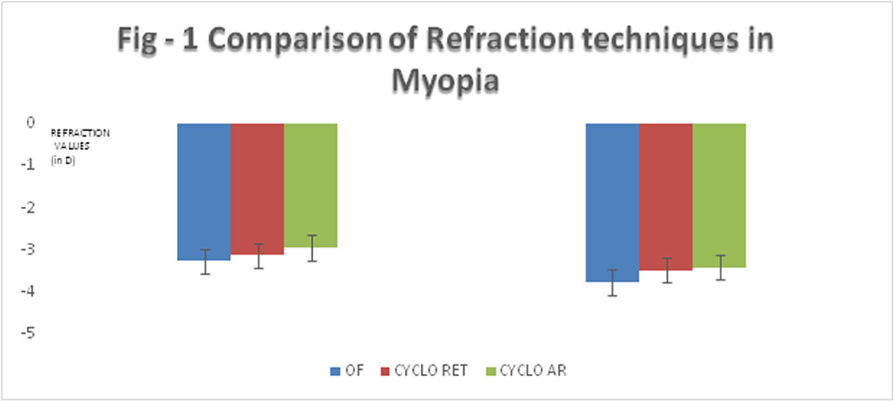
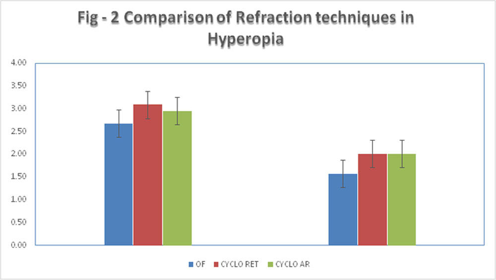
Table 3: Mean (SD) spherical and spherical equivalent of myopia, hyperopia and astigmatism as obtained with the three refraction techniques
| Open Field extended fogging
Sphere Mean (SD) |
Cycloplegic retinoscopy
Sphere Mean (SD) |
Cycloplegic autorefraction
Sphere Mean (SD) |
Open Field extended fogging
Spherical equivalent Mean (SD) |
Cycloplegic retinoscopy
Spherical equivalent Mean (SD) |
Cycloplegic autorefraction
Spherical equivalent Mean (SD) |
|
| Myopia | -3.27 (1.61) | -3.13(1.69) | -2.94 (1.79) | -3.76 (1.8) | -3.48 (2.04) | -3.41 (2.07) |
| Hyperopia | 2.68 (1.8) | 3.09 (1.7) | 2.96 (2.1) | 1.58 (1.7) | 2.01 (1.7) | 2.01 (2.0) |
| Astigmatism | 0.77 (0.5) | 0.79 (0.3) | 0.83 (0.4) | -0.08 (0.8) | -0.2 (0.6) | -0.02 (0.9) |
Discussion
The results of our study suggest that open field autorefraction with extended fogging can be considered as an equivalent for cycloplegic refraction among 11-17 year old children with refractive errors. The results of our study suggest that the difference between cycloplegic retinoscopy and open field autorefraction is well within the clinically agreeable limits of 0.50 D for myopic, hyperopic, and astigmatic refractions.
This is in contrast to the findings of the study by Hopkins et al which show optical fogging to be less effective than cycloplegia for a group of school children aged 6 to 13 years. 14 This difference could be accounted for by the different age group of children of our study. Teenagers are likely to be more compliant to the instructions given while fogging compared to primary school aged children.
Earlier studies show that the fogging technique is a valid method of controlling accommodation for low levels of fogging, 15 but yet another study found varying results with optical fogging.16
Antonio et al’ s study results show that the average spherical equivalent refraction using optical fogging as a method of accommodation control is very similar to that obtained under cycloplegia, while both situations (cycloplegia or fogging lenses) give more positive or less negative values than open-field autorefraction with no additional control of accommodative response. When the average values of spherical equivalent were compared both accommodation control strategies were almost equally successful. 17 Emmetropic and hyperopic groups displayed significant evidence that fogging lenses method was as effective as cycloplegia to prevent myopic shift. Fogging lenses seems to be effective for emmetropes, cycloplegia was more effective for hyperopes; however, differences between both methods were 0.15 D and 0.16 D, respectively.
Another study found that non-cycloplegic refraction done with contralateral fogging technique as effective as the gold standard cycloplegic refraction technique for the measurement of refractive error in healthy, non-strabismic children.18
To conclude, our study has proven that optical fogging can be used effectively as an alternative to cycloplegia in teenage children, thus avoiding the inconvenience associated with the cycloplegic drops. In addition to the absence of adverse response from the administration of the cycloplegic, this methodology could be used by non-medical staff and optometrists not allowed to use diagnostic drugs.
References:
1.Dandona R, Dandona L, Srinivas M, Sahare P, Narsaiah S, Muñoz SR, Pokharel GP, Ellwein LB. Refractive error in children in a rural population in India. Invest Ophthalmol Vis Sci. 2002 Mar;43(3):615-22. PubMed PMID: 11867575.
2.Sheeladevi S, Seelam B, Nukella PB, Modi A, Ali R, Keay L. Prevalence ofrefractive errors in children in India: a systematic review. ClinExp Optom. 2018 Jul;101(4):495-503. doi: 10.1111/cxo.12689. Epub 2018 Apr 22. Review. PubMed PMID: 29682791.
3.Parmar A et al. Int J Community Med Public Health. 2017 Sep;4(9):3376-3379
4.Saxena R, Vashist P, Tandon R, et al. Prevalence of Myopia and Its Risk Factors in Urban School Children in Delhi: The North India Myopia Study (NIM Study). Zhou X, ed. PLoS ONE. 2015;10(2):e0117349. doi:10.1371/journal.pone.0117349.
5.Ojaimi E, Rose KA, Smith W, Morgan IG, Martin FJ, Mitchell P. Methods for a population-based study of myopia and other eyeconditions in school children: the Sydney Myopia Study. Ophthalmic Epidemiol 2005;12:59Y69
6.Twelker JD, Mutti DO. Retinoscopy in infants using a near non- cycloplegic technique, cycloplegia with tropicamide 1%, and cyclo- plegia with cyclopentolate 1%. Optom Vis Sci 2001;78:215Y22.
7..Erdurmus M, Yagci R, Karadag R, Durmus M. A comparison of photorefraction and retinoscopy in children. J AAPOS 2007;11: 606Y11.
8.Williams C, LumbR, HarveyI,Sparrow JM. Screening forrefractive errors with the Topcon PR2000 Pediatric Refractometer. Invest Ophthalmol Vis Sci 2000;41:1031Y7.
9.SteeleG,IrelandD,BlockS. Cycloplegic autorefractionresultsinpre- schoolchildren using the Nikon Retinomax Plus and the Welch Allyn SureSight. Optom Vis Sci 2003;80:573Y7.
10.Junghans BM, Crewther SG. Little evidence for an epidemic of myopia in Australian primary school children over the last 30 years. BMC Ophthalmol 2005;5:1.
11.Twelker JD, Mutti DO. Retinoscopy in infants using a near noncycloplegic technique,
cycloplegia with tropicamide 1%, and cycloplegia with cyclopentolate 1%. Optometry and Vision Science 2001;78:215-22.
12.Chan OYC, Edwards M. Comparison of cycloplegic and noncycloplegic retinoscopy in Chinese pre-school children. Optometry and Vision Science 1994;71:312-8.
13.Suryakumar R, Bobier WR. The manifestation of noncycloplegic refractive state in pre school children is dependent on autorefractor design. Optometry and Vision Science 2003;80:578-86
14. Hopkins S, Sampson GP, Hendicott P, Lacherez P, Wood JM. Refraction inchildren: a comparison of two methods of accommodation control. Optom Vis Sci.2012 Dec;89(12):1734-9. doi: 10.1097/OPX.0b013e318277182c. PubMed PMID: 23142881.
15.Ward PA, Charman WN. An objective assessment of the effect of fogging on 358 accommodation. American Journal of Optometry and Physiological Optics 1987;64:762-7.
16.Reese EE, Fry GA. The effect of fogging lenses on accommodation. American Journal of Optometry and Archives of American Academy of Optometry 1941;18:9-16.
17. Queirós A, González-Méijome J, Jorge J. Influence of fogging lenses andcycloplegia on open-field automatic refraction. Ophthalmic Physiol Opt. 2008Jul;28(4):387-92. doi: 10.1111/j.1475-1313.2008.00579.x. PubMed PMID: 18565095.
18.Yeotikar, Nisha & Chandra Bakaraju, Ravi & S. Roopa Reddy, P & Prasad, Kalyani. (2007). Cycloplegic refraction and non-cycloplegic refraction using contralateral fogging: A comparative study. Journal of Modern Optics. 54. 1317-1324. 10.1080/09500340600855346.
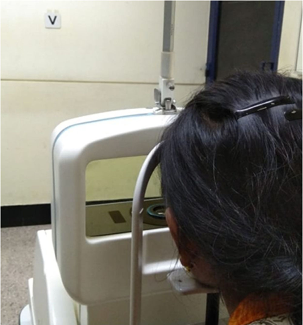
Figure 1: Patient seated on the open field autorefractometer fixing at the letter “V” 3 m away
Leave a Comment