

Dr.TANYA JAIN, Dr.Alok Sen
Introduction-
Vogt-Koyanagi-Harada disease (VKH) is a severe bilateral granulomatous posterior or pan-uveitis associated with serous retinal detachments, disc edema, and vitritis, with eventual development of a sunset glow fundus. Systemically, it may be associated with tinnitus, hearing loss, vertigo, meningismus, poliosis, and vitiligo, although not all patients present with the complete constellation of these extra-ocular findings. 1
VKH appears to have a predilection for pigmented races with significant regional and global variation. 1VKH is not common in the United States and makes up only about 3-4% of referrals to tertiary care centers. 2 VKH is the most common cause of pan-uveitis in India, with a prevalence of 1.5 to 3.5%. 3,4
VKH presents clinically in four different phases: prodromal, acute uveitic, convalescent, and chronic recurrent. 1The acute phase is characterized by a sudden onset, bilateral granulomatous uveitis, with bilateral serous retinal detachments, pockets of sub-retinal fluid and choroidal thickening, blurring of vision, and conjunctival injection. 1
Since October 1999, the diagnosis of VKH disease is based on the Revised Diagnostic Criteria. According to this, the presentations of VKH were described as complete, incomplete, and probable.5
Acute VKH must be treated aggressively with corticosteroids initially, with a minimum treatment duration of 6 months, along with introduction of corticosteroid sparing agents such as cyclosporine A, methotrexate or mycophenolate mofetil in non responders.1
The literature available up till now on Acute VKH describes the various findings on multimodal imaging modalities8,9,10,11,12,13,14or describes the clinical spectrum of the disease in a pre-determined population. The authors have described these findings on multimodal imaging modalities and correlated them with the disease process and also compared and correlated the various imaging modalities.
However after a comphrehensive literature review it becomes apparent that the literature lacks a comprehensive prospective study which correlates all findings commonly found associated with an acute episode of VKH on SD-OCT with the visual acuity of a patient with the treatment protocol remaining unaltered. This study along with the correlation also describes the findings on various imaging modalities and the clinical spectrum of the disease in a tertiary care hospital in Central India. This would be beneficial not only in aiding with the diagnosis, determining disease progression and monitoring therapy but also finds out the prognostic outcomes of patients with Acute VKH disease.
Materials And Methods
We conducted a Longitudinal, Prospective, Non-comparative, observational study of 30 patients (60 eyes) diagnosed as Acute VKH disease At Retina and uvea clinic of Sadguru Netra Chikitsalaya, Jankikund, Chitrakoot from May 2016 to Novemeber 2017. Thesample size was calculated using the N = (Za/2)2 s2 / d2 formula based on outcome variable of Log Mar BCVA in different OCT grading with minimum difference of 0.05 with SD of 0.13 in a published study by Zhou M et al on correlation of visual acuity and OCT changes in late stage VKH .15
Inclusion criteria included all patiends diagnosed with Acute VKH with the Revised International diagnostic criteria and we excluded patients withCo-existing diseases such as diabetic retinopathy, epi-retinal membrane and retinal vein occlusions, refractive error exceeding -8 diopters or +5 diopters, with chronic or chronic recurrent uveitis, in whom appropriate scans could not be obtained, non compliant and patients not adhering to follow-up criteria.
All the patients underwent a complete ophthalmologic examination and the the findings were recorded in the case report file attached at the end. Patients underwent both systemic and ophthalmologic investigations. Ophthalmic investigations included Multimodal imaging colored fundus photography, Spectral Domain-Optical Coherence Tomography (RTvue, Optovue), High Definition-OCT (Avanti, Optovue), fundus fluorescein angiography (Visucam 500 by Zeiss with Color/FA/FAF/ICG), B- scan ultrasonography, indo-cyanine angiography (Visucam 500 by Zeiss with Color/FA/FAF/ICG) and OCT-Angiography (Avanti, Optovue) at presentation.Serial follow-up of the patients with complete ophthalmic examination, follow-up BCVA and HD-OCT and OCT-Angiography were done after the initiation of therapy at 10 days, 1month, 3 months and 6 months. On the HD-OCT scan the following parameters were documented on every serial visit- Central Macular Thickness, Average Macular Thickness, Area of Exudative RD, Hyper-Reflective Dots, RPE Undulation Index, Intra-Membranous Structures, Intra Retinal Cysts and Area of Intra-Retinal Cysts, Pigment Epithelial Detachment , IS-OS Junction and Choroidal Thickness. Area of flow-void areas in the choroidal capillary scans of the OCT-Angiography scans were measured and followed up on each visit.
OCT examination of dilated pupils were captured and scans of 12 mm length along the horizontal and vertical meridians through the foveal center were taken along with a 12mm macular raster scan. The central macular thickness was measured in mm at the fovea using caliper tool of the OCT as the distance between the ILM and the RPE-Bruch’scomplex.Themaximumheightofretinaldetachmentinmm2 at the macula and area of detachment of neurosensory retina was measured using the caliper tool and area tool in Avanti, Optovue. The presence of sub retinal membrane structures and hyper reflective dots was documented at the fovea.
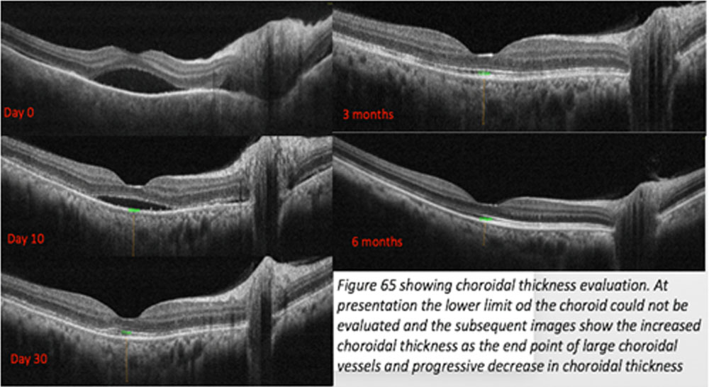
The RPE undulations are defined as wavy protrusions of the RPE more than two peaks and troughs and the RPE undulation index was calculated.The RPE undulation index was measured as the length of the RPE in the horizontal and vertical scans to the total scan length on a foveal centered scan.These measurements were done and repeated on every follow-up visit. Choroidal thickness was defined as the vertical distance between the RPE line and the hyper-reflective line behind the large choroidal vessel layers at the fovea on HD-OCT. When the choroid was so thick that the choroidal-scleral interface was not visible, a thickness value of 500 mm was assigned.The retino-choroidal-screal thickness was measured in mm by the caliper tool in the B-scan.FFA and ICG angiography interpretations were entered in the case report form.
Statistical Methods-
All statistical calculations were done using computer programs Microsoft Excel 2013 (Microsoft Corporation, NY, USA) and SPSS (Statistical Package for the Social Science; SPSS Inc., Chicago, IL, USA) version 21. Results on continuous measurements were presented as mean with standard deviation and results on categorical measurements were represented in number (%). Quantitative variables were analyzed at different intervals using multiple measure ANOVA test and qualitative measures at different intervals were analyzed using Mc. Nemar test. Quantitative measures were compared with each visit using multiple comparison sidak test. For determining the association of visual acuity with OCT variables multiple regression analysis was used.To determine the association of final visual acuity with baseline OCT parameters, unpaired t-test for normally distributed quantitative variables and for comparing categorical data, Chi square test was performed.
Results–
The mean age at presentation was 37.30 (SD=10.4) years with minimum and maximum being 16 and 60 years respectively. Out of the 30 patients, 17 (57%) were females and 13 (43%) were males. The mean duration of disease at which the patients presented was 14.53 (SD=11.50) days with a minimum of 3 and maximum of 60 days. Out of the 30 patients 2 patients were diagnosed cases of hypertension and 1 of diabetes mellitus. 2 patients had a previous history of treated pulmonary tuberculosis. One patient each had a previous history of spondylopathy, anterior scleritis and chikungunya fever. Purely based on clinical history 50% of the patients complained of Simultaneous symptoms in both the eyes. Among the rest of the 15 patients 10 patients complained one eye being involved before the other eye and the remaining 5 noticed symptoms in only one eye.
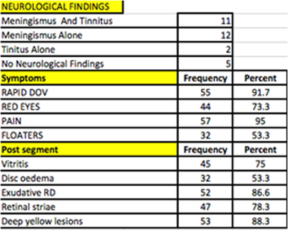
Out of the 30 patients 23 (76.7%) presented with meningismus and 13 (43.3%) presented with tinnitus. 11 patients presented with both meningismus and tinnitus while 2 patients presented with only meningismus. Rest 5 patients did not have any neurological findings and hence were classified as possible VKH (Table 2). None of the patients had alopecia or vitiligo. The mean BCVA (Log MAR) 60 eyes at presentation was .7467 with standard deviation of .67810. The mean IOP mmHg (60 eyes) as measured by applanation tonometry was 13.78mmHg with standard deviation of 2.738. The frequency of signs and symptoms have been given in table1. The most common was pain, dull aching present throughout the day which was associated with redness 73.3% of the times.The most common posterior segment findings were exudative retinal detachment and deep yellowish retinal lesions.
Since we studied only acute cases hence according to the Revised diagnostic criteria 1999 none of the patients qualified into the complete category . Maximum i.e. 25 out of 30 patients (83.3%) were diagnosed as probable VKH and 5 (16.7%) were diagnosed as possible VKH disease (Table 2).
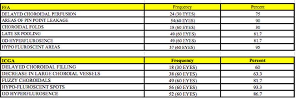
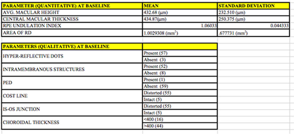
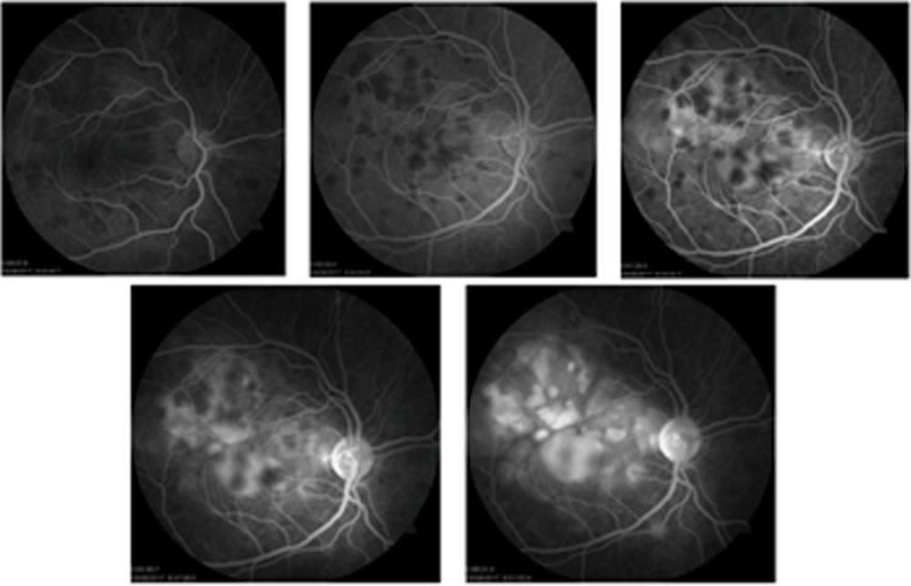
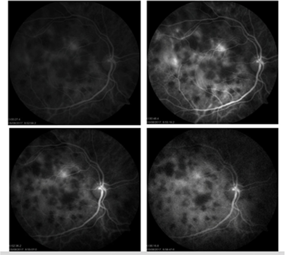
Table 3 shows the FFA and ICGA findings in these patients at baseline and Figure 2 and 3 show the frames of FFA and ICGA respectively demonstrating the typical features. and Table 4 shows the OCT baseline parameters both quantitative and qualitative.
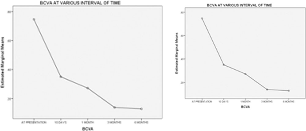
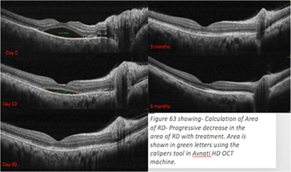
Figure 1 show the trend of visual acuity, RPE undulations, Central macular thickness and area of RD. All the quantitative parameters show a decline with a bump at 1 month due to recurrance in 4 cases. All are statistically significant using repeated measures ANOVA test.
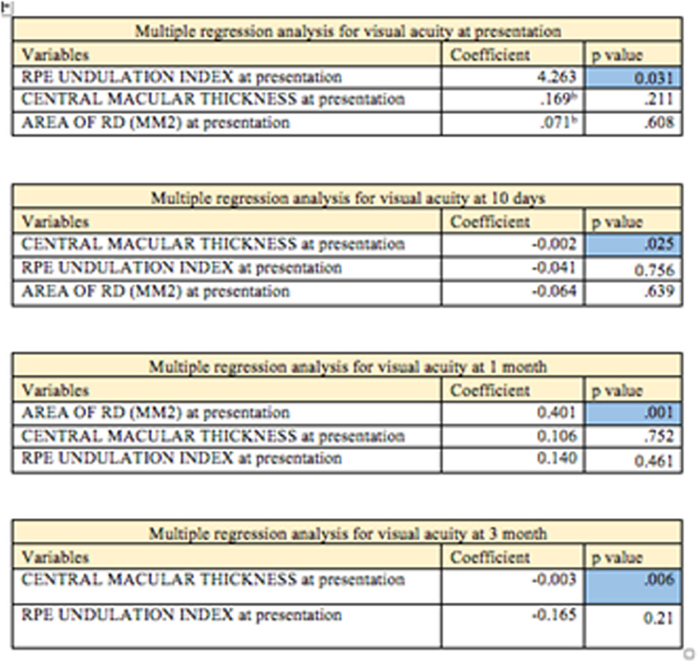
Upon comparing the baseline OCT parameters with the baseline visual acuity using multiple regression analysis RPE undulation index was significantly associated p=0.025 (Table 5) with BCVA. This implies higher RPE undulation index is associated with poor visual acuity at presentation using multiple regression analysis.At 10 days central macular thickness was significantly associated p=0.001 (Table 5) with BCVA. This implies higher CMT is associated with poor visual acuity at 10 days post treatment using multiple regression analysis.At 1 month area of RD was significantly associated p=0.006 (Table 5) with BCVA. This implies higher area of RD is associated with poor visual acuity 1 month post treatment using multiple regression analysis which could be attributed to the recurrences in 8 eyes at 1 month.
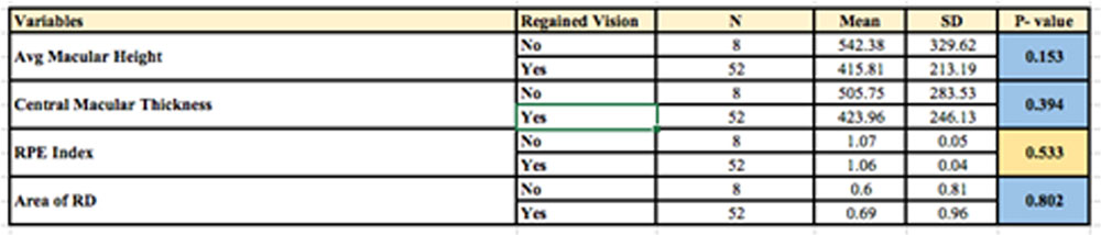
The final visual acuity at 6 months was classified into 2 groups. Those achieving final BCVA better than on equal to 6/9 (Log mar 0.2) and those who did not achieve the final BCVA of better than 6/9 (Log mar 0.2). The baseline OCT parameters both quantitative and qualitative were compared with these 2 groups using chi square test and paired student t test. None of the parameters turned out to be significant. Hence OCT parameters may not help predict the final visual outcome. However RPE undulation index was nearing the significant P value and may be helpful in predicting the final visual outcome along the choroidal thickness (Table 6).
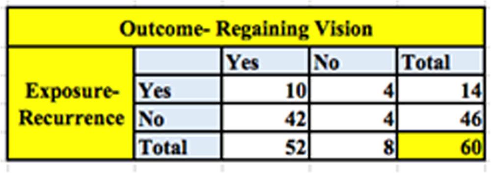
Upon studying the the patients with recurrence, 14 eyes out of 60 eyes i.e. 23% had a recurrence at some time or the other during the 6 months. Out of the 60 eyes 52 (86.6%) eyes regained vision of 6/9 or better (Log Mar 0.2) or better at the end of 6 months (table 7). Upon comparing the patients who regained vision at 6 months with those who had recurrence (table 41), a relative risk of gaining vision better than of equal to 6/9 (Log Mar 0.2) at 6 months was 73%. Hence even if a patient has recurrence the relative risk of patient retaining a good vision is 73%.
Discussion
In this prospective study we describe the demographic distribution of the disease, various multimodal imaging and describe and comprehend the association of visual acuity with various OCT parameters and also determine its clinical significance.
Starting with the basic demographic characteristic, the mean age at presentation in our study was 37.30 years with 87% of the patients between 20-50 years of age. The age at presentation in VKH disease is between 20 to 50 years of age with maximum between 30-40 years and is so in our study which is consistent with other studies in the literature in various other settings.1,11,13,16,6565 Pediatric and elderly patients are rare.
Most studies have documented a Female predisposition to VKH disease in various proportions.1,11,16,65,69 In our study the females were 57% further in agreement with the literature. The mean duration at presentation was about 15 days from the onset of symptoms. This was similar to the known duration of symptoms observed in various studies on patients with acute VKH.1,11,13,59,69
In our study 25 patients had neurological symptoms at presentation out of which 23 patients had meningismus and 13 patients had tinnitus and 5 patients had no neurological features. These are presenting neurological features required for the diagnosis of Acute VKH.5,16,65 As we included only patients with acute VKH 25 patients were diagnosed to have probable VKH while 5 patients were diagnosed to have incomplete VKH disease. No patients were diagnosed as complete VKH as integumentary findings appear only in the convalescent form of the disease. This was consistent with study conducted by Rao et al65 where only 1 patient was diagnosed as complete VKH out of the 20 patients of Acute VKH they studied.
Rao et all65 studied 1147 patients with posterior uveitis and determined the distinguishing features of acute VKH in 20 patients versus 460 patients with non-VKH posterior uveitis. They concluded that upon using multiple positive predictive values and negative predictive value algorithms and various likelihood ratios of various uveitis entities and upon comparing them, the presence of bilateral exudative retinal detachment along with features suggestive of intraocular inflammation strongly suggests acute VKH. Moreover, Arevalo et al69 upon studying 101 patients with 202 eyes of acute VKH disease and found that exudative RD was present in 80% of eyes in patients with acute VKH disease. Multiple other studies have reported the same.1,3,6,7,8,16 In our study also exudative RD was found in 87% of the eyes and was the most common and consistent clinical feature in these patients.
However, in patients with uveitis without clinically apparent exudative retinal detachments, ultrasonography and fluorescein angiography and ICGA can detect the underlying choroidal inflammation. Attia et al11 studied 36 eyes with patients with Acute VKH disease without clinically exudative RD. They found that FFA and ICGA were the key diagnostic modalities in such patients. FA disclosed delayed choroidal perfusion, mild pinpoint leakage, optic disc hyper fluorescence and choroidal folds. In our study 8 eyes did not have a clinically evident exudative RD at presentation and the most common features on FA were pin pint hyper fluorescence, delayed choroidal perfusion and hypo fluorescent areas. optic disc hyper fluorescence was seen in only 2 of these eyes. ICGA findings included delayed choroidal perfusion, decrease in the number of large choroidal vessels, fuzzy choroidal vessels, and hypofluorescent dark dots. In our patients similar findings were seen with hypofluorescent dark dots being the most common followed by fuzzy choroidal vessels.
Foster et al described the echo graphic features in patients with Acute VKHD. They noted diffuse, low to medium reflective thickening of the choroid, Serous retinal detachments, vitreous opacities and thickening of the sclera ns episcleral posteriorly70. Peri-papillary choroidal thickness is also an important parameter to determine and monitor choroidal thickness in patients where the view of the posterior segment is obscured and the appropriate scans cannot be obtained. Since we did not include patients without satisfactory scans the role of B scan ultrasonography in our study was limited. However, it has a significant role in patients where the baseline choroidal thickness could not be obtained due to high RDs and the large choroidal vessels could not be visualized on OCT scans. Increased peri papillary retino-choroidal-screal thickness can be used to document choroidal inflammation in these cases. The mean retino-choroidal-screal thickness in our study at presentation was 2.86mm which significantly decreased following treatment. This has also been observed by Rao et al.1 However, it is relatively unreliable in detecting subclinical recurrences and minor changes in choroidal thickness due to its limited resolution in comparison to OCT 71.
Upon studying the FFA of the all the 60 eyes the most common finding was hypo fluorescent areas seen in 95% of the eyes followed by areas of pin point leakage in 90% of the eyes. Other findings included late SR pooling and optic disc hyper fluorescence in about 80% of the eyes followed by late choroidal perfusion in 75% of the eyes. Least common finding was choroidal folds in 30 % of the eyes. Similar findings have been reported by multiple studies in the literature.13,14 These findings are suggestive of the underlying choroidal inflammation and exudative RD.
ICGA study of the all the 60 eyes the most common finding was hypo fluorescent spots seen in 90% of the eyes followed fuzzy choroidal vessels and Optic disc hyper fluorescence in about 80% of the eyes. Other findings included late choroidal perfusion and decrease in large choroidal vessels. Similar findings were demonstrated by Marwan et al44 where they found that hypofluorescent dark dots (Fig 16) are the most common and most constant angiographic sign that enables the assessment of the extent of choroidal involvement.
These these hypo fluorescent void areas on ICGA have been found to co-relate with the flow void areas on OCT-A by Agarwal et al.10 They concluded that by analyzing the changes in the choriocapillaries layer on OCTA and correlating them with other imaging techniques like Enhanced depth OCT and clinical examination, it could be possible to identify disease resolution, recurrence and persistence. Hence, OCT-A could be used as an additional non-invasive imaging modality in determining disease activity and progression. We also found that the flow void areas on OCTA corresponded to areas of hypo perfusion on ICGA. We also observed that these flow void areas on OCTA disappeared on treatment and the mean rate of disappearance was 3 months. Hence this mandates long term treatment with corticosteroids and immune-suppression and determines the usefulness of OCT-A in monitoring therapy and resolution. These flow-void areas do return on recurrence, however, we observed a more aggressive anterior segment recurrence than extensive choroidal involvement on in these eyes. At the end of 6 months 14 eyes developed recurrences out of which 6 eyes has exudative RDs with choroidal granulomas while the rest had a more severe anterior segment reaction. The co-relation of granulomas on ICGA, EDI and OCT-A requires further studies since simultaneous ICGA scans were not a part of the protocol of our study. These will furthermore lead to the establishment of the use of these non-invasive modalities in predicting recurrences.
SD-Optical coherence tomography (OCT) is a noninvasive imaging method which has increasingly been applied in not only the diagnosis of VKH, but also to monitor treatment response, predict recurrences and also in patients if fluorescein angiography could not be performed in patients with an allergy, pregnancy, or other contraindications. Multiple studies 1,7,8,11,16 have described the diagnostic and distinguishing features of Acute VKH disease. However, in this study we describe the quantitative and qualitative measures of these parameters. Moreover, we have followed up these patients for 6 months’ post treatment and studied these parameters and their resolution and restoration of normal anatomy. We have determined association between visual acuity and OCT parameters at each visit. This has helped us comprehend which parameter has the maximum impact on visual acuity and which is the first to resolve. We also determined the association between baseline OCT characteristics and the final visual acuity.
According to a study conducted by Yao Li et al7 the OCT features suggestive VKH were hyper-reflective dots, sub-retinal membranous structures, high retinal detachments, RPE undulations, and rarely pigment epithelial detachment and intra retinal cysts. The most common feature is hyper-reflective dots followed by intramembranous structures. Along with these, high retinal detachments as well as RPE undulations all have high sensitivities, specificities, positive predictive value, and negative predictive value. Rao et al1, Kato et al68 also studied these eyes and concluded that RPE undulations were a simple and effective way of diagnosing VKH disease. Also, Hososda et alinvestigated the correlation between choroidal and retinal lesions in these eyes and used the parameter of RPE undulation index, quantitatively describing choroidal deformations. The authors found that the eye with acute VKH disease showed an increased RPE undulation index in addition to an increased choroidal thickness.
The quantitative OCT parameters we studied were Central macular thickness, average macular thickness, RPE undulations and Area of RD while Intramembranous structures, hyper-reflective dots, COST line, IS-OS junction and choroidal thickness were the various qualitative features studies.
As mentioned above various studies have studied the usefulness of these in the diagnosis of the disease however literature lacks studies which quantify and follow these patients up and study the resolution of these parameters. Hence we studied the effect of treatment on these parameters and the time to resolution of these parameters.
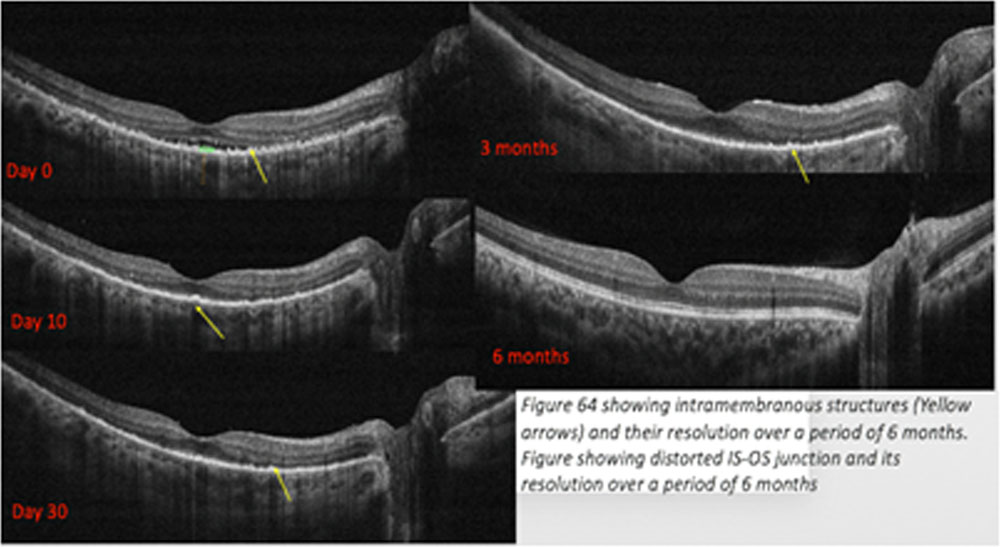
Coming to the baseline parameters, the mean mean average macular height was 432.68mm and mean central macular thickness was 434.87 mm which was comparative to Avg. macular height. RPE undulation index was 1.06. The mean area of retinal detachment was 1.00293 (mm2). Hyper reflective dots Intramembranous structures were documented in 57 and 52 out of 60 eyes respectively. The Cone Outer segment (COST) line and IS-OS junction was disrupted in 55 eyes. The choroidal thickness was masked due to the exudative RD and was documented as being >400 in 44 out of 60 eyes.
Upon following up these patients for 6 months at 10 days, 1 month, 3 months and 6 months post treatment. There was a statistically significant improvement in all the quantitative parameters at different time intervals using the repeated measures anova test. Also, there was a significant improvement when compared with each visit using multiple comparison sidak test. However, there was an increase in the central macular thickness and area of RD and also marginally in RPE undulation index due to recurrences in 4 eyes at 1 month post treatment. The trends have been shown in figure 40,42,44,46. Postulated reason for these recurrences in spite of satisfactory compliance could be inadequate therapy with corticosteroids and immunosuppression. Once treatment was stepped up and continued for adequate amount of time these patients did well and there was no recurrences in the 6 months of follow-up period.
Figure 4,5 and 6 show the the decrease of central macular thickness, Average macular thickness, RPE undulations, Exudative RD, choroidal thickness, Intramembranous structures and IS-OS junction respectively over the 6 months of treatment. These show how RPE undulations are the first to decrease. Since the patients were examined at a pre determined time interval calculating the mean time of resolution was not feasible. However, looking at the trends in RPE undulations are the first to decrease followed by Central macular thickness and Area of RD. Intra- membranous structures and choroidal thickness are the last to normalise.
Upon comparing the baseline OCT parameters with the baseline visual acuity using multiple regression analysis RPE undulation index was significantly associated p=0.025 with BCVA. This implies that higher RPE undulation index is associated with poor visual acuity at presentation. At 10 days central macular thickness was significantly associated p=0.001 with BCVA. This implies higher CMT is associated with poor visual acuity at 10 days post treatment. Hence, higher RPE undulation index is associated with a worse visual acuity at presentation. A similar observation was reported by Hasoda et al9 where they upon studying 42 eyes in patients with acute VKH RPE undulation index was associated with a poor visual acuity at presentation. However Hasoda et al9 did not include the other quantitative parameters which could serve as a confounding factor unlike in our study where a multi variant analysis was done to determine the same. Hence we conclude that RPE Undulation index could be used as a marker to predict disease severity at presentation.
In order to determine the prognostic factors we divided the patients into two groups. Those with final BCVA (at 6 months) better than or equal to 6/9 (Snellen Visual acuity) and those with final BCVA less than 6/9. We tried to find the association between baseline OCT parameters and final BCVA using Chi square test and paired T test. Although we did not find any significant association between the same, RPE undulation index was weekly associated (P value nearing 0.05)with poor visual acuity. Since this study was not designed around these parameters and may be inadequately powered and hence such results.
Hashizume et al8 studied 61 eyes of 31 patients with acute VKH and divided RPE undulations into slight, moderate and severe and concluded that presence of RPE undulations was associated with poor visual acuity and could predict recurrences in such patients. However, this study did not quantify the RPE undulations and used a uni-variate analysis without taking other OCT parameters into consideration. Therefore , higher RPE undulations could be a marker for poor visual outcomes post treatment.
In our study in the 6 months of follow-up, 14 eyes out of 60 eyes i.e. 23% had a recurrence at some time or the other during the 6 months. Out of the 60 eyes 52 (86.6%) eyes regained vision of 6/9 or better (Log Mar 0.2) or better at the end of 6 months. Upon comparing the patients who regained vision at 6 months with those who had recurrence (table 41), a relative risk of gaining vision better than of equal to 6/9 (Log Mar 0.2) at 6 months was 73%. Hence even if a patient has recurrence the chance of patients retaining a good vision is 73%. This is in accordance to a study published by Aravelo et al69 in 2015 to determine the clinical outcomes in patients with VKH disease. They studied 202 eyes with acute VKH and 107 out of those had recurrences within 12 years of follow-up. They concluded that with adequate therapy, more than 70% of eyes maintain visual acuity of 20/50 or better over the long term. Hence the prognosis in recurrent cases is relatively good. However the follow up of these patients was 12 years. Which definitely presents with a bigger picture on recurrences.
Conclusion
From this prospective study of 30 patients (60 eyes) of acute VKH disease upon studying the clinical characteristics, multimodal imaging and association of visual acuity with various OCT parameters we found that Acute VKH disease presents most commonly in patients between 30-40 years with mean age being 37.30 years with a female preponderance. Patients usually present between within 15 days of onset of symptoms. Since integumentary findings present in the convalescent phase of the disease maximum patients present with neurological symptoms and are diagnosed as probable VKH disease. The most common symptom is rapid diminution of vision and the most common sign is sub retinal fluid fluid pockets in these patients. On ICGA the most common and diagnostic finding was hypo florescent areas in early phase suggestive of choroidal granulomas which determine the extent of the disease. On FFA the most common findings include hypo fluorescent areas, pin point hyper fluorescence and Sub retinal pooling of dye. There is a statistically significant increase in BCVA post treatment and maximum patients achieve a final BCVA of 6/9 (Snellen) or better. There is a statistically significant improvement in all OCT parameters post treatment and up to 6 months of follow-up. RPE undulation is the first parameter to normalize post treatment. Intra-membranous structures and choroidal thickness are the last to stabilize post treatment. The flow void areas on OCT-A seem to represent the underlying choroidal granulomas and disappear by a mean period of 3 months and hence this modality holds a potential to used as a non invasive modality to monitor treatment and even predict recurrences. RPE undulations are associated with poor visual acuity at presentation and could serve as a marker of disease severity and the RPE undulation index is weakly associated with worse final visual acuity and thus could be used to predict the same. Eyes which develop recurrence have a 70% relative risk to achieve a final BCVA of 6/9 (Snellen) or better.

References
- O’Keefe GAD, Rao NA, Vogt-Koyanagi-Harada Disease, Survey of Ophthalmology (2016), 10.1016/j.survophthal.2016.05.002
- Ohno S, Char DH, Kimura SJ, et al. Vogt-Koyanagi-Harada syndrome. American Journal of Ophthalmology. 1977;83(5):735-740
- Martin T, Rathinam S, Cunningham E. 086: Prevalence, clinical characteristics, and causes of vision loss in children with Vogt-Koyanagi-Harada disease in South India. Journal of American Association for Pediatric Ophthalmology and Strabismus. 2009;13(1):e22
- Damico F, Cunha-Neto E, Goldberg A, Iwai L, Marin M, Hammer J Et al. T-Cell Recognition and Cytokine Profile Induced by Melanocyte Epitopes in Patients with HLA-DRB1*0405-Positive and -Negative Vogt-Koyanagi-Harada Uveitis. Investigative Ophthalmology & Visual Science. 2005;46(7):2465.
- Read R, Holland G, Rao N, Tabbara K, Ohno S, Arellanes-Garcia L Et al., Revised diagnostic criteria for Vogt-Koyanagi-Harada disease: report of an international committee on nomenclature, American Journal of Ophthalmology. 2001;131(5):647-652.
- Yamaguchi Y, Otani T, Kishi S. Tomographic Features of Serous Retinal Detachment with Multilobular Dye Pooling in Acute Vogt-Koyanagi-Harada Disease. American Journal of Ophthalmology. 2007;144(2):260-265
- Liu X, Peng X, Wang S, You Q, Li Y, Xiao Y et al. Features of Optical Coherence Tomography for the Diagnosis of Vogt–Koyanagi–Harada Disease. Retina. 2016;1.
- Hashizume K, Imamura Y, Fujiwara T, Machida S, Ishida M, Kurosaka D. Retinal Pigment Epithelium Undulations in Acute Stage of Vogt-Koyanagi-Harada Disease. Retina. 2016;36(2):415-421.
- Hosoda Y, Uji A, Hangai M, Morooka S, Nishijima K, Yoshimura N. Relationship Between Retinal Lesions and Inward Choroidal Bulging in Vogt-Koyanagi-Harada Disease. American Journal of Ophthalmology. 2014;157(5):1056-1063.e1.
- Aggarwal, Kanika et al. “The Role Of Optical Coherence Tomography Angiography In The Diagnosis And Management Of Acute Vogt–Koyanagi–Harada Disease”. Ocular Immunology and Inflammation (2016): 1-12. Web
- Attia S, Khochtali S, Kahloun R, Ammous D, Jelliti B, Ben Yahia S et al. Clinical and multimodal imaging characteristics of acute Vogt–Koyanagi–Harada disease unassociated with clinically evident exudative retinal detachment. International Ophthalmology. 2015;36(1):37-44.
- Arellanes-García L, Hernández-Barrios M, Fromow-Guerra J, Cervantes-Fanning P. Fluorescein fundus angiographic findings in Vogt–Koyanagi–Harada syndrome. International Ophthalmology. 2007;27(2-3):155-161.
- Fardeau C, Tran T, Gharbi B, Cassoux N, Bodaghi B, LeHoang P. Retinal fluorescein and indocyanine green angiography and optical coherence tomography in successive stages of Vogt-Koyanagi-Harada disease, International Ophthalmology. 2007;27(2-3):163-172.
- Abouammoh M, Gupta V, Hemachandran S, Herbort C, Abu El-Asrar A. Indocyanine green angiographic findings in initial-onset acute Vogt-Koyanagi-Harada disease. Acta Ophthalmologica. 2016;94(6):573-578.
- Zhou M, Jiang C, Gu R, Sun Z, Huynh N, Chang Q. Correlation between Retinal Changes and Visual Function in Late-Stage Vogt-Koyanagi-Harada Disease: An Optical Coherence Tomography Study. Journal of Ophthalmology. 2015; 2015:1
- Baltmr A, Lightman S, Tomkins-Netzer O. Vogt Koyanagi Harada syndrome; current perspectives. Clinical Ophthalmology. 2016;Volume 10:2345-2361.
- Nakayama M, Keino H, Okada A, Watanabe T, Taki W, Inoue M et al. Enhanced Depth Imaging Optical Coherence Tomography of The Choroid In Vogt–Koyanagi–Harada Disease. Retina. 2012;32(10):2061-2069.
- Jabs DA, Nussenblatt RB, Rosenbaum JT, et al., Standardization of uveitis nomenclature for reporting clinical data. Results of the First International Workshop, Am J Ophthalmol, 2005;140: 509–16
- Pattison E. Uveomeningoencephalitic Syndrome (Vogt-Koyanagi-Harada). Archives of Neurology. 1965;12(2):197-205.
- Vogt A ,Frühzeitiges Ergrauen der Zilien, und Bemerkungen über den, sogenannten plötzlichen Eintritt , dieser Veränderung, Early graying of cilia and remarks about the so-called sudden occurrence of these change, Klin Monatsbl Augenheilkd, 1906;44:228–242.
- Koyanagi YD, bei schwerer, nicht traumatischen Ursprungs, Dysacusis, alopecia and poliosis in severe uveitis without traumatic origin, Klin Monatsbl Augenheilkd, 1929;82: 194–211.
- Harada E, Clinical study of non suppurative choroiditis: a report of acute diffuse choroiditis, Acta Society of Ophthalmology Japan, 1926;30:356–378.
- Herbort C, Mochizuki M. Vogt–Koyanagi–Harada disease: inquiry into the genesis of a disease name in the historical context of Switzerland and Japan. International Ophthalmology. 2007;27(2-3):67-79.
- Biswas J, Narain S, Das D, Ganesh S. Pattern of uveitis in a referral uveitis clinic in India. International Ophthalmology. 1997;20(4).
- Chi W, Yang P, Li B, Wu C, Jin H, Zhu X et al. IL-23 promotes CD4+ T cells to produce IL-17 in Vogt-Koyanagi-Harada disease. Journal of Allergy and Clinical Immunology. 2007;119(5):1218-1224.
- Yamaki K, Gocho K, Hayakawa K, Kondo I, Sakuragi S (2000) Tyrosinase family proteins are antigen specific to Vogt–Koyanagi–Harada disease. J Immunol 165:7323–9
- Kobayashi H, Kokubo T, Takahashi M, et al. Tyrosinase epitope recognized by an HLA-DR-restricted T-cell line from a Vogt-Koyanagi-Harada disease patient. Immunogenetics. 1998;47(5):398-403.
- Ng JY, Luk FO, Lai TY, et al. Influence of molecular genetics in Vogt- Koyanagi- Harada disease. July 2014:1-12. doi:10.1186/s12348-014-0020-1.
- Chu M, Yang P, Hu R, Hou S, Li F, Chen Y Et al. Elevated Serum Osteopontin Levels and Genetic Polymorphisms of Osteopontin Are Associated with Vogt-Koyanagi-Harada Disease. Investigative Ophthalmology & Visual Science. 2011;52(10):7084.
- Rao N. Pathology of Vogt–Koyanagi–Harada disease. International Ophthalmology. 2007;27(2-3):81-85.
- Inomata H, Rao N. Depigmented atrophic lesions in sunset glow fundi of Vogt-Koyanagi-Harada Disease. American Journal of Ophthalmology. 2001;131(5):607-614.
- Font R, Fine B, Messmer E, Rowsey J. Light and Electron Microscopic Study of Dalén-Fuchs Nodules in Sympathetic Ophthalmia. Ophthalmology. 1983;90(1).
- Sakamoto T. Class II Major Histocompatibility Complex on Melanocytes of Vogt-Koyanagi-Harada Disease. Archives of Ophthalmology. 1991;109(9).
- 34.Inomata H, Sakamoto T. Immunohistochemical studies of Vogt-Koyanagi-Harada disease with sunset sky fundus. Current Eye Research. 1990;9(sup1).
- Sukavatcharin S, Tsai J, Rao N. Vogt–Koyanagi–Harada disease in Hispanic patients. International Ophthalmology. 2007;27(2-3).
- Damico F, Kiss S, Young L. Vogt-Koyanagi-Harada Disease. Seminars in Ophthalmology. 2005;20(3).
- Tsui E, Bottini A, Ghadiali Q, Balaratnasingam C, Barbazetto I. Unilateral Ocular Manifestations of Vogt–Koyanagi–Harada Disease. Ocular Immunology and Inflammation. 2017.
- Keino H, Goto H, Mori H, Iwasaki T, Usui M. Association Between Severity of Inflammation in CNS and Development of Sunset Glow Fundus in Vogt-Koyanagi-Harada Disease. American Journal of Ophthalmology. 2006;141(6).
- Lira R, Oliveira C, Marques M, Silva A, Pessoa C. Adverse reactions of fluorescein angiography: a prospective study. Arquivos Brasileiros de Oftalmologia. 2007;70(4):615-618.
- Kalogeromitros D, Makris M, Aggelides X, Mellios A, Giannoula F, Sideri K et al. Allergy skin testing in predicting adverse reactions to fluorescein: a prospective clinical study. Acta Ophthalmologica. 2009;89(5):480-483.
- Yanoff M, Duker J. Ophthalmology. 4th ed. Pittsburgh: Elsevier/Saunders; 2014.
- Chee S, Jap A, Cheung C. The Prognostic Value of Angiography in Vogt-Koyanagi-Harada Disease. American Journal of Ophthalmology. 2010;150(6):888-893.e1.
- Herbort CP, LeHoang P, Guex-Crosier Y (1998) Schematic interpretation of indocyanine green angiography in posterior uveitis using a standard protocol. Ophthalmology 105:432–440
- Bouchenaki N, Herbort C. The contribution of indocyanine green angiography to the appraisal and management of Vogt-Koyanagi-Harada disease. Ophthalmology. 2001;108(1):54-64.
- Herbort C, Mantovani A, Bouchenaki N. Indocyanine green angiography in Vogt–Koyanagi–Harada disease: angiographic signs and utility in patient follow-up. International Ophthalmology. 2007;27(2-3).
- Chee S, Jap A. The outcomes of indocyanine green angiography monitored immunotherapy in Vogt–Koyanagi–Harada disease. British Journal of Ophthalmology. 2012;97(2):130-133.
- Fardeau C, Tran T, Gharbi B, Cassoux N, Bodaghi B, LeHoang P. Retinal fluorescein and indocyanine green angiography and optical coherence tomography in successive stages of Vogt-Koyanagi-Harada disease. International Ophthalmology. 2007;27(2-3):163-172.
- Costa R, Skaf M, Melo L, Calucci D, Cardillo J, Castro J et al. Retinal assessment using optical coherence tomography. Progress in Retinal and Eye Research. 2006;25(3):325-353.
- Chee S, Jap A, Bacsal K. Spectrum of Vogt-Koyanagi-Harada disease in Singapore. International Ophthalmology. 2006;27(2-3):137-142.
- Miyanaga M, Kawaguchi T, Shimizu K, Miyata K, Mochizuki M. Influence of early cerebrospinal fluid-guided diagnosis and early high-dose corticosteroid therapy on ocular outcomes of Vogt–Koyanagi–Harada disease. International Ophthalmology. 2007;27(2-3):183-188.
- Ohguchi M, Sugiura S,Corticosteroid therapies in ophthalmic diseases, Japan journal of clinical Ophthalmology. 1976;30:527–53.
- Rubsamen P. Vogt-Koyanagi-Harada Syndrome. Archives of Ophthalmology. 1991;109(5):682.
- Andreoli C, Stephen Foster C. Vogt-Koyanagi-Harada Disease. International Ophthalmology Clinics. 2006;46(2):111-122.
- Rao N. Treatment of Vogt-Koyanagi-Harada Disease by Corticosteroids and Immunosuppressive Agents. Ocular Immunology and Inflammation. 2006;14(2):71-72.
- Sasamoto Y, Ohno S, Matsuda H. Studies on Corticosteroid Therapy in Vogt-Koyanagi-Harada Disease. Ophthalmologica. 1990;201(3):162-167.
- Errera M, Fardeau C, Cohen D, Navarro A, Gaudric A, Bodaghi B et al. Effect of the duration of immunomodulatory therapy on the clinical features of recurrent episodes in Vogt-Koyanagi-Harada disease. Acta Ophthalmologica. 2011;89(4):e357-e366.
- Paredes I, Ahmed M, Foster C. Immunomodulatory Therapy for Vogt-Koyanagi-Harada Patients as First-Line Therapy. Ocular Immunology and Inflammation. 2006;14(2):87-90.
- Kondo Y, Fukuda K, Suzuki K, Nishida T. Chronic non infectious uveitis associated with Vogt–Koyanagi–Harada disease treated with low-dose weekly systemic methotrexate. Japanese Journal of Ophthalmology. 2011;56(1):104-106.
- Cuchacovich M, Solanes F, Díaz G, Cermenati T, Avila S, Verdaguer J et al. Comparison of the Clinical Efficacy of Two Different Immunosuppressive Regimens in Patients with Chronic Vogt-Koyanagi-Harada Disease. Ocular Immunology and Inflammation. 2010;18(3):200-207.
- Andrade R, Muccioli C, Farah M, Nussenblatt R, Belfort R. Intravitreal triamcinolone in the treatment of serous retinal detachment in Vogt-Koyanagi-Harada syndrome. American Journal of Ophthalmology. 2004;137(3):572-574.
- Hosoda Y, Hayashi H, Kuriyama S. Posterior subtenon triamcinolone acetonide injection as a primary treatment in eyes with acute Vogt–Koyanagi–Harada disease. British Journal of Ophthalmology. 2015;99(9):1211-1214.
- Jeroudi A, Angeles-Han S, Yeh S. Efficacy of Adalimumab for Pediatric Vogt-Koyanagi-Harada Syndrome. Ophthalmic Surgery, Lasers and Imaging Retina. 2014;45(4):332-334.
- Zmuda M, Tiev K, Knoeri J, Héron E. Successful Use of Infliximab Therapy in Sight-threatening Corticosteroid-resistant Vogt-Koyanagi-Harada Disease. Ocular Immunology and Inflammation. 2013;21(4):310-316.
- Jap A, Chee S. Imaging in the Diagnosis and Management of Vogt-Koyanagi-Harada Disease. International Ophthalmology Clinics. 2012;52(4):163-172.
- Rao N, Gupta A, Dustin L, Chee S, Okada A, Khairallah M et al. Frequency of Distinguishing Clinical Features in Vogt-Koyanagi-Harada Disease. Ophthalmology. 2010;117(3):591-599.e1.
- Rathinam SR, Vijayalakshmi P, Namperumalsamy P, et al. Vogt–Koyanagi–Harada syndrome in children, Ocular Immunology Inflammation, 1998;6:155–161.
- Ikeda N, Hayasaka S, Hayasaka Y. Uveitis and pseudo uveitis Presenting for the First Time in Japanese Elderly Patients. Ophthalmologica. 2005;219(5):263-266.
- Kato Y, Yamamoto Y, Tabuchi H, Fukushima A. Retinal pigment epithelium folds as a diagnostic finding of Vogt-Koyanagi-Harada disease. Japanese Journal of Ophthalmology. 2012;57(1):90-94.
- Arevalo J, Lasave A, Gupta V, Kozak I, Al Harbi M, Al Rushood A et al. Clinical Outcomes of Patients with Vogt–Koyanagi–Harada Disease Over 12 Years at a Tertiary Center. Ocular Immunology and Inflammation. 2015;24(5):521-529.
- Forster D. Echographic Features of the Vogt-Koyanagi-Harada Syndrome. Archives of Ophthalmology. 1990;108(10):1421.
- Maruko I, Iida T, Sugano Y, Oyamada H, Sekiryu T, Fujiwara T et al. Subfoveal Choroidal Thickness after treatment of Vogt–Koyanagi–Harada Disease. Retina. 2011;31(3):510-517.
Leave a Comment