

Dr. Soumen Mondal
Aim:
To describe the characteristics of screened babies in two tertiary referral units in Eastern India, who have severe retinopathy of prematurity (ROP), to provide information regarding lacuna in screening.
Methods:
Information on consecutive babies referred to 2 tertiary eye care centres who had Type I ROP as well as babies who had Stage 4 and Stage 5 ROP between October 2014 to September 2017 were retrieved from medical records
Results:
Data of 52 babies out of 1020 babies screened was analyzed. Amongst these 52 babies, 21 (40.4%) had Stage 3 ROP, 11 (21.1%) had Stage 4 ROP and 20 babies (38.5%) had Stage 5 ROP. Median age of presentation was 5.5 months (1 – 23 months). In subgroup analysis, median age of presentation of the Stage 5 babies was 6.5 months. All the babies with Stage 5 had missed screening at month 1 of life; 16 (80%) of whom had no advice from neonatologist for screening.
Conclusion:
Comprehensive screening programs for ROP are urgently needed with a task force comprising of neonatologists and ophthalmologists. Importance of timely referral of babies to an ophthalmologist should be emphasized to the neonatology community. More ophthalmologists need to be trained for ROP screening.
Introduction
ROP is fast emerging as an important cause of childhood blindness in India.[1‑3] In spite of recommendations for screening of preterm babies to rule out ROP,[4] there is no evidence of universal ROP screening programs in India. ROP blindness in India is increasing due to the highest number of preterm births in the world (3,519,100),[5] suboptimal neonatal care, lack of awareness, screening and treatment programs not in place, and increasing numbers of Neonatal Intensive Care Units and Special New Born Care Units opening all over the country. Severe ROP is a potentially vision threatening disease which, if left undetected and hence untreated, might lead to bilateral blindness. The present study was undertaken to define the characterestics of babies who had severe ROP with special emphasis on the babies having Stage 5 ROP thereby trying to explore the factors responsible for lacuna in proper and timely screening of babies in need.
Methods
Medical records of babies seen for ROP screening in two tertiary care eye centres in Eastern India from October 2014 to September 2017were analysed retrospectively. Inclusion criteria for being eligible for analysis were babies who had Type 1 ROP,as well as babies who had Stage 4 or Stage 5 ROP. The group of babies with Type 1 ROP (without retinal detachment) was named Group A, babies with Stage 4 ROP was called Group B and those with Stage 5 ROP was called Group C. Type 1 ROP is defined as high risk prethreshold disease ie either ROP in Zone I with plus disease, or, Stage 3 ROP in Zone I, or, Stage 3 ROP in Zone II or Zone III with plus disease.[6]Stage 4 ROP signifies that retinal detachment has started to occur due to the traction of the fibrous and vascular tissue over the ridge. It is called 4A when the retinal detachment spares the macula, and stage 4B when retinal detachment involves the macula.Stage 5 ROP signifies that total retinal detachment has occurred.Differences in age at presentation among the three stages were analysed using the Kruskal-Wallis test using the SPSS software.
Results
A total number of 1020 babies were seen in the Out Patients Department of 2 eye care centres of Eastern India within the study period. Amongst them eyes of 52 babies fulfilled the inclusion criteria of whom, 28 were male babies and 24 female.
Birth weight ranged from 750 grams to 2300 grams, with a mean of 1365 grams.
Gestational age of the babies at birth, as per parent’s recall or as per birth records, ranged from 24 weeks to 35 weeks;with a mean of 31 weeks.
Among these 52 babies, 20 were discharged from medical colleges, 19 from government run SNCUs and 13 babies were from private NICUs.
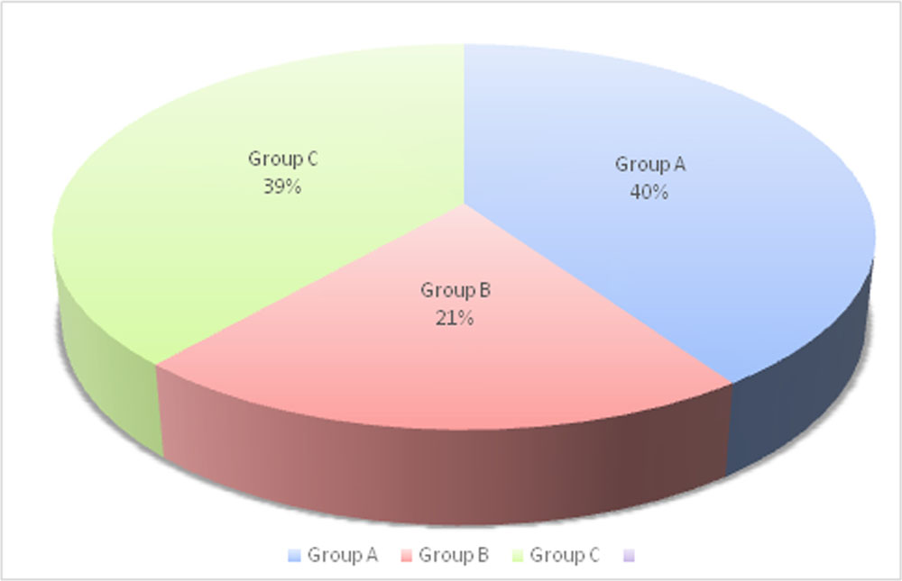
21 babies (40.4%) had Type 1 ROP without any retinal detachment, which were designated Group A in the study, 11 (21.1%) had Stage 4 ROP (Group B) and 20 babies (38.5%) had Stage 5 ROP (Group C).
Median age of presentation was 5.5 months (1 – 23 months). In subgroup analysis, median age of presentation of Group A was 2.1 months and that of Group B was 4.3 months. Median age of presentation of babies in Group Cwas 6.5 months (4 – 23 months). Difference of outcome of stages of ROP between the groups when the median age of presentation was taken into account was found to be statistically significant (p<0.01).
Mean birth weight of Group A babies was 1433 grams (1250 – 1600 grams), for Group B babies, 1375 grams (1050 – 1550 grams). Mean birth weight of Group C babies was 1253 grams (850 – 1575 grams).
In Group C, none of the babies had been seen in the first month of life. 16 out of 20 babies (80%) had no advice from their neonatologists for retina examination at the time of discharge from NICU. Among these 16, 8 babies were from government SNCUs, 6 from medical college NICUs and 2 from private NICUs. Of the 4 babies who had been referred for eye examination in their discharge advice, the parents did not turn up as they felt that the distance from their home to the eye hospital was far. All these 4 babies were discharged from medical colleges. All these 20 babies came for eye check up when their parents or their pediatricians noticed a white pupillary reflex.
Discussion:
The present study highlights the point that the babies who had worse prognosis were not screened at the recommended time. All infants weighing ≤2000 g or ≤34 weeks of gestation need to be screened [7] and the first screening should be within day 30 of lifeaccording to the most recent Indian screening guidelines [8]. In our study, it was observed that the babies who had the best prognosis, that is babies belonging to Group A were seen earlier at a median age of presentation of 2.1 months whereas median age of presentation of babies in Group C, who had the worst prognosis, was 6.5 months.This difference in the median time of presentation with reference to their Stage of ROP was found to be statistically significant.
The babiesin Group C did not have a single ROP screening examination. Out of 20 babies 16 (80%) have not been advised for screening by the neonatologist or pediatrician. The rest 4 babies had been recommended, but the parents could not bring the babies for check up at the right time. The reason given by the parents was that the eye hospital was too far from their residence and it was difficult for them to travel with the neonate.If the number of ophthalmogists who are trained to screen ROP were more, it would have been easier for the parents to find one nearer at home, and the ROP screening for these now-blind babies would not have been missed; thereby this needless blindness could have been prevented.
It was also noted in our study that neonatologists and paediatricians (including those working in teaching hospitals) should be more vigilant regarding advice of ROP screening of vulnerable neonates at proper time.
Our finding corroborates with a study from Northern India which demonstrated that the “likely reasons for non-screening include lack of awareness among pediatricians and ophthalmologists, non-availability of trained ophthalmologists, and a poor knowledge of the screening criterion.”[9]
Conclusion
Eastern India is urgently in need of comprehensive screening programs for ROP with a task force comprising of neonatologists and ophthalmologists. Timely referral of babies to an ophthalmologist is of utmost importance to prevent the babies from devastating visual outcome, and this should be emphasized to the neonatology community. We need more trained ophthalmologists willing to go for neonatal retina screening for ROP so that parents do not have to travel great distances to get the screening done.
Reference
- Chawla D, Agarwal R, Deorari AK, Paul VK. Retinopathy of prematurity. Indian J Pediatr2008;75:73‑76.
- Jalali S, Anand R, Kumar H, Dogra M, Azad R, Gopal L. Programme planning and screening strategy in retinopathy of prematurity. Indian J Ophthalmol2003;51:89‑99
- Gilbert C, Fielder A, Gordillo L, Quinn G, Semiglia R, Visintin P, et al. Characteristics of babies with severe retinopathy of prematurity in countries with low, moderate and high levels of development: Implications for screening programmes. Pediatrics 2005;115:518‑525
- National Neonatology Forum of India: Evidence Based Guidelines; available at www.nnfi.org
- Blencowe H, Cousens S, Oestergaard MZ, Chou D, Moller AB, Narwal R, et al. National, regional, and worldwide estimates of preterm birth rates in the year 2010 with time trends since 1990 for selected countries: A systematic analysis and implications. Lancet. 2012;379:2162–72.
- Early Treatment of Retinopathy of Prematurity Cooperative Group. Revisedindications for thetreatment of retinopathy of prematurity. Arch Ophthalmol 2003;121:1684-96.
- Vinekar A, Dogra MR, Sangtam T, Narang A, Gupta A. Retinopathy of prematurity in Asian Indian babies weighing greater than 1250 grams at birth: Ten year data from a tertiary care center in a developing country. Indian J Ophthalmol. 2007;55:331–6
- National Neonatology Forum (NNF) Clinical Practice Guidelines. www.nnfpublication.org
- Sanghi G, Dogra M, Katoch D, Gupta A. Demographic profile of infants with stage 5 retinopathy of prematurity in North India: implications for screening .Ophthalmic Epidemiol.2011 Apr;18(2):72-4.
Leave a Comment