

Dr.Subhash Prasad,Dr.ABHISHEK RANJAN,Dr.VIDHATA
Introduction
One of the late sequelae of open globe injury involving the anterior segment is formation of iris cyst.Though not common,can cause significant morbidity of the eye,if it occurs. These cysts develop from epithelial cells of the cornea or conjunctiva that have been implanted or transplanted on the iris through the wound [1]. These cells in the anterior chamber then grow into a sheet or form a cyst.
The small and asymptomatic iris cyst can be just be followed up. Larger iris cysts can grow in size, and cause pupillary block and secondary glaucoma, uveitis, corneal decompensation and eventually, a painful, blind eye. These cysts require intervention.Various modalities of treatment, ranging from minimally invasive techniques like aspiration of the cyst and laser therapy, to aggressive surgical procedures like block excision have been described for the management of iris cysts [2]. The purpose of this work is to describe the management of a post-traumatic iris cyst by cutting the cyst with vitrectomy cutter through two side port incision.
Methods:
In a prospective study of seven cases of traumatic iris cyst which presented to us between January 2013 and December 2017 were included. Detailed work up included proper history/evaluation of records of previous repair whenever available,time since primary repair,visualacuity,intraocular pressure(IOP),slit lamp examination,fundusexamination.Yag laser iridotomy was tried in all.It recurred in all from3-6 months.Cystectomy was done under peribulbaranesthesia or GA.Two side ports were made in limbus.Corneal endothelium was coated with viscodispersive OVD containing chondroitin sulphate and viscodissection of peripheral anterior synechia was done.Irrigation was maintained using a cannula through one side port and other port was used to do cystectomy of anterior wall was done with 23 G vitrectomy cutter using infinity or centurion vision system.All patients received topical steroid,cycloplegic and antiglaucoma.Patients were followed up on next day,oneweek,onemonth,three month and finally 6 months.In each follow up patients were evaluated for visual acuity,IOP,status of cornea,iris and for any recurrence of cyst.Patients with initial rise in IOP were followed more regularly.
Results:
Out of seven,six patients had penetrating coneal/sclerocorneal injury which were repaired and one patient gave history of self sealingwound.All presented from 6months to 3 years of primary repair. Age ranged from 3-15 years with average age of 7 years.4 patients less than 5 years,2 between 5-10 years and one was 15 years of age.Four patients presented within first year rest from 1-3 years of injury.Six patients were male and onefemale.Three patients had injury of cornea only, whereas four had injury involving coneaosclera or limbus.four patients had already undergone cataract surgery for traumatic cataract following trauma.Visual acuity ranged from finger count close to face to 6/18.Cyst was present in one quadrant in threepatients,Two quadrants in three and all four quadrant in one.Cyst was obscuring pupil in 2 patients and partly coming in pupillary area in 2.IOP was raised in three patients with corneal haze in one.All patients with cyst involving more than two quadrants had rise in IOP.While IOP of one patient was controlled after the procedure,two required antiglaucomatreatment.Pupillary area was clear in all patients after cystectomy.Visual acuity ranged from 6/60 to 6/12 at the end of six months.Patching was started in better eye for amblyopia treatment of effected eye.Post cystectomy none of the patients had recurrence in six months follow up.
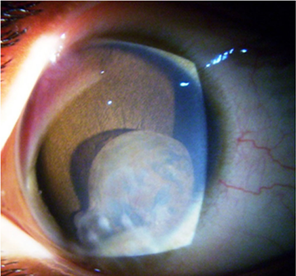
Fig-1 Cyst involving one quadrant
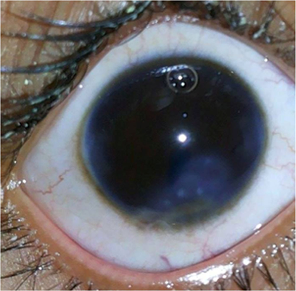
Fig-2 First post op day after cystectomy
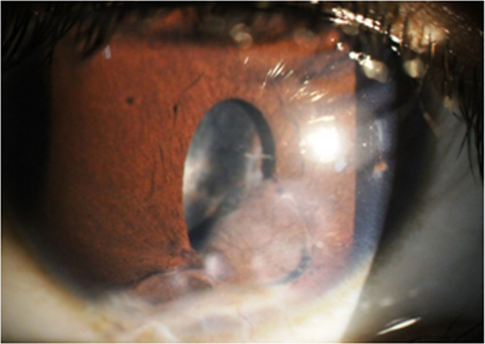
Fig-3 After 6 months
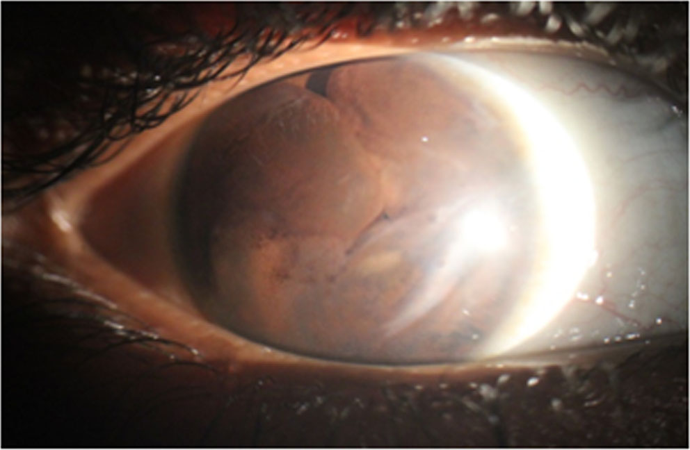
Fig-4 Cyst involving four quadrant
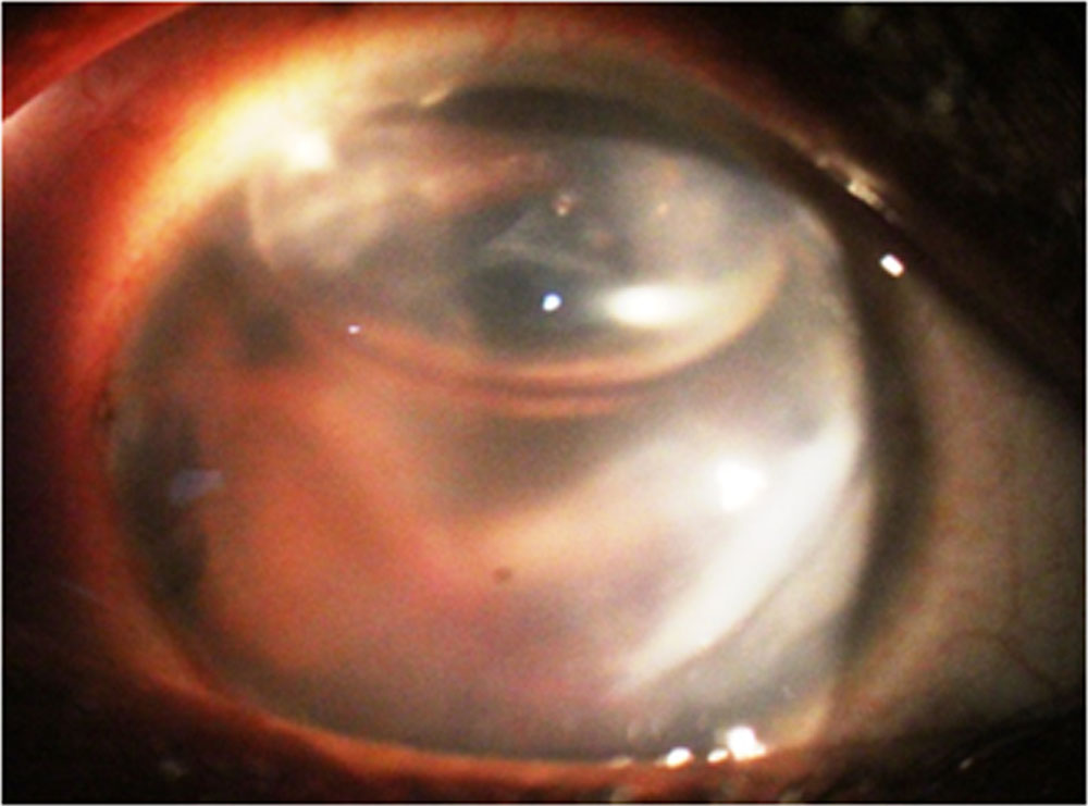
Fig-5 First post op day after cystectomy
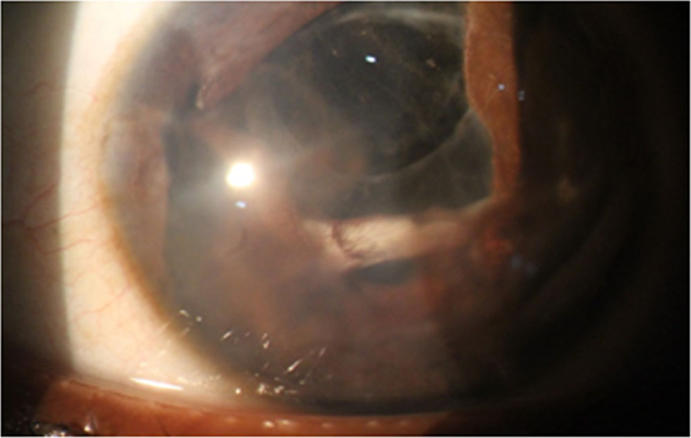
Fig-6 . After 6 months
Discussion:
Secondary iris cysts may occur as a result of penetrating corneal injury. Small and stable cysts may be closely observed for signs of progression or complications like intraocular inflammation.The larger cysts require surgical management.
Many procedures for the management of iris cysts with varying outcomes have been described. Traditionally treatment for iris cysts aimed at aggressive surgical excision and devitalization of the epithelial tissues. These require complete block excision of all epithelial layers and adjacent cornea, iris, anterior chamber angle and ciliary body [3,4].However, these procedures, while generally successful in eradicating the epithelial tissue, involved extensive surgical procedures with the possibility of significant collateral damage to adjacent ocular structures, with resultant poor functional outcome [5]. More recently, many conservative surgical approaches to the management of iris cysts have been reported, including aspiration with or without cauterization, diathermy or iridectomy, injection of sclerosing agents, electrocautery and photocoagulation [3,6,7, ]. Viscodissection followed by cystectomy has also been described.Argon laser photocoagulation of cysts have also been described [8,9]. However, procedures like aspiration of the cyst have been reported to have a high rate of recurrence. Laser treatment with Nd-YAG laser to rupture the cyst has also been reported, but with a poor outcome, with violent post-operative inflammatory reaction and high recurrence rates [10,11].
Marsupialization is the surgical technique of cutting a slit into cyst and suturing the edges of the slit or remove part of it to form a continuous surface from the exterior surface to the interior surface of the cyst.This is quite useful in Bartholin and dentigerouscyst.Removingpart of iris cyst wall serve the same purpose preventing collection of fluid inside and further progression.This can be achieved by using scissor,but certainly requires larger incision and more collateral damage particularly corneal endothelium.Using 23 vitreous cutter requires small paracentasis incision in limbus and removes cyst wall in more cotrolled and precise manner.In this study it was possible to remove anterior wall of cyst in almost all cases.There was no recurrence during the follow up period in any eye.There is no report of management of iris cyst by vitreous cutter in literature.
Conclusion:
Anterior wall cystectomy(marsupialization) by vitrectomy cutter is effective and least invasive technique of surgical management of traumatic iris cyst
References:
- Rizzuti A.B. Traumatic implantation cysts of the iris; with special emphasis on surgical aspects. Am. J. Ophthalmol. 1955;39(1):13–20. doi: 10.1016/0002-9394(55)92648-9.
- Moreno-López M., Arruabarrena C., Regueras A., Noval S. [Conservative surgical management of a post-traumatic iris cyst]. Arch. Soc. Esp. Oftalmol
- Naumann G.O., Rummelt V. Block excision of cystic and diffuse epithelial in growth of anterior chamber. Arch. Ophthalmol. 1992;110:223–227. doi:
- Forster R.K. Corneoscleralblock excision of postoperative anterior chamber cysts. Trans. Am. Ophthalmol. Soc. 1995;93:83–97.
- Haller J.A., Stark W.J., Azab A., Thomsen R.W., Gottsch J.D. Surgical management of anterior chamber epithelial cysts. Am. J. Ophthalmol. 2003;135(3):309–313. doi: 10.1016/S0002-9394(02)01960-8
- Bruner W.E., Michels R.G., Stark W.J., Maumenee A.E. Management of epithelial cysts of the anterior chamber. Ophthalmic Surg. 1981;12(4):279–285
- Tsai J.C., Arrindell E.L., O’Day D.M. Needle aspiration and endodiathermy treatment of epithelial inclusion cyst of the iris. Am. J. Ophthalmol. 2001;131(2):263–265. doi: 10.1016/S0002-9394(00)00707-8
- Cleasby G.W. Photocoagulation of iris-ciliary body epithelial cysts. Trans. Am. Acad. Ophthalmol. Otolaryngol. 1971;75(3):638–642
- Scholz R.T., Kelley J.S. Argon laser photocoagulation treatment of iris cysts following penetrating keratoplasty. Arch. Ophthalmol. 1982;100(6):926–927. doi: 10.1001/archopht.1982.01030030934006
- Gupta V., Rao A., Sinha A., Kumar N., Sihota R. Post-traumatic inclusion cysts of the iris: a longterm prospective case series. ActaOphthalmol. Scand. 2007;85(8):893–896. doi: 10.1111/j.1600-0420.2007.00975.x
- Xiao Y., Wang Y., Niu G., Li K. Transpupillary argon laser photocoagulation and Nd:YAG laser cystotomy for peripheral iris pigment epithelium cyst. Am. J. Ophthalmol. 2006;142(4):691–693. doi: 10.1016/j.ajo.2006.04.050
Leave a Comment