

Dr.Randhir Kumar, Dr.Satya Prakash Tiwary,Dr.Nimmi Rani
Abstract
Large-angle esotropiaposes a lot of challenge in getting a satisfactory alignment. Veryoften we have to offer a bilateral surgery, which patients may deny at times, as they are reluctant to be operated in the dominant eyein the presence of amblyopia in affected eye. A patient with large-angle esotropia, measuring more than 80 pd base out ,not amenable to standard recession and resection procedures, underwent primary true muscle transplantation, wherein a fragment of resected lateral rectus muscle was transplanted to the medial rectus muscle with a non-absorbable suture. Now the elongated medial rectus was recessed to the desired length by usual technique. Postoperative deviation was within 10 PD of orthophoria and was maintained at follow-up of 6 months. There was mild underaction of the weakened muscle. True muscle transplantation is a good option in large-angle esotropia with good alignment and long term stability,
Keywords: esotropia, true muscle transplantation, medial rectus ,lateral rectus muscle
Large-angle esotropia poses a lot of challenge in getting a satisfactory alignment. Very often we have to offer a bilateral surgery, which patients may deny at times, as they are reluctant to be operated in the dominant eye in the presence of amblyopia in affected eye. We present a case of such a large angle esotropiabeing managed with true muscle transplantation
Methods
A patient presented to us with an esotropia of more than 80 pdbase out deviation
in primary position . the patient underwent thorough preoperative orthoptic checkup and refraction. The patient was followed up on day 1, day 30, and at 6 months. the patients underwent true muscle transplantation,in which the resected extra stump of lateral rectus (LR) was transplanted to the medial rectus (MR) using 6‑0 prolene. The new medial rectus(MR)was recessed by a standard recession technique.
Surgical procedure
The MR muscle was dissected and separated through aVonNoorden’slimbal incision, Anonabsorbable6‑0 prolene suture was placed at the muscle insertion. Next, the LR muscle was hooked. Two single arm 6‑0 Vicryl sutures were placed at a
Distance of 9 mm from the insertion as is done in a routine resection of rectus muscle and another pair of 6‑0 Vicryl was placed at the insertion. The LR muscle was cut at the distal end and finally from itsoriginal insertion, and the posteriorly (distally) placed 6‑0 Vicryl sutures were passed through the original insertion as in a routine rectus muscle resection. The excess muscle stump was then placed at the MR site, and the distal end of this stump was sutured with the proximal end of MR with the 6‑0 prolenealready placed on the MR . The original MR was incised from the insertion. The now elongated muscle was sutured at 6 mm from the original insertion site ofMR as is done in a routine rectus muscle recession.
Results.
The preoperative angle was 90pd base out. Themean postoperative angle at the end of6 months follow‑up was10pd. The amount of deviation was more or less constantover a period of 6 months. There was mild underaction of the MR muscle. The abduction was normal in all the patients.
Discussion
Correction of deviationby transplanted muscle,is a known entity, and the fact that the tissue remains viable but would lose its contractile properties isalso a known fact.[5] Jethani J et al have published a case where theycombined it with a loop myopexy for an abnormally largeesotropia and hypotropia.[1,2] Muscle transplantation procedure has been describedearlier though not much popularized.[3‑5] .A few cases have been reported by Diamondet al. and Amitavaet al.[4,5]
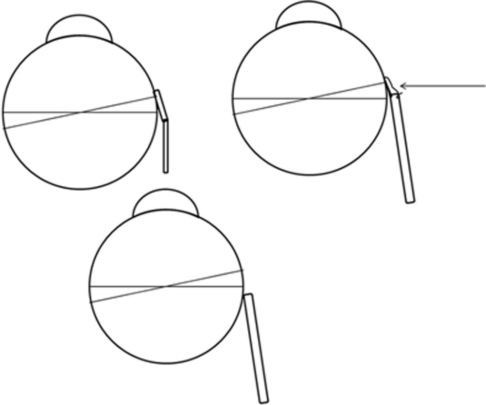
Figure 1: Schematic diagrams representing the muscle transplantation (a), the hang back recession (b) and the conventional recession or the long‑term result of hang back recession (c). It is important to understand here that the segment used (a) could be the muscle segment or any expander [8]
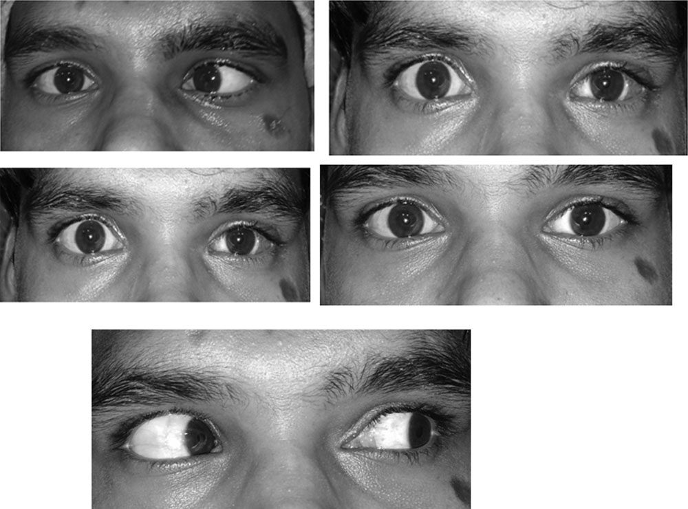
Figure 2: The photographs show the preoperative (a), the immediate postoperative (b), and the later postoperative motility of the operated eye. Note that the motility (c) is very good in the left eye (muscle transplantation) postsurgery.
Bhambhwaniet al.[6] have raised some concern regarding the stability of this result and argued that the surgery is similar to a hemihang‑back sutures. For such a large angle our option include a hemihang‑back, very large unconventional recessions of the MR muscle or very large unconventional resection of LR muscle.Such large recessions should not be done for comitant squints, since the surgeon may induce severe restriction and therefore incomitancyeven after getting a good primary position result.
The other option could be an elongation of muscle. This could be achieved by marginal myotomies or adding a silicone band or nonabsorbable sutures. Silicone bands and chicken sutures have been used successfully in Brown’s syndrome for muscle elongation.[7,8] However, these bands are used as spacers and between the cut ends of the tendon and not at the insertion. Using it close to the insertion under the conjunctiva could lead to extrusion. Considering this fact, we feel it is much safer to use the muscle stump for such a procedure.
We could had an option of operating upon three muscles of two eyes, provided the patient had consented for the same. The muscle transplantation effectively gives us the opportunity to correct very large angle cases with a single eye surgery [Fig. 1].
With the help of muscle transplantation, the effective length of the muscle could be increased and therefore the results could be improved. The chances of any rejection of
the transplanted muscle are virtually nil. We believe that themovement restriction in the extreme gaze on the side of the muscle transplantation is seen as a result of the excessive weakening of the muscle, but the muscle is still anterior to the equator, and therefore the motility restriction is minimal. This, however, cannot be compared with a large conventional recession which would place the new insertion posterior to
the functional equator. The postoperative pictures clearly show minimal adduction loss
even after muscle transplantation [Figs. 2].
A hemihang‑back recession will also have two major problems, first, the muscle may creep forward with such a large recession on hang back (we are doing close to 14–15 mm from the insertion and almost 20 mm from the limbus), and the second obvious problem that it would attach to a point which would be posterior to the arc of contact, and hence, the restriction of ocular motility in the ipsilateral direction would
make it a big incomitant procedure [Fig. 1].
Conclusion
True muscle transplantation is a good option in large-angle esotropia with good alignment and long term stability,speciallywhwre we want to correct a large angle esotropia by operating upon a s ingle eye
References
- Jethani J, Amin S. Loop myopexy with true muscle transplantation for very large angle heavy eye syndrome patient. Indian J Ophthalmol 2015;63:71‑2.
- Jethani J. The muscle transplantation and loop myopexy in so‑called heavy eye syndrome. Indian J Ophthalmol 2015;63:558‑9.
- Hiatt RL. Extraocular muscle transplantation. Trans Am OphthalmolSoc 1973;71:426‑58.
- Amitava AK, Goswami AK, Mishra A. Large‑angle strabismus and primary true muscle transplantation. J PediatrOphthalmol Strabismus 2005;42:211‑5.
- Diamond GR. True transposition procedures. J PediatrOphthalmol Strabismus 1990;27:153‑6.
- Bhambhwani V, Kadav M, Aparnaa C, Pandey PK. Heavy eye syndrome: Role of recessions, resections, loop myopexy, and transplants. Indian J Ophthalmol 2015;63:558.
- Wright KW. Results of the superior oblique tendon elongation procedure for severe Brown’s syndrome. Trans Am Ophthalmol Soc 2000;98:41‑8.
- Jethani JN, Shah N, Amin S, Jethani M. Stability and effects of muscle transplantation for very large angle esotropia: A study of 22 patients. Indian Journal of Ophthalmology. 2017;65(7):607-609. doi:10.4103/ijo.IJO_54_17.
Leave a Comment