

Dr. V S Vijitha, V20290, Dr.Anasua Ganguly
Abstract
Objectives: To demonstrate utility of image processing softwares as a tool to objectively assess ocular surface squamous neoplasia (OSSN). To identify & compare parameters aiding clinical differentiation of conjunctival intraepithelial neoplasia (CIN) and invasive squamous cell carcinoma (SCC).
Methods: This case-control study included all biopsy-proven cases of OSSN presenting as an ocular surface nodule. Lesions with dysplasia and carcinoma in situ were classified as controls and invasive SCC as cases. Exclusion criteria included predominantly corneal, diffuse and nodulo-ulcerative lesions. Slit lamp image analysis was performed using Matlabs and Image J softwares. The effect of predictors like demography, seropositive status, dimensions of the lesions, presence of keratin, pigmentation, corneal involvement, vascularity and feeder vessels on the final histopathologic grade were assessed
Results:A total of 39 patients with OSSN comprising 19 cases and 20 controls were included. Mean age in cases and controls was 46 ± 16.3 years and 41±12.8 years. 74% of cases and 60% of controls worked outdoors. Cases had more prevalence of sero-positivity than controls (p=0.006). Longest diameter (7 vs. 5 mm, OR 2.55; p= 0.04), height (1.98 vs. 1.2 mm, OR 2.85; p= 0.04) and number of clock hours involved (2.24 vs 1.94 mm, OR 0.17; p= 0.06) were found to be positive predictors of invasive OSSN on multivariate analysis.
Conclusion: Histopathology remains the gold standard to distinguish CIN from invasive SCC. Consideration of clinical characteristics like largest diameter, height, clock hours involved can favor the diagnosis of invasive SCC. This is the first study demonstrating image processing software as an objective tool to measure ocular surface lesions.
Key words: OSSN, CIN, Digital image analysis
Introduction
Squamous cell neoplasia of the conjunctiva can be confined to the surface epithelium (conjunctival intraepithelial neoplasia or dysplasia) or may invade the basement and stroma in which case it is termed as invasive squamous cell carcinoma. In conjunctival intraepithelial neoplasia (CIN) (which includes dysplasia and carcinoma in situ), the abnormal cellular proliferation involves only partial thickness of the epithelium and is classified based on severity as mild, moderate or severe with the latter involving full thickness epithelium and it differs from carcinoma in situ by the presence of an intact surface layer of cells. In carcinoma in situ there are no longer normal surface cells and when the basement membrane and stroma are breached it is termed invasive carcinoma. Histopathology has remained the gold standard to distinguish CIN from invasive SCC. 1-8 However, there is a need for less invasive but objective options of predicting the course and determining the prognosis of the ocular surface squamous neoplasia. Digital image analysis has been employed for ophthalmic clinical and surgical use for assessment of eyelid contour in ptosis surgery, objective assessment of size of the ostium following dacryocystorhinostomy. 9-10 This study was designed to explore the potential of utility of image processing softwares as a tool to objectively assess OSSN and also determine the morphological parameters aiding clinical differentiation of conjunctival intraepithelial neoplasia (CIN) and invasive squamous cell carcinoma (SCC).
Methods
Electronic medical records of all cases diagnosed as OSSN from May 2015 to March 2018 at a single tertiary care center in Southern India were reviewed. Cases which presented with a limbal or scleral nodule, with a biopsy proven diagnosis within the spectrum of OSSN were included for analysis. Predominantly corneal, nodulo-ulcerative or ulcerative, diffuse lesions and lesions without photographic documentation were excluded.Socio-demographics, seropositive status, clinical characteristics and histopathological features were assessed. Lesions with a histopathological diagnosis of carcinoma in situ and invasive squamous cell carcinoma were classified as cases and those within the spectrum of mild to severe dysplasia were classified as controls. Objective assessment of the dimensions of the lesions and feeder vessels was also performed using image processing softwares (Matlabs and Image J). Contour software developed in the Matlab (Mathworks, Natick, MA) and Image J software (National in- stitute of health, Bethesda, MD) were employed to analyze all images. The first step involved standardization of the photograph with the help of Contour software (Fig. 1a, step 1). In the next step, the user subjectively determined the centre of the lesion with the point tool. This was followed by automatic dropping of lines, 15° apart, by the software. Following this, with the help of the pentagon tool, the maximum diameter of the lesion and its perpendicular distance was measured (Fig. 1b, step 2). A scale was then set for the standardized photograph in the Image J software (Fig 1c, step 3) and the area of the lesion was measured by outlining the lesion with a drawing tool (Fig 1d, step 4). Thus an objective measurement of the maximum length, breadth and are were obtained. Data was entered in Microsoft Excel and logistic regression was used to access the effect of predictors such as age, sex, occupation, duration of symptoms, seropositive status, dimensions of the lesions, extent of involvement and presence of surface keratin, pigmentation, corneal involvement, vascularity and feeder vessels.
Results
A total of 39 lesions with best captured slit lamp images of OSSN were included for the study, 19 of them being cases and 20 of them controls. The socio-demographic details of the corresponding patients are depicted in Table 1.74% of cases and 60% of controls worked outdoors. With respect to seropositive status, cases had a higher prevalence of seropositivity than controls (p=0.006). Amongst the clinical characteristics, longest diameter (7 vs. 5 mm, OR 2.55; p= 0.04), height (1.98 vs. 1.2 mm, OR 2.85; p= 0.04) and number of clock hours involved (2.24 vs 1.94 mm, OR 0.17; p= 0.06) were found to be positive predictors of invasive OSSN on multivariate analysis. (Table 2).
Discussion
The clinical presentation of OSSN can be extremely varied and confirmation of the diagnosis in a variety of atypical cases depends on the histopathological examination of the lesion. With mild dysplasia on end of the spectrum and invasive squamous carcinoma at the other, it is clinically impossible to differentiate between them. Moreover, variants of squamous cell carcinoma like mucoepidermoid carcinoma, spindle cell carcinoma and OSSN associated with HIV infection can have aggressive clinical. Surgery, chemotherapy and immunotherapy are the various treatment options available for OSSN which may be used singly or in combination.3 However, lacuna exists in literature regarding the rate of progression of dysplasia and what fraction of dysplastic OSSN eventually evolve into invasive carcinoma. This is because by and large the same management protocol is adopted for the entire spectrum of OSSN.
Assessment and documentation of OSSN lesions is not only important for the diagnosis but also for the long term follow up. While recording the dimension can be highly subjective, photographic documentation is more precise and avoids inter observer variability. Therefore, analysing these digital images to provide objective dimensions can be very useful. This technique is more likely to give an accurate estimate as the manually drawn contour line accurately maps the exact surface area of the lesion. Going a step further this study explores the potential of utilizing the clinical parameters to distinguish dysplasia from carcinoma.
As OSSN can have a myriad presentation, certain helpful hints can aid in the diagnosis which include: limbal location, interpalpebral region of the lesion, feeder vessels, keratin etc. One important fact to note is that thickness of the lesion is not always an indication of invasive SCC as even reasonably thick tumours tend to be confined within the epithelium. The presentation of CIN and invasive SCC is very similar thus making clinical differentiation difficult. In the present study, it was noted that longest diameter (7 vs. 5 mm, OR 2.55; p= 0.04), height (1.98 vs. 1.2 mm, OR 2.85; p= 0.04) and number of clock hours involved (2.24 vs 1.94 mm, OR 0.17; p= 0.06) were positive predictors of invasive OSSN on multivariate analysis. By using a combination of the red flag signs it should be possible to extrapolate and provide an objective score to identify invasive carcinoma.1-2
Limitations of this study are its relatively small sample size and being a cross sectional study. With large numbers a predictive score can be devised to hint for or against the diagnosis of dysplasia versus carcinoma. Further advances in the biomedical field can bring forth an automated and user friendly software to analyze digital images at the time of capture itself. Though the current practice pattern does not vary with the degree of dysplasia as long as the lesion is confined to ocular surface this may have a bearing on the treatment plan in future and further elucidate the natural course of dysplasia and invasive carcinoma
Conclusion: Histopathology remains the gold standard to distinguish CIN from invasive SCC. Cumulative consideration of certain clinical characteristics like largest diameter, height, clock hours involved can objectively favour the diagnosis of invasive SCC and thus affect theprognosis. This is the first study demonstrating image processing software as a simple and objective tool to measure ocular surface lesions.
References
- Shields CL, Shields JA. Tumors of the conjunctiva and cornea. Surv Ophthalmol. 2004 Jan-Feb;49(1):3-24.
- Mittal R, Rath S, Vemuganti GK. Ocular surface squamous neoplasia – Review of etio-pathogenesis and an update on clinico-pathological diagnosis. Saudi Journal of Ophthalmology. 2013;27(3):177-86.
- Brownstein S: Mucoepidermoid carcinoma of the conjunctiva with intraocular invasion. Ophthalmology1981; 88:1126–30.
- Cha SB, Shields CL, Shields JA, et al: Massive precorneal extension of squamous cell carcinoma of the conjunctiva.Cornea1993; 12:537–40.
- Gunduz K, Shields CL, Shields JA, et al: Intraocular neoplastic cyst from mucoepidermoid carcinoma of the conjunctiva. 1998; Arch Ophthalmol 116:1521–3.
- McKelvie PA, Daniell M, McNab A, et al: Squamous cell carcinoma of the conjunctiva: a series of 26 cases. Br J Ophthalmol2002; 86:168–73.
- Shields JA, Shields CL, Gunduz K, Eagle RC Jr: The 1998 Pan American Lecture. Intraocular invasion of conjunctival squamous cell carcinoma in five patients. Ophthal Plast Reconstr Surg1999; 15:153–60.
- Tunc M, Char DH, Crawford B, Miller T: Intraepithelial and invasive squamous cell carcinoma of the conjunctiva: analysis of 60 cases. 1999; Br J Ophthalmol 83:98–103.
- Flynn TH, Rose GE, Shah-Desai SD. Digital image analysis to characterize the upper lid marginal peak after levator aponeurosis repair. Ophthalmic Plast Reconstr Surg. 2011; 27(1):12-4.
- Ganguly A, Kaza H, Kapoor A, Sheth J, Ali MH, Tripathy D et al. Comparative Evaluation of the Ostium After External and Nonendoscopic Endonasal Dacryocystorhinostomy Using Image Processing (Matlabs and Image J) Softwares. Ophthalmic Plast Reconstr Surg. 2017;33(5):345-349.
Figure1:
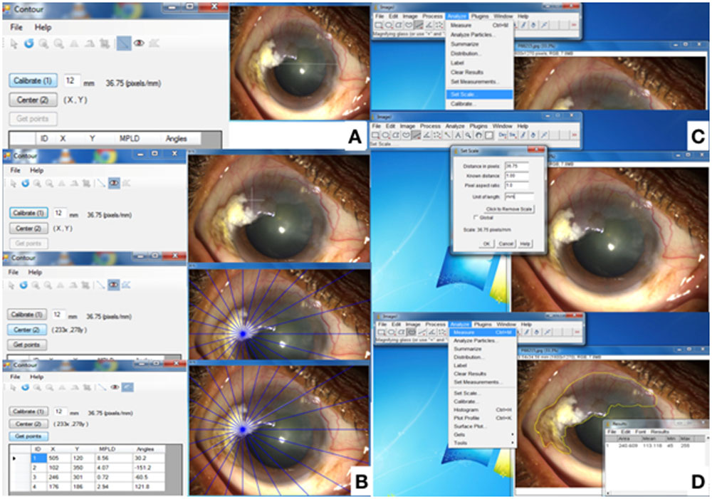
Table 1: Demographics and clinical characteristic of cases and controls
| Cases
(n=19) |
Controls
(n=20) |
|
| Definition | Carcinoma in situ or Invasive carcinoma | Mild, moderate and severe dysplasia |
| Gender
Male Female |
15 4 |
15 5 |
| Mean Age | 46 ± 16.3 years | 41±12.8 years |
Table 2: Logistic regression analysis performed to assess the effect of predictors of final histo-pathological grade
| Univariate | Multivariate | |||
| OR(95%CI) | P value | OR(95%CI) | P value | |
| Age | 1.03 (0.98,1.08) | 0.234 | 0.1 (0.01,1.35) | 0.082 |
| Sex | 0.4 (0.1,1.66) | 0.206 | ||
| Occupation | 0.36 (0.09,1.37) | 0.134 | ||
| Duration of symptoms | 1.07 (0.78,1.47) | 0.678 | ||
| Sero-positivity | 0.12 (0.03,0.5) | 0.004 | 0.03 (0,0.37) | 0.006 |
| Longest diameter | 1.32 (1.01,1.71) | 0.039 | 2.55 (1.01,6.42) | 0.047 |
| Height | 1.86 (0.98,3.54) | 0.057 | 2.85 (1.1,7.37) | 0.031 |
| Area | 1.0018 (0.9976,1.006) | 0.397 | ||
| Clock hours | 1.11 (0.66,1.88) | 0.692 | 0.17 (0.03,1.08) | 0.06 |
| Keratin | 0.88 (0.24,3.18) | 0.839 | ||
| Pigmentation | 2.17 (0.59,7.99) | 0.246 | ||
| Corneal involvement | 0.89 (0.25,3.16) | 0.855 | ||
| Extent of Corneal involvement | 1.17 (0.6,2.28) | 0.643 | ||
| Intrinsic vascularity | 0.86 (0.23,3.25) | 0.821 | ||
Leave a Comment