

Dr.Ajay Khanna,Dr.Rohit Om Parkash,Dr. Tushya om parkash,Dr.Shruti Mahajan
31-year-old male came to our hospital on 3rd may 2018 for Lasik
- Refractive Error:
OD -1.75D Spherical/-0.75D Cylindrical Axis 70o -6/6
OS -1.75D Spherical/-0.75D Cylindrical Axis 65o -6/6
- Corneal Topography: Normal – OU
- Thinnest Pachymetry:
OD- 518 Microns
OS- 522 Microns
- Slit lamp Examination: Normal-OU
- Fundus Examination: Normal-OU
- Pre-Operative antibiotics: Gatif loxacin 0.5% eye drops & Tobramycin 0.3% eye drops 4 times a day were started OU two days before Lasik.
- Lasik was Planned on 21st May 2018.
- Slit Lamp Examination was done by our surgeon (fellow) before taking the patient to Lasik OT.
- First Case on list.
- Lasik surgery performed by same surgeon(fellow) using Moria Microkeratome.
- Surgery was uneventful.
- After about one and half hour, the patient was feeling more pain, irritation, and watering in right eye as compared to left eye.
- On Slit Lamp Examination: many small whitish round unusual spots were seen on front surface of cornea in right eye only. Left eye was normal.
- Immediately, I examined the video of Lasik surgery and found that there were few white round spots were visible over the front surface of corneal f lap in the right eye. Left eye was normal.
- May be these spots were missed during Slit Lamp Examination done just before surgery and also during surgery by our surgeon (fellow).
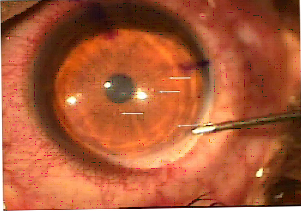
- Gatifloxacin 0.5% eye drops, Tobramycin 0.3% eye drops & Lubricant eye drops were given to be instilled one hourly on day of surgery till next follow-up after 24
- Next day, whitish spots were very faint and the patient was feeling much better, almost asymptomatic.
- We started routine Topical eye Drops:
– Gatiquin –P(gatifloxacin 0.3% + prednisolone 1%)eye drops four hourly – Tobramycin 0.3% eye drops four hourly
– Systane Ultra(lubricant) eye drops four hourlies in both eyes.
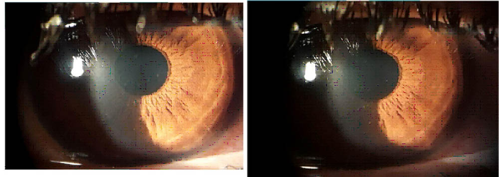
Again, we re-examined the patient after 48 hours of starting steroid drops, there was:
- Pain
- Redness
- Watering
- Blurred Vision
- Whitish round spots again reappeared on anterior corneal surface in R/E only
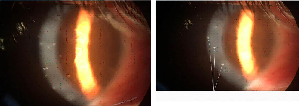
Superficial round white spots
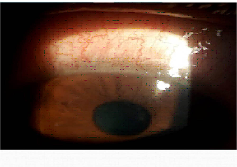
Conjunctival congestion
Initial impression was of non-specific viral keratoconjunctivitis but unilateral, very superficial
appearance of keratitis spots involving just superficial aspect of epithelial layer of cornea led to suspicion.
Occupational History: Gym Owner with frequent exposure to steam bath and Sauna in his premises.
We decided to take corneal scrapings for study under microscope by microbiologist.
In Operation Theatre, under microscope:
- Whitish round superficial spots were gently scraped with hockey stick knife and scrapings were sent to Laboratory for microscopic examination by Microbiologist.
- Corneal surface was thoroughly cleaned with BSS.
- Microbiologist found microsporidia with Gram staining.
- Treatment started:
– Tab. Albendazole 400mg BD orally
Topically: -Voriconazole 1%eye drops two hourlies
-Homatropine 2%eye drops eight hourlies
-Lubricant eye drops two hourlies
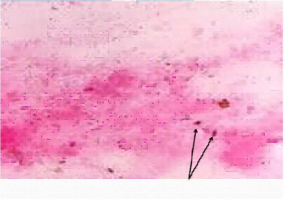
MICROSPORIDIA
After that we examined the patient after 3 days
He started improving. Whitish spots became very faint.
- Homatropine 2% eye drops were discontinued. Rest of the treatment was continued for two weeks on examination after two weeks of treatment:
- All White spots disappeared.
- No redness, watering, irritation and pain
- Visual acuity: 6/9 in R/E and 6/5 in L/E
We started Gatilox-DM(gatif loxacin 0.3%+dexamethasone 0.1%) eye drops and stopped voriconazole 1% eye drops. Gatilox-DM eye drops were continued in R/E three times a day for one week.Visual acuity improved to 6/6 in R/E after one week. After that only lubricant eye drops were continued and we examined the patient every month up to three months. Final visual acuity improved to 6/5 in right eye after 3 months.
Discussion
- Microsporidia –Intracellular parasites causing human infections. These are usually waterborne pathogens usually affecting immunocompromised individuals.
- Recently, there is increase in incidence of Microsporidial
- KeratoConjunctivitis (MKC) affecting normal immunocompetent individuals worldwide.
- Presentation is usually Unilateral Superficial KeratoConjunctivitis with punctate superficial keratitis in immunocompetent individuals.
- Usually younger age group with history of exposure to rain water, swimming pool water, mud, sauna, steam bath and hot springs.
- Usually self-limiting in immunocompetent individuals resolving in 1-2 weeks.
- May take more time to resolve when steroids are used.
- Bilateral and deeper involvement usually occurs in immunocompromised individuals.
- Responds very well to antifungal and antiparasitic agents especially in immunocompetent individuals.
- Topical steroids and bandage contact lenses should be avoided during active phase as these may cause deeper involvement especially in immunocompromised individuals.
Leave a Comment