

Dr.RAMYA APPANRAJ , Dr.Saravanan V R
Introduction: Retinal detachment occurs when fluid accumulates between the sensory retina and the retinal pigment epithelium. The most common type is the rhegmatogenousretinal detachment in which fluid from the vitreous cavity enters the potential subretinal space through a break in the retina and dissects the neurosensory retina from the retinal pigment epithelium. The annual incidence of RRD has been reported to be between 6.3 and 17.9 per 100,000 population, with a lifetime risk (up to 60 years of age) of 0.6%. The risk of developing a retinal detachment(RD) in the fellow eye ranges between 5% and 15%. The incidence of RD increases after cataract surgery: the cumulative 6-year risk of RD increases by a factor of between 6 and 8 and continues to increase linearly for 20 years after cataract surgery. Retinal detachment is also associated with increasing age, myopia, and certain vitreoretinal degenerations.1
Surgical approaches to RRD include scleral buckling, pneumatic retinopexy and pars plana vitrectomy(PPV).Treatment is often individualized depending on factors that include the number and location of breaks, amount of subretinal fluid, macular status, presence of proliferative vitreoretinopathy (PVR), lens status, and medical or ocular comorbidity. Traditionally, scleral buckling (SB) was viewed as the gold standard treatment for uncomplicated RRD. Pars plana vitrectomy (PPV) was traditionally reserved for treatment of eyes with complications, such as those showing giant retinal tears or exhibiting significant proliferative vitreoretinopathy (PVR). In the 1980s, the indications for PPV in RRD patients were broadened to include less complicated instances, and the term “primary vitrectomy” was introduced by Kloti.2
As the necessary instrumentation for, and safety of, PPV continue to improve with developments in microscope technology, intensified endoillumination, and wide-angle viewing systems, the indications for vitrectomy in RRD have been further expanded to include most patients with RRD.Studies have shown that primary vitrectomy may offer reattachment rates and visual acuity outcomes similar to those of scleral buckling surgery for primary repair of detachments, without some of the complications associated with scleral buckling.3-7 Several publications have shown that scleral buckling is not essential to achieve retinal reattachment.4, 8-11, Indeed, PPV is more useful than SB in eyes requiring simultaneous cataract extraction or those withpseudophakic status.12-18
Compared to SB, PPV offers several advantages. The view of the retinal periphery is enhanced, identification of retinal breaks is rendered easier, achievement of complete intraoperative retinal attachment is possible, the risks of hemorrhage or retinal incarceration inherent to the external drainage procedure applied during SB is eliminated, and the technique is less likely to cause a refractive change. In addition, the recent introduction of small-gauge vitrectomy has shifted the paradigm of standard vitreous surgery to Microincision vitrectomy surgery(MIVS) that is less invasive, affords fast recovery, and is sutureless.
Transconjunctivalsutureless 27-gauge PPV has emerged as an important advancement in vitreoretinal surgery instrumentation. Initially, 27 gauge TPPV was utilized in cases with noncomplex conditions such as vitreous hemorrhage and for macular indications. There has been an emergence of 27-gauge PPV in wider variety of cases with more complex conditions.Recently one study19 reported the outcome of 27 gauge microincision vitrectomy in posterior segment diseases. They found that the use of the 27- gauge system appeared safe and effective, with favorable visual and anatomic outcomes across a variety of surgical indications. Surgical indications were varied, including not only macular cases but also more complicated cases such as diabetic tractional retinal detachment and rhegmatogenous retinal detachment, and some with PVR requiring silicone oil.No studies regarding outcomes of 27 gauge PPV surgery in RRD has been published in India since the commercial introduction of this technology. The purpose of our study is to review the initial experience, clinical outcomes, and safety profile of a 27 gauge MIVS system without scleral buckling in cases of rhegmatogenous retinal detachment.
AIMS AND OBJECTIVES
AIM
The aim of our study is to evaluate the effectiveness and safety of 27-gauge transconjunctival pars plana vitrectomy without scleral buckling in the treatment of primary RRD.
OBJECTIVE
Primary Objective
- To measure the single surgery anatomical success rate judged by the retinal reattachment in the absence of tamponade agent.
- Mean improvement in visual acuity at 3 month postoperatively.
Secondary Objective
To analyseintraoperative and post-operative complications of the surgery and surgeon’s comfort during the surgery.
MATERIALS AND METHODS
Study Area
The study was conducted at Aravind Eye Hospital, Coimbatore. It is a tertiary eye care hospital with high volume of patients coming from over area of radius 100 kilometers and referrals from other hospital.It has all the eye speciality departments separately, equipped with all essential and advanced instruments performing all basic and advanced surgeries.
Study Population
All the patients coming to Out-patient Retina department of Aravind eye hospital, Coimbatore were studied.There was no gender restriction. All age groups were included except less than 18 years. The patients were from the urban and rural population of south India.
Study Design
A prospective observational study.
Sample Size
Theobjective of this thesis is to evaluate the efficacy of 27-gauge Vitrectomy surgery outcome through prospective observational study. The mean outcome and its standard deviation are observed from the previous study. Hence, the sample size is calculated based on the power analysis with the following parameters; power (80%), alpha (5%), m (0.79), m0 (1.25), standard deviation is 0.93. The estimated sample size is 32 in this case which is obtained from the formula given below.
Study Duration
June 2015 –May 2016 Inclusion Criteria
All cases of primary RRD (no previous surgery) in pseudophakic and phakic eyes including eyes with horseshoe tear, retinal hole associated with RRD.
Exclusion Criteria
- Evidence of Proliferative Vitreoretinopathy (PVR).
- Eyes with retinal dialysis.
- Patients younger than 18 years, with severe systemic disease, pregnancy.
- Complex RD requiring silicon oil tamponade is needed.
- Physically / mentally disabled patients who cannot comply with postoperative positioning.
Data Collection Technique and Tools
An informed consent was obtained in accordance with the Helsinki Declaration prior to inclusion in the study. Ethics committee approval was obtained and allthe possible merits and risks of the treatment were explained to the patient.At the presentation of the patient, a detailed medical and ocular history was taken, including the duration of visual loss and the presence of any ocular pathologies or prior interventions. All patients underwent following ophthalmic examination:
1.Uncorrected and Best-corrected visual acuity measured by Snellen’s chart and converted into logMAR units for statistical analysis.
2.Intraocular pressure (IOP) measurement was done by Goldmannapplanation tonometry.
3.Anterior segment evaluation was done by Slit lamp Examination.
4.Posterior segment evaluation was done with indirect ophthalmoscope and 20D lens with scleral indentation with special care taken to identify all retinal breaks and their location and to describe the configuration, location and extension of the RD in clock hours and any presence of PVR.
All the patients were subjected to 27G TPPV surgery. The surgery was done by one of the three surgeons of retina department.
Preoperative Preparations
Any systemic diseases like diabetes mellitus or hypertension were controlled. Systemic antibiotics and antibiotic drops were started. The pupil was dilated with instillation of tropicamide 0.5% with phenyephrine 5%, thrice at 15 minute intervals an hour prior to surgery. In hypertensive patients, phenylephrine was avoided.
Surgical Technique
The procedure was based on 27G transconjucntivalsutureless vitrectomy(Alcon constellation) in which 27G cannulas were placed using a 1-step nonbevelled technique with a trocar-cannula system. The conjunctiva was displaced anteriorly from the intended sclerotomy site with forceps. A trocar was inserted at an angle of approximately90° to the sclera, facing directly toward the vitreous cavity, 3.5 or 4.0mm away and parallel to the limbus depending on the lens status.
The angle was kept perpendicular to the surface as the cannula gets inserted into the eye and a scleral tunnel incision made. The cannula was held in place with forceps and the trocar was removed. The first sclerotomy was made in the inferotemporal region, and a 27-gauge infusion cannula was attached. After verifying its position inside the vitreous cavity using a 27-gauge light pipe, the infusion line was opened and cannulas were placed in the superotemporal and superonasal region.Core vitrectomy was done and induction of PVD was achieved when necessary. Perfluorocarbon liquid (PFCL) was used in all cases to stabilize the retina.
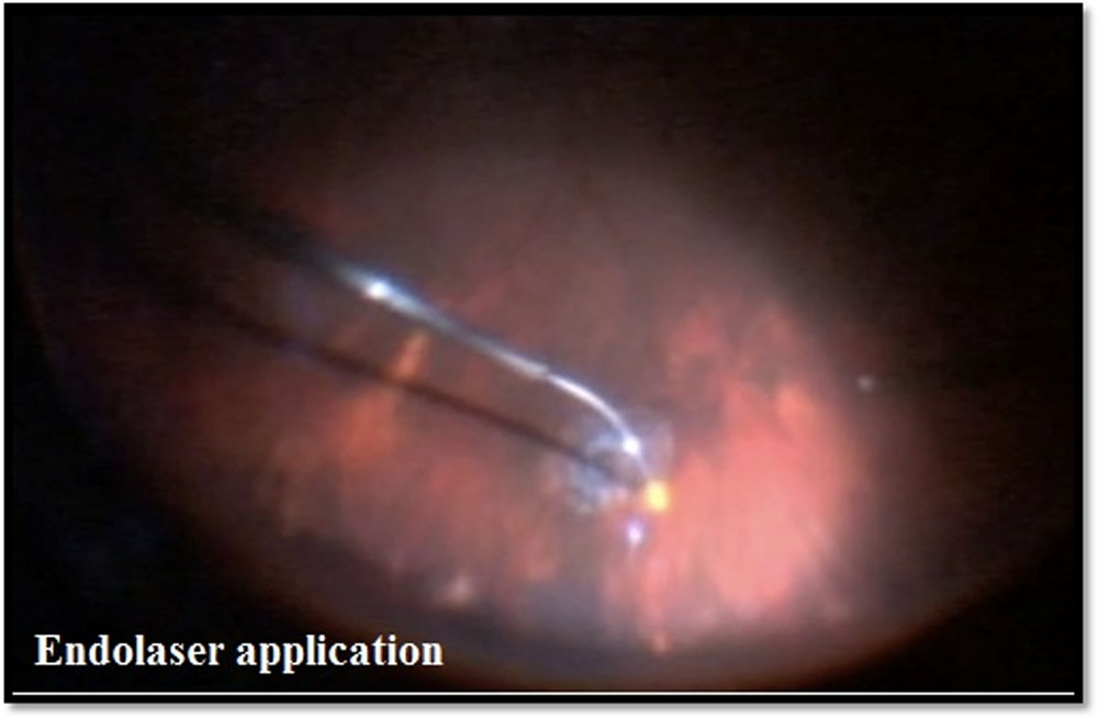
Then shaving of the peripheral vitreous gel was done and retinal break identified. Subretinal fluid was removed through the existing retinal tears. Endolaser photocoagulation of all retinal lesions and prophylactic 360° peripheral laserwas done after the fluid–air exchange. Finally air was exchanged for gas.Tamponade agent used in all the surgeries was C3F8 gas. The patients were advised to maintain prone position postoperatively. All the patients were treated postoperatively with topical steroid eye drops in a tapering dose down for 8 weeks, topical antibiotics for 1 week, and cycloplegic drops for 2 weeks.
Postoperative follow-up
Follow-up was done on 1 week, 2 weeks, 6 weeks and 3 months to assess the following:
- Anatomical and functional outcome
- Post operative Intraocular pressure
- Post surgical complications
- Redetachment and management.
An optical coherence tomography(OCT) scan (Spectralis OCT, HeidelburgInc, Germany) of the macula was done at 3 month follow up to detect any persistent subretinal fluid, retained subretinal perfluorocarbon liquid, epiretinal membranes, or other complications.
Figure 1: Introduction of ports
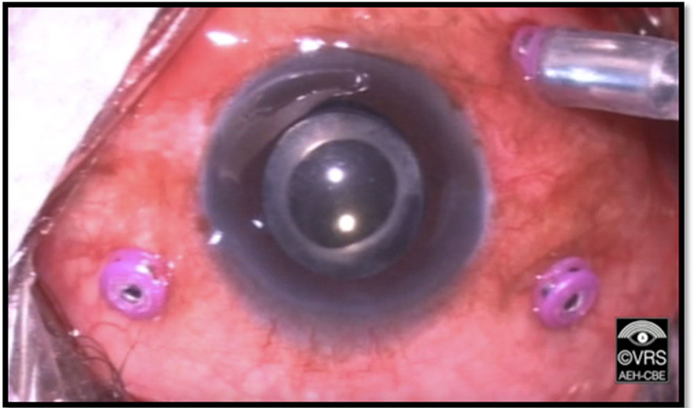
Figure 2: Core Vitrectomy
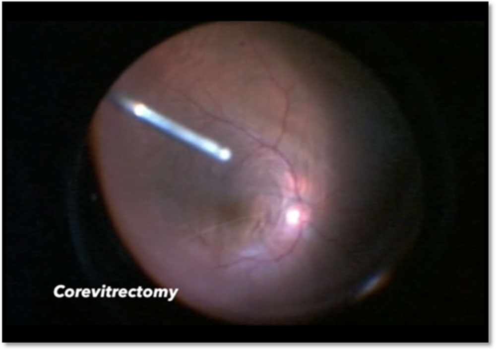
Figure 3: Endolaser application
Surgeon’s comfort in performing surgery was analysed by interviewng them at the end of surgery regarding how much comfortable they were with 27G TPPV and asked them to grade the comfort level as below:
Table 1 Grade of Surgeon’s comfort
| Grade | Comfort level |
| 0 | Extremely difficult |
| 1 | Very difficult |
| 2 | Difficult |
| 3 | Mild difficulty |
| 4 | Comfortable |
| 5 | Very comfortable |
Outcome measuresof our study were single-operation anatomical success rate, final anatomical success rate, postoperative(3 month) best corrected visual acuity, surgeon’s comfort, and surgical complications.
The anatomical reattachment was judged successful when retina remain attached in the absence of any tamponade agent.
IOP spike was defined as IOP of more than 21mmHg at any of the follow up visitsand was treated with the appropriate anti glaucoma medication and brought under control.
Statistical Methods
Statistical tools applied in this study have been evaluated using SPSS (version 16) software. There are two statistical measures used in this study known as descriptive analysis and paired t-test analysis. The descriptive analysis is a basic analysis to measure the data using percentage analysis or mean and standard deviation. The paired t-test analysis is used to compare two populations, in which one population contains preoperative data and another population contains postoperative data. The effectiveness of paired t-test is observed using the p-value (p£0.05) known as significant.
RESULTS AND OBSERVATIONS
32 patients having rhegmatogenous retinal detachment were taken for this clinical study. Their demographic and clinical details are as follows:
The mean of the age of patients in our study was 57.03(SD= 10.16). The distribution in various age groups is shown in table.The youngest patient was 24 years and oldest patient 81 years of age. Maximum patients were of more than 60 years of age.Males were predominantly affected (72%) than females (28%) in our study.Right eye involvement(68%) was more than left eye involvement(38%) out of the 32 patients studied.Systemic history revealed out of 32 patients, 3 patients(9%) had DM and 8 patients(25%) had hypertension and 6 patients(19%) had both DM and hypertension. 15 patients(47%) had no systemic history. Hence DM was present in 9 patients(28%) and hypertension was present in 14 patients(43.75%).The mean duration of presentation of the patient was 17.34(SD 34.88) days. Maximum number of patients(66%) presented within a week.
The percent of patients who presented between 8-30 days were 25%. 9% of the patients presented after a month.21 patients (66%) out of 32 who presented with RRD were pseudophakic patients and one had dislocated IOL while 10 patients were phakic (31.25%) out of which 7 patients had immature cataract. The patient having dislocated IOL underwent vitrectomy with scleral fixation intraocular lens.Out of 32 patients 19 patients(59%) had detachment in one or two quadrants while 13 patients(41%) had detachment in three or four quadrants. All the patients had attached retina postoperatively except one patient.31% (n=10) of the total patients presented with macula on RRD while 69%(n=21) presented with macula off RRD. 5 out of 10 patients presented with BCVA better than 6/12.
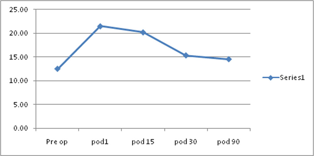
In 18 patients out of 32(56%) break were located in the superior part of retina. 8 patients out of 32(25%) had break located in the inferior part of retina. While remaining 6 patients(19%) had breaks both in superior and inferior part of retina. More than half of the patients (56%) in our study had breaks located in superior half of retina and two third had single break(75%).There were 8 patients who had or were found to have other associated pathology apart from RRD whose details is shown in table. 3 patients who had macular hole and one who had vasculitis had final BCVA less than 6/12.The mean IOP preoperatively was 12.47(SD 3.08) and mean IOP postoperatively at 3 months was 14.5 (SD 2.70). The difference was statistically significant(p<0.05).
The mean IOP at each visit is shown in the table. The trend of IOP change at each postoperative visit is shown in figure 17. The number of patients who had IOP spike at any postoperative visit was about 40.63%. All of them got controlled by antiglaucoma medical treatment except 2 patients who required C3F8 gas release.
Table 2: Distribution of age group
| Age Group | N | |
| <40 | 1 | |
| 41-50 | 8 | |
| 51-60 | 10 | |
| >60 | 13 | |
| Total | 32 |
Figure 6: Distribution of age group
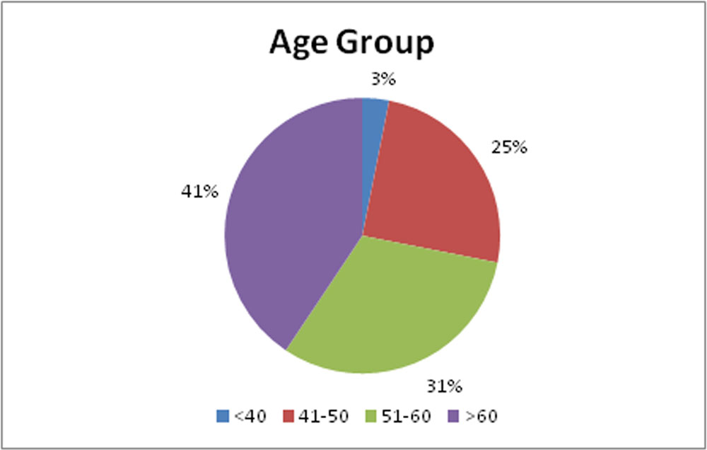
Table 3: Sex Distribution
| Gender | No. of Patient | Percentage |
| Male | 23 | 72 |
| Female | 9 | 28 |
| Total | 32 | 100 |
Figure 7: Sex Distribution
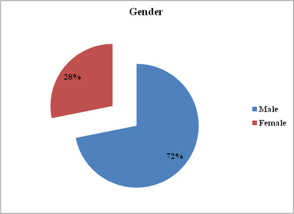
Table 4: Laterality
| Eye | No. of Patients | Percentage |
| RE | 20 | 63 |
| LE | 12 | 38 |
| Total | 32 | 100 |
Figure 8: Laterality
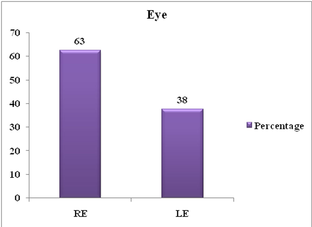
Table 5: Systemic Association
| Systemic association | No. of Patients | Percentage |
| DM | 3 | 9 |
| DM, HTN | 6 | 19 |
| HTN | 8 | 25 |
| Nil | 15 | 47 |
| TOTAL | 32 | 100 |
Figure 9: Systemic Association
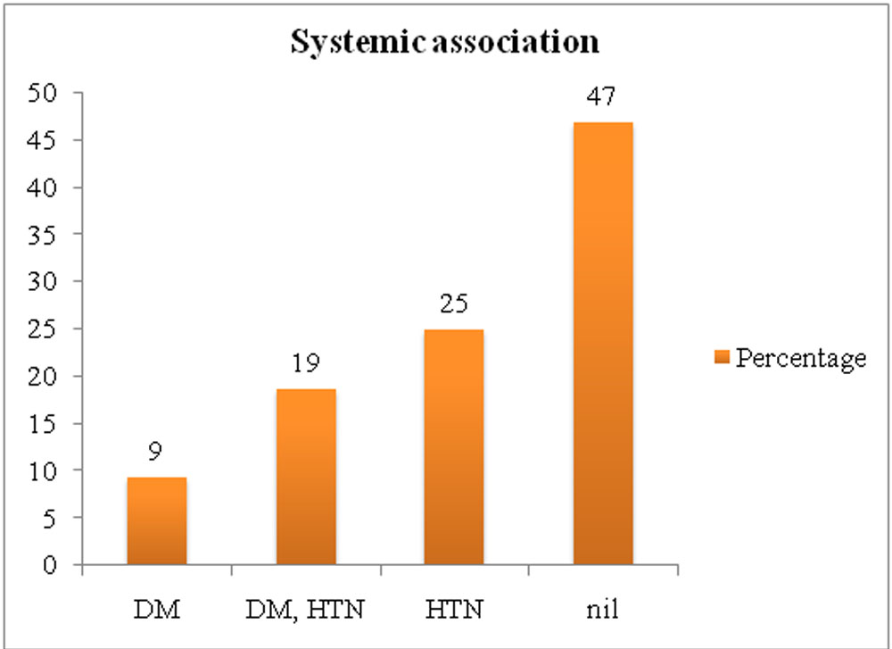
Table 6: Distribution of duration of visual loss
| Duration of visual loss | n | Percent |
| <= 7 days | 21 | 65.63 |
| 8-30 days | 8 | 25 |
| >30 days | 3 | 9.37 |
Figure 10:Distribution of duration of Visual Loss
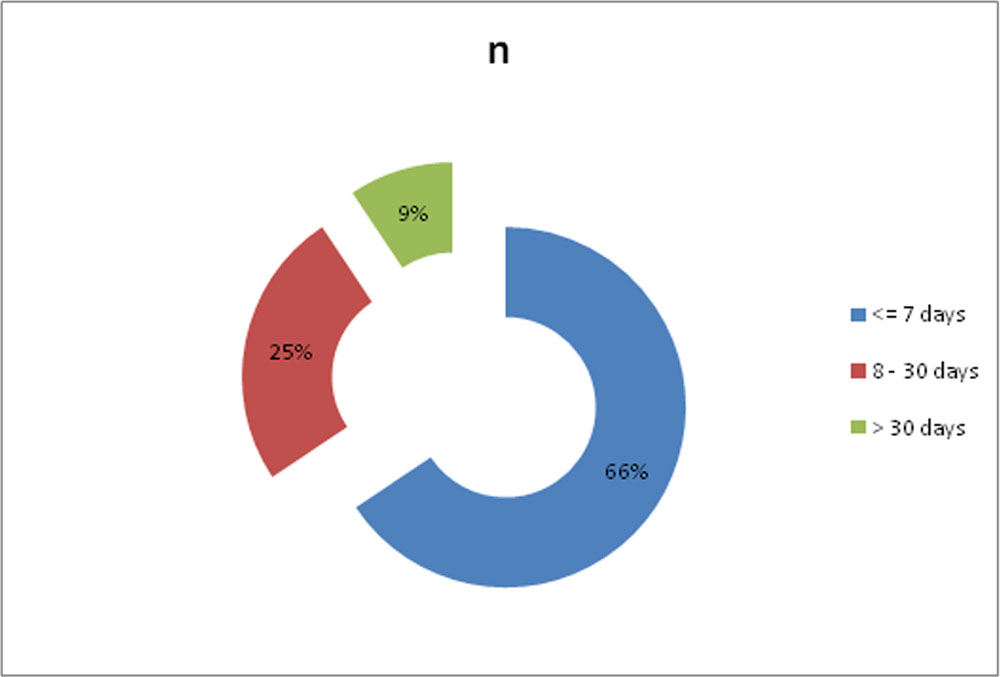
Table 7: Pre Operative Lens Status
| Lens status- Pre op | n | Percentage |
| Pseudophakic | 21 | 66 |
| clear lens | 2 | 6 |
| IMC | 7 | 22 |
| dislocated IOL | 1 | 3 |
| early imc | 1 | 3 |
| TOTAL | 32 | 100 |
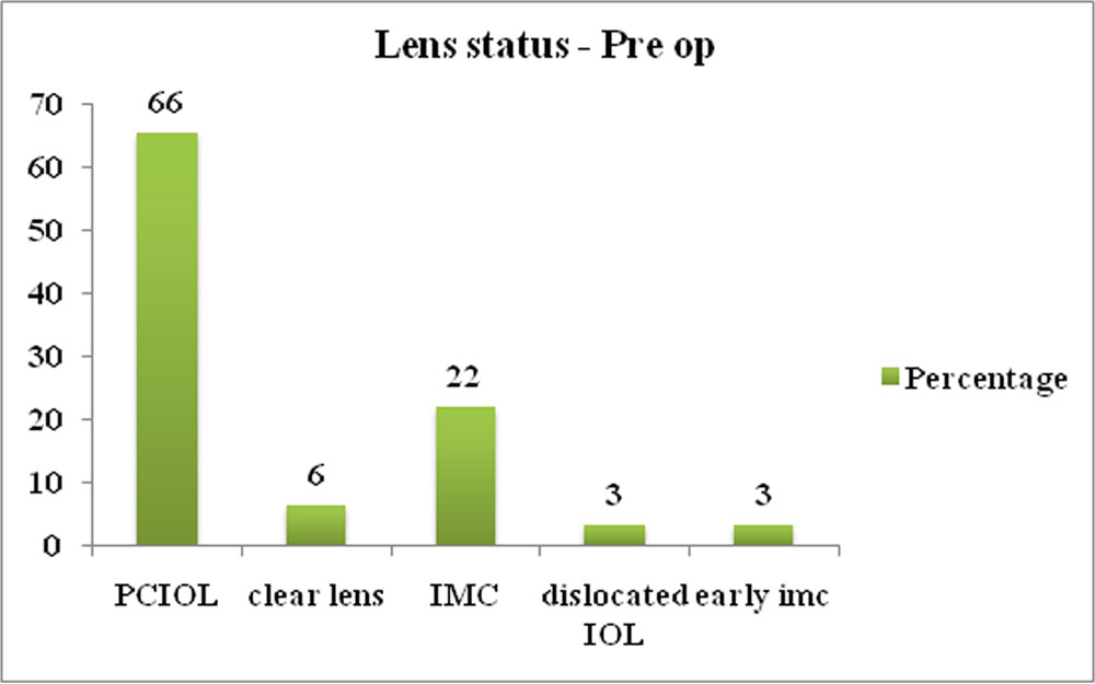
Figure 11:Pre operativeLens Status
Table 8: Extent of retinal involvement in quadrants
| Extent of retinal detachment | Number of patients | Percent |
| 1 quadrant | 4 | 12 |
| 2 quadrant | 15 | 47 |
| 3quadrant | 9 | 28 |
| 4 quadrant(total RD) | 4 | 13 |
| Total | 32 | 100 |
Figure 12: Extent of retinal involvement in quadrants
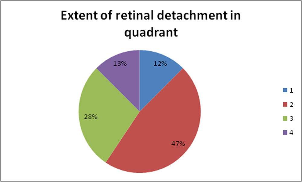
Table 9: Preoperative Macula Status
| Preoperative Macula status | n | Percent (%) |
| On | 10 | 31 |
| Off | 22 | 69 |
| Total | 32 | 100 |
Figure 13:Preoperative Macula Status
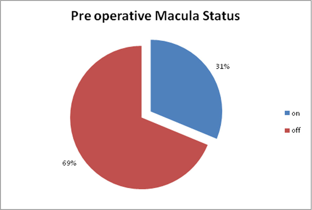
Table 10:Location of retinal break
| Retinal break-location | n | Percentage |
| I (Inferior part of retina) | 8 | 25 |
| S (superior part of retina) | 18 | 56 |
| C(Both) | 6 | 19 |
| TOTAL | 32 | 100 |
Figure 14: Location of retinal break
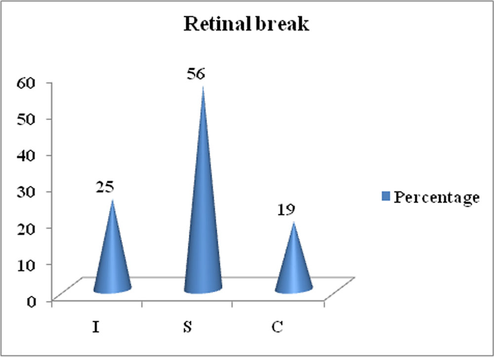
Table 11: Number of Retinal Break
| Number of retinal break | n | Percent |
| 1 | 24 | 75 |
| 2 | 5 | 16 |
| >2 | 3 | 9 |
Figure15 Number of Retinal Break
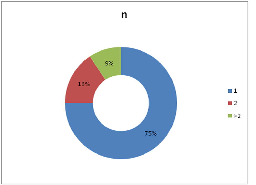
Table 12:Asociated pathology
| Associated Pathology | n | Percentage |
| Nil | 24 | 75 |
| Macular hole | 3 | 9 |
| High myopia | 2 | 6 |
| Vitreous haemorrhage (inferiorly) | 1 | 3 |
| Dislocated IOL | 1 | 3 |
| Vasculitis, pale disc | 1 | 3 |
| Total | 32 | 100 |
Figure 16: Percentage of Associated Pathology
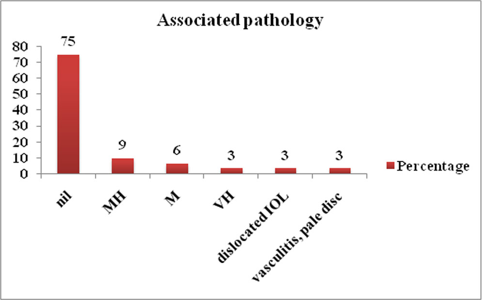
Table 13: Mean BCVA and mean IOP
| Paired Samples Statistics | |||||
| Mean | N | Std. Deviation | Std. Error Mean | ||
| BCVA (PRE-OP) | 1.1562 | 32 | .97879 | .17303 | |
| BCVA (POD 90DAYS) | .4500 | 32 | .23827 | .04212 | |
| IOP-PRE OP | 12.4688 | 32 | 3.07943 | .54437 | |
| IOP-POD1 | 21.4688 | 32 | 7.85343 | 1.38830 | |
| IOP-POD15 | 20.1875 | 32 | 7.54957 | 1.33459 | |
| IOP- 6 wk | 15.3125 | 32 | 3.86412 | .68309 | |
| IOP-POD90 | 14.5000 | 32 | 2.70006 | .47731 | |
Figure :17: Trend of mean IOP in Post Operative Visits.
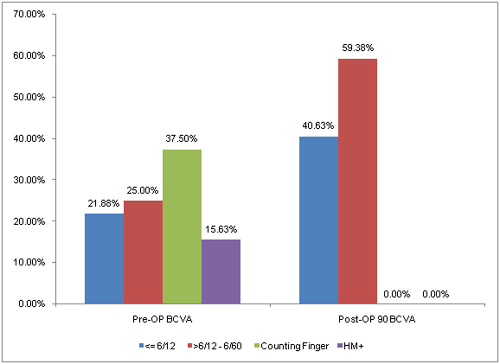
The mean of the preoperative logMAR visual acuity was 1.16(SD 0.98) and the mean of 3 month postoperative logMAR visual acuity was 0.45(SD 0.24). The difference was statistically significant.The preoperative visual acuity equal to or better than 6/60 was found to be in 15 patients(46.88%) while 17 patients(53.12%) presented with visual acuity less than 6/60. At 3 month follow up all the patients had best corrected visual acuity of 6/60 or better among which 40.63% of the patients had visual acuity of 6/12 or better.
Table 14: Mean BCVA
| Pre operative meanlogMAR visual acuity (SD) | Post operative mean logMAR visual acuity (SD) | P value |
| 1.16 (0.98) | 0.45 (0.24) | <0.01 |
Table 15: Visual acuity range vs BCVA
| Visual acuity range | Pre-OP BCVA | Post-OP 90 BCVA | ||
| No. of Patients | Percent | No. of Patients | Percent | |
| 6/12 or better | 7 | 21.88% | 13 | 40.63% |
| 6/12 – 6/60 | 8 | 25.00% | 19 | 59.38% |
| Counting Finger
5mt-1mt |
12 | 37.50% | 0 | 0.00% |
| HM+ | 5 | 15.63% | 0 | 0.00% |
| Total | 32 | 100.00% | 32 | 100.00% |
Figure 18:Visual acuity range vs BCVA
Single surgery anatomical success rate in our study was 97% with only one patient requiring resurgery, who developed redetachment due to PVR. He underwent Re-TPPV with 25G and retina was settled at the end of 3 months. So final anatomical success rate at last follow up was 100%.
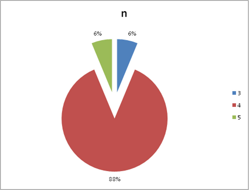
Surgeons were comfortable(grade 4) in majority of the cases(88%). Only in 2 cases they were uncomfortable with mild difficulty. None of the patients required conversion into 25G or 23G.
Table 16: Anatomical Outcome
| Re Surgery required | n | Percentage |
| No | 31 | 97 |
| Yes | 1 | 3 |
| TOTAL | 32 | 100 |
Figure 19Anatomical Outcome
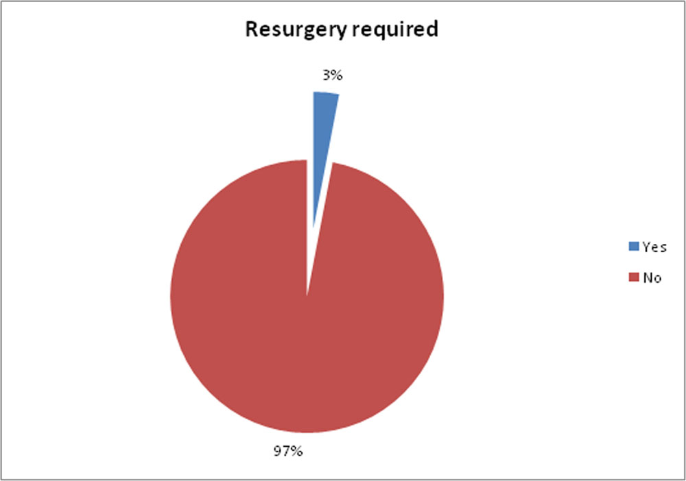
Table 17: Comfort Level Analysis
| Surgeon’s comfort grade | N | Percent |
| 0 | 0 | 0 |
| 1 | 0 | 0 |
| 3 | 0 | 0 |
| 3 | 2 | 6 |
| 4 | 28 | 88 |
| 5 | 2 | 6 |
| Total | 32 | 100 |
Figure 20Comfort Level Analysis
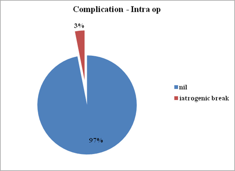
Intra operative complication:No major intraoperative complication was encountered except for iatrogenic peripheral retinal break in 1 eye.
Table 18: Intra Operative Complications
| Complication – Intra op | n | Percentage |
| Nil | 31 | 97 |
| iatrogenic break | 1 | 3 |
| TOTAL | 32 | 100 |
Figure 21:Intra Operative Complications
Post-Operative Complication:
The most common complication in our study was IOPspike (40.63%) followed by postoperative uveitis(34.38%).In 15.63% of the patients there was development or progression of cataract. 6.25 %(2 patients) of the cases developed shallow AC one of which required C3F8gas release. 1 patient developed PVR postoperatively causing the opening of old break and redetachment. The retina was successfully attached by 2nd surgery in that case.
Table 19: Post-Operative Complications
| Post op Complications | No. of Patients | Percent |
| Post operative uveitis | 11 | 34.38% |
| IOP spike | 13 | 40.63% |
| Cataract | 5 | 15.63% |
| PVR | 1 | 3.13% |
| Shallow AC | 2 | 6.25% |
| Nil | 8 | 25.00% |
OCT Findings:
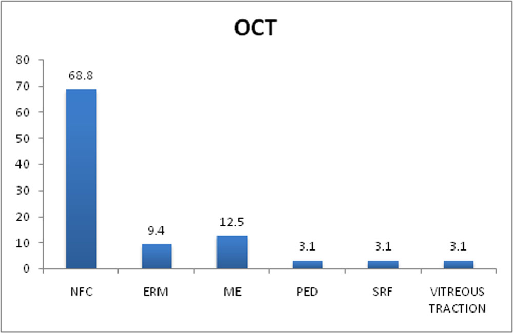
Table 20: OCT findings at 3 month postoperatively
| OCT at pod 90 | N | Percent |
| NFC (Normal foveal contour) | 22 | 68.8 |
| ERM | 3 | 9.4 |
| ME | 4 | 12.5 |
| PED | 1 | 3.1 |
| SRF | 1 | 3.1 |
| Vitreous traction | 1 | 3.1 |
| TOTAL | 32 | 100 |
3 patients out of 32(3%) developed ERM (Epiretinal membrane) detected by OCT. The other complications detected by OCT were macular edema in 4 patients (12.5%), shallow SRF (subretinal fluid) in 1 patient (3.1%), pigment epithelial detachment(3.1%), vitreous traction in 1 patient (3.1%).
Figure 22: OCT findings
CORRELATION WITH VISUAL ACUITY OUTCOME
- Age
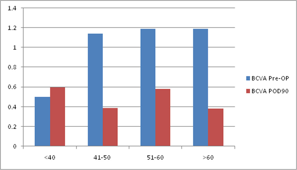
Maximum improvement in mean visual acuity occurred in more than 60 years age group followed by 41-50 age group.
Table 21: Comparison of age group vs mean BCVA
| Age Group | N | BCVA Pre-OP | BCVA POD90 | p-value | ||
| Mean | SD | Mean | SD | |||
| <40 | 1 | 0.5 | — | 0.6 | — | |
| 41-50 | 8 | 1.14 | 1.07 | 0.39 | 0.26 | 0.054 |
| 51-60 | 10 | 1.19 | 0.84 | 0.58 | 0.22 | 0.062 |
| >60 | 13 | 1.19 | 1.11 | 0.38 | 0.22 | 0.016 |
| Total | 32 | 1.16 | 0.98 | 0.45 | 0.24 | |
Figure 23: Comparison of age group vs mean BCVA
- Duration of Symptoms:
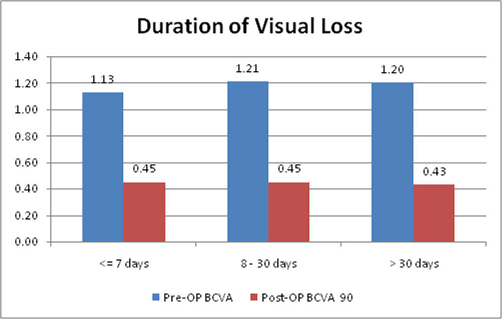
Mean improvement in visual acuity was more in the group who presented early- less than 7 days and was statistically significant. The group which presented between 8-30 days also had same mean visual acuity improvement but was not statistically significant. Thus patients who presented early in the course of RRD had better visual prognosis than those who presented late.
Table 22: Comparison of duration of visual loss vs mean BCVA
| Duration of visual loss | n | Pre-OP BCVA(logMAR) | 90 day Post-OP BCVA (logMAR) | p-value | ||
| Mean | SD | Mean | SD | |||
| <= 7 days | 21 | 1.13 | 0.88 | 0.45 | 0.24 | p < 0.01 |
| 8 – 30 days | 8 | 1.21 | 1.16 | 0.45 | 0.19 | 0.124 |
| > 30 days | 3 | 1.20 | 1.56 | 0.43 | 0.40 | 0.400 |
Paired t-test
Figure 24: Comparison of duration of visual loss vs mean BCVA
- Extent of Detachment
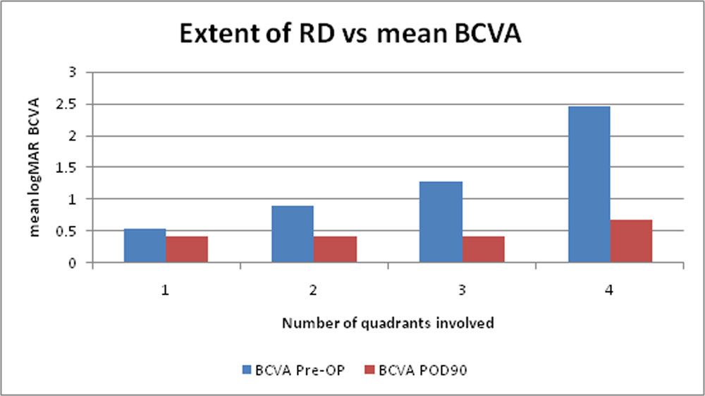
Preoperatively extent of retinal detachment was directly proportional to mean visual acuity and mean improvement in visual acuity at 90 day was better and almost similar in all group except in total RD(4 quadrant involvement) in which it was little less.
Table 23: Comparison ofextent of retinal detachment Vs mean BCVA
| Quadrants | N | BCVA Pre-OP | BCVA POD90 | p-value | ||
| Mean | SD | Mean | SD | |||
| 1 | 4 | 0.55 | 0.49 | 0.43 | 0.15 | 0.715 |
| 2 | 15 | 0.89 | 0.81 | 0.41 | 0.26 | 0.028 |
| 3 | 9 | 1.29 | 0.93 | 0.42 | 0.24 | 0.025 |
| 4 | 4 | 2.45 | 1.1 | 0.68 | 0.15 | 0.058 |
| Total | 32 | 1.16 | 0.98 | 0.45 | 0.24 | |
Figure 25: Comparison ofextent of retinal detachment Vs mean BCVA
- Location of break and number of break
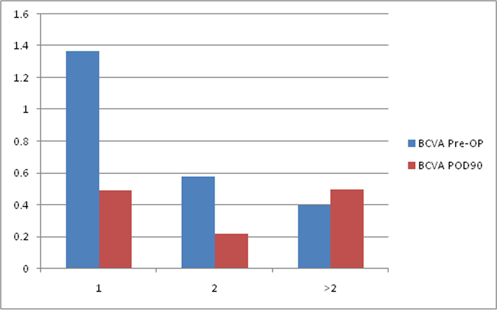
Visual acuity improvement was found to be less in cases of more than 2 breaks and in superiorly located breaks.
Table 24: Comparison of location of breakvs mean BCVA
| Location | N | BCVA Pre-OP | BCVA POD90 | p-value | ||
| Mean | SD | Mean | SD | |||
| S | 16 | 1.59 | 1.15 | 0.53 | 0.19 | 0.001 |
| I | 9 | 0.9 | 0.42 | 0.4 | 0.26 | 0.008 |
| C | 7 | 0.5 | 0.56 | 0.34 | 0.28 | 0.600 |
| Total | 32 | 1.16 | 0.98 | 0.45 | 0.24 | |
Figure 26: Comparison of location of breakvs mean BCVA
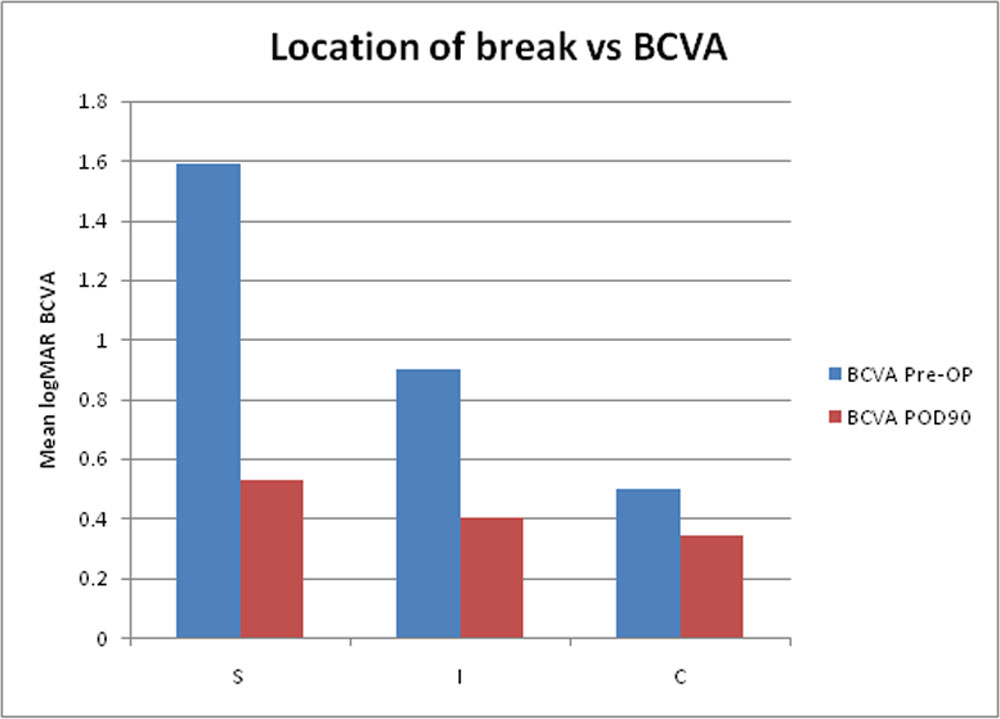
Table 25: Comparison of number of retinal breaksVs mean BCVA
| N | BCVA Pre-OP | BCVA POD90 | p-value | |||
| Mean | SD | Mean | SD | |||
| 1 | 24 | 1.37 | 1.01 | 0.49 | 0.22 | <0.001 |
| 2 | 5 | 0.58 | 0.62 | 0.22 | 0.19 | 0.185 |
| >2 | 3 | 0.4 | 0.4 | 0.5 | 0.3 | 0.225 |
| Total | 32 | 1.16 | 0.98 | 0.45 | 0.24 | |
- Macula status
In detached macula group meanlogMAR BCVA improved significantly from 1.58 to 0.45 and in attached macula group it improved from 0.39 to 0.32. Since the patient of attached macula group had better preoperative vision mean improvement in BCVA was not significant statistically.
Table 26: Comparison of preoperative macula statusvs mean BCVA
| Macula Status | N | Pre-OP BCVA(logMAR) | 90 dayPost-OP BCVA (logMAR) | p-value | ||
| Mean | SD | Mean | SD | |||
| Attached | 10 | 0.39 | 0.40 | 0.32 | 0.19 | 0.629 |
| Detached | 22 | 1.50 | 0.97 | 0.51 | 0.24 | p < 0.01 |
| Total | 32 | 1.16 | 0.98 | 0.45 | 0.24 | p < 0.01 |
Paired t-test
Figure 28: Comparison of preoperative macula statusVs mean BCVA
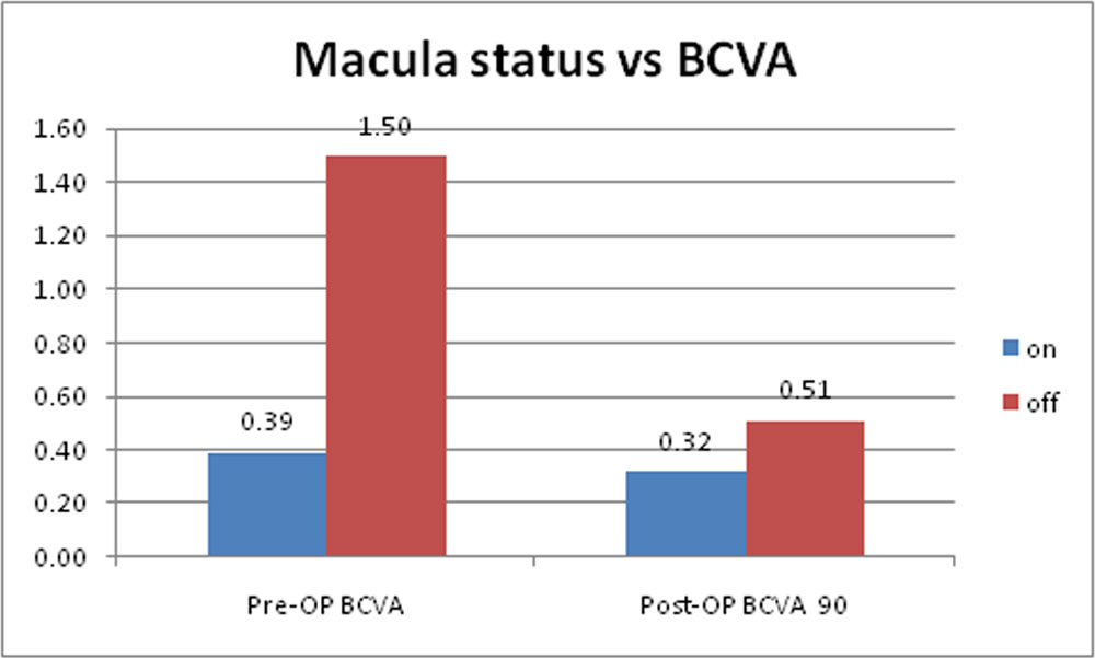
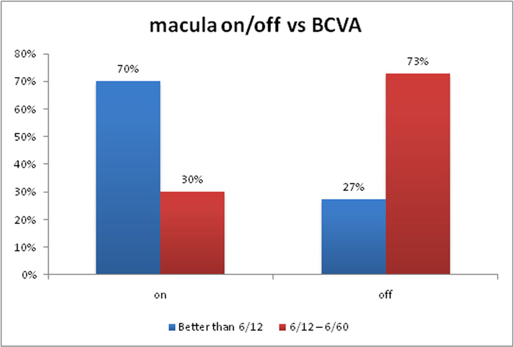
Table 27: Comparison of preoperative macula status vs BCVA range in Snellens
| Post op BCVA range (Snellen’s) | macula on/off | Total | p-value | |
| on | off | |||
| Better than 6/12 | 7 | 6 | 13 | 0.029 |
| 70% | 27% | 41% | ||
| 6/12 – 6/60 | 3 | 15 | 19 | |
| 30% | 73% | 59% | ||
| Total | 10 | 22 | 32 | |
Figure 29: Comparison of preoperative macula status vs BCVA range in Snellens
- Lensstatus
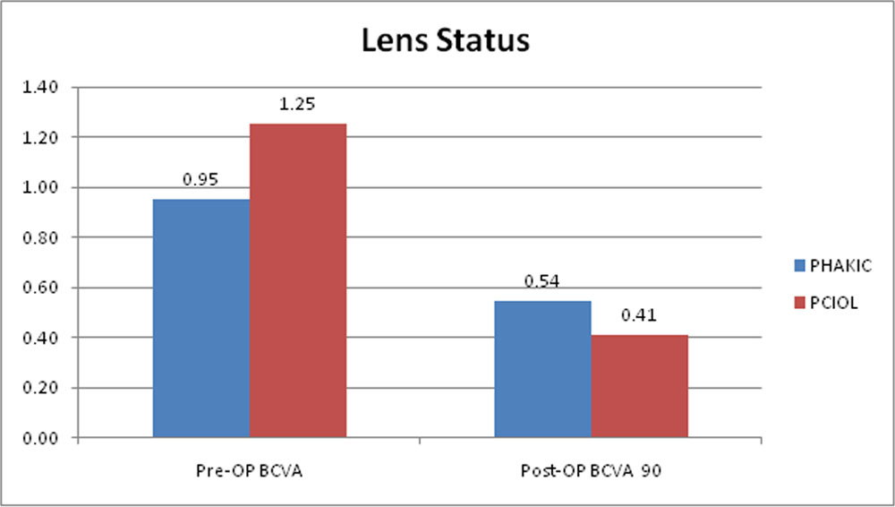
Pseudophakic patients were found to have better improvement in mean BCVA as compared to phakic group and the difference was statistically significant.
Table 28: Comparison of preoperative lens statusvs mean BCVA
| Lens | N | Pre-OP BCVA(logMAR) | 90 day Post-OP BCVA (logMAR) | p-value | ||
| Mean | SD | Mean | SD | |||
| Phakic | 10 | 0.95 | 1.04 | 0.54 | 0.19 | 0.231 |
| Pseudophakia | 22 | 1.25 | 0.96 | 0.41 | 0.25 | p < 0.01 |
Paired t-test
Figure 30: Comparison of preoperative lens statusvs mean BCVA
DISCUSSION
Many studies have been done showing superiority of primary vitrectomy in RRD with 20,23 and 25G over scleral buckle especially in pseudophakic patients. We conducted a prospective study to evaluate the efficacy and complications of 27G vitrectomy in uncomplicated RRD cases. We compared our results with previous studies of 23G and 25G vitrectomy in RRD.The mean age group in our study was 57.03 (SD 10.16) years which was near to the study by Figueroa et al53 of 55.8 (SD 16.78)years.
72% of the patients were males which was comparable to 73.7% found in the study by Marta S. Figueroa et al53.
In our study 63% of the patient presented with RE involvement and 37% with LE. Miller et al55 had 42.9% of the patient who presented with RE involvement and 57.1% with LE.
The mean time of the duration of visual loss of the patients in our study was 17.34 (SD 34.88, range 1-180) days with the minimum of 1 day and maximum of 180 days. Figueroa et al53 reported mean duration of visual loss of 6.89 (SD 6.32, range 1-40) days and Lai et al reported 8 days (range1-35).
The mean preoperative best corrected visual acuity in our study was 1.16(SD 0.98) while it was 0.81(SD 0.66) in the study by Figueroa et al53 and 0.70 in Miller et al55 study.
Most patients in our study had undergone prior cataract surgery i.e. pseudophakic(68.75%) but 31.25% of the patients had phakic RRD at the time of diagnosis. While these values were 62.4% and 37.6% for Marta S. Figueroaet al 53, 85.7% and 14.3 in Miller et al 55, 62% and 38% in Michael M Lai et al44 respectively.
Out of 32 patients in our study 19 patients(59.38%) had retinal detachment in one or two quadrants while 13 patients (40.62%)had more extensive RD which was near to the values of 71.4% and 28.6% in the study by Miller et al.55
Macula was attached in 10 patients(31.25%) and was detached in remaining 22 patients(68.75%). In the study conducted by Marta S Figueroa et al 53 macula was attached in 52% of patients and 48% had detached macula. Miller et al 55 reported 66.7% of the cases with attached macula and 33.30% of cases with detached macula. Michael M Lai44 reported 38% of the cases with attached macula and 62% with detached macula.
More than half of the breaks were located in superior retina in our study as was the case in the study by Miller et al.55 Three-fourth of the eyes had single break(75%) unlike Miller et al 55 who reported 2 or more breaks in more than one half of eyes.
The primary anatomical outcome in our study was 97%. Out of 32 eyes only one eye developedredetachmentwho presented at 3 month. The redetachment was due to PVR and was successfully operated second time to give 100% final anatomical outcome. The single surgery anatomical success rate of 97% found in our study done with 27G PPV is comparable with the results found in other studies. The single surgery anatomical success rate reported for 20 G ranges from 74.3% to 89.3% and for 23 G between 80% and 96.2%.42,43, 59Marta S. Figueroa et al 53 reported single surgery anatomical success rate with 23 G PPV to be 96.2%. Miller et al55 reported 92.9% success with 25 G PPV while Michael Lai et al 44 reported74% single surgery anatomical successwith 25 G PPV. The final reattachment rates were 100% for all three studies.53, 55, 44
In our study the meanlogMARbest corrected visual acuity improved from 1.16(SD 0.98) preoperatively to 0.45(SD 0.24, p<0.01) postoperatively at 3 months (p<0.01). Marta S. Figueroa et al53 reported mean improvement inlogMAR BCVA from 0.81 (SD 0.66) to 0.21(SD 0.29).Michael M. Lai et al44 reported mean improvement in visual acuity from 20/100(0.7 logMAR) to 20/60(0.50logMAR, p-0.0001).Hence our results in this aspect were comparable to Lai et al44 study. The less improvement in mean BCVA in our study compared to the study by Figueroa et al 53 can be due to less preoperative mean BCVA and more number of macula off patients (68.75%) in our study as compared to Figueroa et al53 (48%).
In the group of attached macula mean logMARBCVA improved from 0.39 (SD0.40) to0.32 (SD 0.19, p=0.629) in our study and in detached macula group mean BCVA improved from 1.50 (SD 0.97) to 0.51 (SD 0.24, p<0.01).Miller et al 55 reported mean improvement inlogMAR BCVA in attached macula group from 0.43 to 0.23 (p=0.24) and in detached macula group from 1.60 to 0.23 (p=0.001). Lai et al 44 reportedthat in patients with attached macula, the mean visual acuity remained unchanged at 20/40(0.3 logMAR). While in detached macula group mean visual acuity improved from 20/300(1.2logMAR) to 20/70(0.55logMAR, p<0.0001). Thus in this regard, our results were comparable to Lai et al 44 but not to Miller et al55.
Overall in our study the number of patients who achieved final BCVA of 6/12 or better was 41%. This was 78.2% reported in study by Figueroa et al53and 55% in the study by Lai et al 44. In attached macula group in our study,the number of patients whose final BCVA was 6/12 or better was 70% while it was 27% in detached macula group. In the study by Lai et al44 these values were 75% and 42% respectively.Thus our results on this aspect were almost comparable to the results of Lai et al44. The decrease in overall number of patients attaining BCVA of 6/12 or better in our study compared to Figueroa et al53 can be explained on the basis of increased number of patients in macula off group in our study(68.75%) compared to Figueroa et al.53
The mean improvement in BCVA in our study was better in pseudophakic patients(0.41logMAR (SD 0.25); p <0.01) compared to phakic patients(0.54logMAR(SD 0.19;p=0.231) which is consistent with the result of Figueroa et al53. They reported pseudophakic eyes had mean improvement in BCVA to 0.19logMAR (SD 0.25) while in phakic patients it was 0.25logMAR (SD 0.35).
Intraoperative retinal break, scleral suture requirement, hypotony, cataract, macular pucker, cystoid macular edema, vitreous hemorrhage, retinal redetachment, choroidal detachment, endophthalmitis and glaucoma have been previously reported in several studies with 20-G PPV, 23 and 25-G TSV.7, 10, 43, 4 ,58,60In our study no major intraoperative complication was encountered except for iatrogenic retinal break that was observed in 1 eye (3.0%). Postoperative complications were glaucoma(40.63%) controlled by topical medicationmedications except 2 eyes which required C3F8 gas release for control of IOP,postoperative uveitis controlled by topical medication(34.83%),cataract formation or progression (15.63%), macular edema (12.5%), ERM (9%), shallow AC(6.25%), SRF (3%),second PPV(3%). The post operative complications reported by Figueroa et al53 were cataract (24%), IOP spikes (20.3%), ERM (7%), retained perfluoro-N-octane (PFO, 4.5%),hemovitreous (0.8%), redetachment (3.8%), second PPV (5.3%), macular edema(0.8%), diplopia (1.5%). Thus complications in our study were almost similar compared to Figueroa et al53 study exceptthat the percentage of patients having IOP spikes and macular edema were more in our study and post-operative uveitis was not found in Figueroa et al53 study andwe didn’t had any complication of hemovitreous, retained PFO or diplopia.
One of the concerns regarding MIVS is the higher rate of postoperative hypotony due to wound leakage. This could also increase the risk of endophthalmitis.In a case series of more than 900 cases of 23-G vitrectomy, Parolini et al described only 3.3% of eyes with transient hypotony and 3.9% of sclerotomy leakage and no cases of endophthalmitis.60 In other reports, the incidence rates of endophthalmitis have been shown to be similar with 20-G, 23-G, and 25-G vitrectomy, ranging from 0.02% for 20-G vitrectomy to 0.13% for 25-G vitrectomy.61In our study none of the eyes presented with hypotony or endophthalmitis.Thus, microincisional vitrectomy does not appear to increase the rates of postvitrectomyendophthalmitis.
All surgeons were comfortable with 27G vitrectomy system in most of uncomplicated RRD patients in our study recommending its use in those patients. Because of smaller wound size, simplified wound construction, improved microarchitecture design 27G offers several advantages over 23G and 25G and with increased experience its use is expanding to even
complicated cases of RD and other posterior segment diseases.19,62
SUMMARY
This is a prospective observational study which was conducted at Aravind Eye Hospital, Coimbatore between June 2015 and May 2016. 32 cases were included in the study.
- Patients were in the age group of 24-81 years and the mean age was 57.03 (SD 10.16) years with predominant male patients in our study group.
- The mean duration of presentation of the patient was 17.34(SD 34.88) days. Maximum number of patients(66%) presented within a week.
- 21 out of 32 patients were pseudophakic, 1 had dislocated IOL and rest 10 patients were phakic.
- More than half patients had retinal detachment in one or two quadrants with 22 out of 32 patients having detached macula. Most cases had a single break and located superiorly.
- The single surgery anatomical success rate in our study was 97% with one patient developing redetachment. The final anatomical success rate was 100%.
- The mean of the preoperative logMAR visual acuity was 1.16(SD 0.98) and the mean of 3 month postoperative logMAR visual acuity was 0.45(SD 0.24). The difference was statistically significant. The number of patients attaining postoperative visual acuity better than 6/12 was 40.63%.
- Surgeons were comfortable in operating with 27G vitrectomy in 88% of the patients, very comfortable in 6% of patients and had mild difficulty in 6% of the patients.
- No major intraoperative complication was encountered except for iatrogenic peripheral retinal break in 1 eye.
- The most common postoperative complication in our study was IOP spike(40.63%) followed by postoperative uveitis (34.38%).In 15.63% of the patients there was development or progression of cataract. 6.25%(2 patients) of the cases developed shallow AC one of which required C3F8gas release. 1 patient developed PVR postoperatively causing the opening of old break and redetachment.
- 3 patients out of 32(3%) developed ERM (Epiretinal membrane) detected by OCT. The other complications detected by OCT were macular edema in 4 patients (12.5%), shallow SRF (subretinal fluid) in 1 patient (3.1%), pigment epithelial detachment (3.1%), vitreous traction in 1 patient (3.1%).
CONCLUSIONS
- Patients who present early in the course of disease have better visual outcome.
- Primary vitrectomy without scleral buckling is an effective technique in treating patients with uncomplicated RRD without PVR, both anatomically and functionally, with anatomical success rate comparable to all otherstudies done with 25G and 23G.
- 27G TPPV is safe and effective for uncomplicated RRD cases.
- 27G TPPV is comfortable to surgeon in majority of the patients in treating uncomplicated RRD.
- Complications were few and comparable to those described for 23 and 25G TPPV for treating RRD patients.
LIMITATIONS
- Being a time bound study we could not include large number of patients, i.e. our sample size is small.
- We observed anatomical and visual outcome in 27G TPPV only. We didn’t compare its results with 25G or 23G TPPV system. Hence a comparative study is needed to show the superiority of 27G over 25/23G vitrectomy in uncomplicated RRD cases.
- The follow up period in our study was small (3 months). A long term study is needed to evaluate long term complications.
- Since we excluded the cases having PVR associated with RRD the results cannot be justified for complex RRD.
RECOMMENDATIONS
Primary vitrectomy in primary RRD cases is safe and effective.
- In uncomplicated RRD cases 27G TPPV has excellent anatomical success rate and comparable visual outcome to 25G, 23G and 20G. Thus we recommend its use should be extended to uncomplicated RRD cases apart from macular indications.
- Since complex RRD were not included in our study there is a need to study the efficacy of 27G vitrectomy in complex RRD.
- Because of the absence of availability of 27G silicon oil cannula in India, there is a need to manufacture it so that 27G vitrectomy can be used for complex RRD.
- Since we excluded patients of Age less than 18 years, role of 27G vitrectomy in RRD in paediatric population could not be determined. The use of 27G vitrectomy in ROP (Retinopathy of prematurity) has started, so we recommend to study the outcome of 27G vitrectomy in paediatric RRD patients.
- We recommend a large prospective comparative study for the use of 27G TPPV system in RRD cases to show its efficacy and safety over other gauge vitrectomy.
BIBLIOGRAPHY
- Mitry D, Charteris DG, Fleck BW, Campbell H, Singh J. The epidemiology of rhegmatogenous retinal detachment: geographical variation and clinical associations. Br J Ophthalmol. 2010 Jun;94(6):678–84.
- Klöti R. Amotio-ChirurgieohneSkleraeindellung. PrimäreVitrektomie. KlinischeMonatsblätterfürAugenheilkunde. 1983 May;182(5):474–8.
- Heimann H, Hellmich M, Bornfeld N, Bartz-Schmidt KU, Hilgers RD, Foerster MH. Scleral buckling versus primary vitrectomy in rhegmatogenous retinal detachment (SPR Study): design issues and implications. SPR Study report no. 1. Graefes Arch ClinExpOphthalmol. 2001 Aug;239(8):567–74.
- Weichel ED, Martidis A, Fineman MS, McNamara JA, Park CH, Vander JF, et al. Pars plana vitrectomy versus combined pars plana vitrectomy-scleral buckle for primary repair of pseudophakic retinal detachment. Ophthalmology. 2006 Nov;113(11):2033–40.
- Saw S-M, Gazzard G, Wagle AM, Lim J, Au Eong K-G. An evidence-based analysis of surgical interventions for uncomplicated rhegmatogenous retinal detachment. ActaOphthalmol Scand. 2006 Oct;84(5):606–12.
- Feltgen N, Weiss C, Wolf S, Ottenberg D, Heimann H, SPR Study Group. Scleral buckling versus primary vitrectomy in rhegmatogenous retinal detachment study (SPR Study): recruitment list evaluation. Study report no. 2. Graefes Arch ClinExpOphthalmol. 2007 Jun;245(6):803–9.
- Ahmadieh H, Moradian S, Faghihi H, Parvaresh MM, Ghanbari H, Mehryar M, et al. Anatomic and visual outcomes of scleral buckling versus primary vitrectomy in pseudophakic and aphakic retinal detachment: six-month follow-up results of a single operation–report no. 1. Ophthalmology. 2005 Aug;112(8):1421–9.
- Figueroa MS, López-Caballero C, Contreras I. Anatomical and functional outcomes of vitrectomy for the treatment of pseudophakicregmatogenous retinal detachment. Archivos de la Sociedad Española de Oftalmología (English Edition). 2010 Jan 1;85(2):59–63.
- Heimann H, Bartz-Schmidt KU, Bornfeld N, Weiss C, Hilgers R-D, Foerster MH, et al. Scleral buckling versus primary vitrectomy in rhegmatogenous retinal detachment: a prospective randomized multicenter clinical study. Ophthalmology. 2007 Dec;114(12):2142–54.
- Martínez-Castillo V, Zapata MA, Boixadera A, Fonollosa A, García-Arumí J. Pars plana vitrectomy, laser retinopexy, and aqueous tamponade for pseudophakicrhegmatogenous retinal detachment. Ophthalmology. 2007 Feb;114(2):297–302.
- Stangos AN, Petropoulos IK, Brozou CG, Kapetanios AD, Whatham A, Pournaras CJ. Pars-plana vitrectomy alone vs vitrectomy with scleral buckling for primary rhegmatogenouspseudophakic retinal detachment. Am J Ophthalmol. 2004 Dec;138(6):952–8.
- Sodhi A, Leung L-S, Do DV, Gower EW, Schein OD, Handa JT. Recent trends in the management of rhegmatogenous retinal detachment. SurvOphthalmol. 2008 Feb;53(1):50–67.
- Heimann H, Zou X, Jandeck C, Kellner U, Bechrakis NE, Kreusel K-M, et al. Primary vitrectomy for rhegmatogenous retinal detachment: an analysis of 512 cases. Graefes Arch ClinExpOphthalmol. 2006 Jan;244(1):69–78.
- Pastor JC, Fernández I, Rodríguez de la Rúa E, Coco R, Sanabria-Ruiz Colmenares MR, Sánchez-Chicharro D, et al. Surgical outcomes for primary rhegmatogenous retinal detachments in phakic and pseudophakic patients: the Retina 1 Project–report 2. Br J Ophthalmol. 2008 Mar;92(3):378–82.
- Schwartz SG, Flynn HW. Pars plana vitrectomy for primary rhegmatogenous retinal detachment. ClinOphthalmol. 2008 Mar;2(1):57–63.
- Mendrinos E, Dang-Burgener NP, Stangos AN, Sommerhalder J, Pournaras CJ. Primary vitrectomy without scleral buckling for pseudophakicrhegmatogenous retinal detachment. Am J Ophthalmol. 2008 Jun;145(6):1063–70.
- Colyer MH, Barazi MK, von Fricken MA. Retrospective comparison of 25-gauge transconjunctivalsutureless vitrectomy to 20-gauge vitrectomy for the repair of pseudophakic primary inferior rhegmatogenous retinal detachment. Retina (Philadelphia, Pa). 2010 Dec;30(10):1678–84.
- Kunikata H, Nishida K. Visual outcome and complications of 25-gauge vitrectomy for rhegmatogenous retinal detachment; 84 consecutive cases. Eye (Lond). 2010 Jun;24(6):1071–7.
- Khan MA, Shahlaee A, Toussaint B, Hsu J, Sivalingam A, Dugel PU, et al. Outcomes of 27 Gauge Microincision Vitrectomy Surgery for Posterior Segment Disease. Am J Ophthalmol. 2016 Jan;161:36-43-2.]
- Ghazi NG, Green WR. Pathology and pathogenesis of retinal detachment. Eye (Lond). 2002 Jul;16(4):411–21.
- Sebag J. Anomalous posterior vitreous detachment: a unifying concept in vitreo-retinal disease. Graefes Arch ClinExpOphthalmol. 2004 Aug;242(8):690–8.
- Byer NE. What happens to untreated asymptomatic retinal breaks, and are they affected by posterior vitreous detachment? Ophthalmology. 1998 Jun;105(6):1045-1049-1050.
- Goldberg MF. Clear lens extraction for axial myopia. An appraisal. Ophthalmology. 1987 May;94(5):571–82.
- Javitt JC, Street DA, Tielsch JM, Wang Q, Kolb MM, Schien O, et al. National outcomes of cataract extraction. Retinal detachment and endophthalmitis after outpatient cataract surgery. Cataract Patient Outcomes Research Team. Ophthalmology. 1994 Jan;101(1):100–105; discussion 106.
- Duker JS. Steinert RF. Cataract surgery: technique, complications, and management. Philadelphia: WB Saunders; 1995.
- Tielsch JM, Legro MW, Cassard SD, Schein OD, Javitt JC, Singer AE, et al. Risk factors for retinal detachment after cataract surgery. A population-based case-control study. Ophthalmology. 1996 Oct;103(10):1537–45.
- Austin KL, Palmer JR, Seddon JM, Glynn RJ, Rosenberg L, Gragoudas ES, et al. Case-control study of idiopathic retinal detachment. Int J Epidemiol. 1990 Dec;19(4):1045–50.
- Chapter 136 – Rhegmatogenous Retinal Detachment [Internet]. Free Medical Textbook. 2010 [cited 2016 Jun 28]. Available from: https://medtextfree.wordpress.com/2010/12/31/chapter-136-rhegmatogenous-retinal-detachment/
- Ashrafzadeh MT, Schepens CL, Elzeneiny II, Moura R, Morse P, Kraushar MF. Aphakic and phakic retinal detachment. I. Preoperative findings. Arch Ophthalmol. 1973 Jun;89(6):476–83.
- Byer NE. Changes in and prognosis of lattice degeneration of the retina. Trans Am AcadOphthalmolOtolaryngol. 1974 Apr;78(2):OP114-125.
- Benson WE, Morse PH. The prognosis of retinal detachment due to lattice degeneration. Ann Ophthalmol. 1978 Sep;10(9):1197–200.
- Lincoff H, Gieser R. Finding the retinal hole. Arch Ophthalmol. 1971 May;85(5):565–9.
- Kazahaya M. Prophylaxis of retinal detachment. SeminOphthalmol. 1995 Mar;10(1):79–86.
- Trikha R, Telander NB and DG. Small Gauge Pars Plana Vitrectomy [Internet]. InTech; 2012 [cited 2016 Jun 26]. Available from: https://www.intechopen.com/books/vitrectomy/small-gauge-pars-plana-vitrectomy
- Machemer R, Buettner H, Norton EW, Parel JM. Vitrectomy: a pars plana approach. Trans Am AcadOphthalmolOtolaryngol. 1971 Aug;75(4):813–20.
- O’Malley C, Heintz RM. Vitrectomy with an alternative instrument system. Ann Ophthalmol. 1975 Apr;7(4):585–8, 591–4.
- Lois N, Wong D. Pseudophakic retinal detachment. SurvOphthalmol. 2003 Oct;48(5):467–87.
- American Society of Retina Specialists. Annual Preferences and Trends Survey. 2009. Online. Available at: https://www.asrs.org, access for members
- Fujii GY, De Juan E, Humayun MS, Pieramici DJ, Chang TS, Awh C, et al. A new 25-gauge instrument system for transconjunctivalsutureless vitrectomy surgery. Ophthalmology. 2002 Oct;109(10):1807–1812; discussion 1813.
- Eckardt C. Transconjunctivalsutureless 23-gauge vitrectomy. Retina (Philadelphia, Pa). 2005 Mar;25(2):208–11.
- Oshima Y, Wakabayashi T, Sato T, Ohji M, Tano Y. A 27-gauge instrument system for transconjunctivalsuturelessmicroincision vitrectomy surgery. Ophthalmology. 2010 Jan;117(1):93–102.e2.
- Lewis SA, Miller DM, Riemann CD, Foster RE, Petersen MR. Comparison of 20-, 23-, and 25-gauge pars plana vitrectomy in pseudophakicrhegmatogenous retinal detachment repair. Ophthalmic Surg Lasers Imaging. 2011 Apr;42(2):107–13.
- Tsang C-W, Cheung BT-O, Lam RF, Lee GK-Y, Yuen CY-F, Lai TY-Y, et al. Primary 23-gauge transconjunctivalsutureless vitrectomy for rhegmatogenous retinal detachment. Retina (Philadelphia, Pa). 2008 Oct;28(8):1075–81.
- Lai MM, Ruby AJ, Sarrafizadeh R, Urban KE, Hassan TS, Drenser KA, et al. Repair of primary rhegmatogenous retinal detachment using 25-gauge transconjunctivalsutureless vitrectomy. Retina (Philadelphia, Pa). 2008 May;28(5):729–34.
- Von Fricken MA, Kunjukunju N, Weber C, Ko G. 25-Gauge sutureless vitrectomy versus 20-gauge vitrectomy for the repair of primary rhegmatogenous retinal detachment. Retina (Philadelphia, Pa). 2009 Apr;29(4):444–50.
- Bourla DH, Bor E, Axer-Siegel R, Mimouni K, Weinberger D. Outcomes and complications of rhegmatogenous retinal detachment repair with selective sutureless 25-gauge pars plana vitrectomy. Am J Ophthalmol. 2010 Apr;149(4):630–634.e1.
- Acar N, Kapran Z, Altan T, Unver YB, Yurtsever S, Kucuksumer Y. Primary 25-gauge sutureless vitrectomy with oblique sclerotomies in pseudophakic retinal detachment. Retina (Philadelphia, Pa). 2008 Oct;28(8):1068–74.
- Nam Y, Chung H, Lee JY, Kim JG, Yoon YH. Comparison of 25- and 23-gauge suturelessmicroincision vitrectomy surgery in the treatment of various vitreoretinal diseases. Eye (Lond). 2010 May;24(5):869–74.
- Brazitikos PD, Androudi S, Christen WG, Stangos NT. Primary pars plana vitrectomy versus scleral buckle surgery for the treatment of pseudophakic retinal detachment: a randomized clinical trial. Retina (Philadelphia, Pa). 2005 Dec;25(8):957–64.
- Kinori M, Moisseiev E, Shoshany N, Fabian ID, Skaat A, Barak A, et al. Comparison of pars plana vitrectomy with and without scleral buckle for the repair of primary rhegmatogenous retinal detachment. Am J Ophthalmol. 2011 Aug;152(2):291–297.
- Siqueira RC, Gomes CVMC, Dalloul C, Jorge R. Vitrectomy with and without scleral buckling for retinal detachment. Arq Bras Oftalmol. 2007 Apr;70(2):298–302.
- Romano MR, Das R, Groenwald C, Stappler T, Marticorena J, Valldeperas X, et al. Primary 23-gauge sutureless vitrectomy for rhegmatogenous retinal detachment. Indian J Ophthalmol. 2012 Feb;60(1):29–33.
- Figueroa MS, Contreras I, Noval S, PACORES Study Group. Anatomic and visual outcomes of 23-G vitrectomy without scleral buckling for primary rhegmatogenous retinal detachment. Eur J Ophthalmol. 2013 Jun;23(3):417–22.
- Kapran Z, Acar N, Altan T, Unver YB, Yurttaser S. 25-Gauge sutureless vitrectomy with oblique sclerotomies for the management of retinal detachment in pseudophakic and phakic eyes. Eur J Ophthalmol. 2009 Oct;19(5):853–60.
- Miller DM, Riemann CD, Foster RE, Petersen MR. Primary repair of retinal detachment with 25-gauge pars plana vitrectomy. Retina (Philadelphia, Pa). 2008 Aug;28(7):931–6.
- Mura M, Tan SH, De Smet MD. Use of 25-gauge vitrectomy in the management of primary rhegmatogenous retinal detachment. Retina (Philadelphia, Pa). 2009 Oct;29(9):1299–304.
- Yanyali A, Celik G, Dincyildiz A, Horozoglu F, Nohutcu AF. Primary 23-gauge vitreoretinal surgery for rhegmatogenous retinal detachment. Int J Ophthalmol. 2012;5(2):226–30.
- Campo RV, Sipperley JO, Sneed SR, Park DW, Dugel PU, Jacobsen J, et al. Pars plana vitrectomy without scleral buckle for pseudophakic retinal detachments. Ophthalmology. 1999 Sep;106(9):1811–1815; discussion 1816.
- Albrieux M, Rouberol F, Bernheim D, Romanet J-P, Chiquet C. Comparative study of 23-gauge vitrectomy versus 20-gauge vitrectomy for the treatment of rhegmatogenous retinal detachment. Graefes Arch ClinExpOphthalmol. 2011 Oct;249(10):1459–68.
- Parolini B, Prigione G, Romanelli F, Cereda MG, Sartore M, Pertile G. Postoperative complications and intraocular pressure in 943 consecutive cases of 23-gauge transconjunctival pars plana vitrectomy with 1-year follow-up. Retina (Philadelphia, Pa). 2010 Jan;30(1):107–11.
- Wu L, Berrocal MH, Arévalo JF, Carpentier C, Rodriguez FJ, Alezzandrini A, et al. Endophthalmitis after pars plana vitrectomy: results of the Pan American Collaborative Retina Study Group. Retina (Philadelphia, Pa). 2011 Apr;31(4):673–8.
- Rizzo S, Barca F, Caporossi T, Mariotti C. Twenty-seven-gauge vitrectomy for various vitreoretinal diseases. Retina (Philadelphia, Pa). 2015 Jun;35(6):1273–8.
Leave a Comment