

 FP1739 : Aqueous venting for Aurolab Aqueous Drainage Implant (AADI) ; Is it effective ?:An in vitro study

Dr.FAISAL T. T. ,Dr.Ajay Jurangal,Dr.Natasha Gautam Seth,Dr.Pandav Surinder Singh
Purpose:To study the aqueous venting efficacy of a new indigenously manufactured non-valved glaucoma drainage device, Aurolab Aqueous Drainage Implant (AADI) tube puncture or slit.
Method: Multiple AADI tubes were collected (extra length cut off after the implantation), same was punctured to sizes like 0.38 mm (5-0 needle), double puncture (20G MVR blade) and slits of size (mm) 1, 2, 3 and 4. One end of the tube was ligated water tight and other end was connected to a pressure gated syringe pump to measure the Opening Pressure (OP) through the puncture or slit. OP was measured in vitro with or without breaking the fluid column.
Results: Mean initial Opening pressure (mmHg) was 87.16, 45.5, 54.16,26.33 and 17 for slit sizes of 1mm, double puncture (MVR blade),2,3 and 4mm respectively. On subsequent flow the mean Opening pressure (mm Hg) for same slit sizes was 78.16, 34.00, 25.66,13.33 and 7.66 respectively and difference was significant(P<.05). There was no flow through single or multiple 5-0 needle puncture even at 90 mmHg pressure.
Conclusions: Needle puncture, double slit with MVR or single 1-2mm slit is not likely effective for venting of aqueous to manage initial high intraocular pressure (IOP) following a non-valved glaucoma drainage device implantation. A slit size between2-3 mm is required for venting of aqueous, which may be effective in vivo also.
Introductions:
Glaucoma drainage devices have been widely used in themanagement of refractory glaucoma because of a highrisk for failure (e.g., failed trabeculectomy, neovascularand uveitic glaucoma, and traumatic glaucoma) withstandard filtration surgery 1, 2. Glaucoma drainage devices (GDD) have now assumed an important role in the treatment of refractory glaucomas. They are used either as a primary or a secondary procedure where trabeculectomy with or without adjunctive antimetabolite therapy has either failed or is known to get poor outcomes3. In the past two decades, several types of glaucoma drainage implants have been developed 4. Two of the most commonly implanted aqueous drainage devices are the Ahmed glaucoma valve (AGV) and the Baerveldt implant. The AGV device incorporates a one-way valve to prevent postoperative hypotony and shallow anterior chambers 5,6. However, several studies reported that it was associated with high rates of encapsulation and inadequate intraocular pressure reduction, often requiring postoperative glaucoma medications. The Baerveldt implants (BGI) provide a greater surface area in the end plate for aqueous reabsorption, resulting in better IOP control, a reduction in glaucoma medication,and less encapsulation in the long term 7, 8.
In India, BGI is unavailable and AGV is available at an approximate price of US$250; this is not affordable to many of our patients. A prototype of the Baerveldt 350 design, the Aurolab Aqueous Drainage Implant (AADI, Aurolabs, Madurai, India), has been made available since 2013 and is being manufactured indigenously at a relatively lower price of US$50. Being a low-cost device, it has the potential to break the cost barrier in developing countries like ours in the quest to prevent blindness in glaucomas that are difficult to treat byconventional techniques like trabeculectomy3.
Studies have showed that overall outcome in terms of IOP reduction and number of antiglaucoma medication was better in AADI as compared to AGV3. The main disadvantage of BGI/AADI is initial high IOP following surgery for a period of 4-6 weeks, which corresponds to spontaneous absorption of absorbable ligature. Therefor patients have to continue antiglaucoma medication for a period of initial 4-6 weeks. In most of refractory glaucoma IOP would be high even with medication and a period of 4-6 weeks are quiet long especially in secondary glaucomas.
To tackle the initial high IOP techniques like needle puncture or making of slits in the AADI tubes were tried, but there is no data available in literature regarding the effectiveness of tube puncture or slit3. To address theseissues,we have studied the effectiveness of AADI tube puncture or slit by using highly accurate pressure gated syringe pump system in vitro
Materials and method:
This is an in vitro experimental study and was conducted at Advanced Eye Centre, Postgraduate Institute of Medical Education and Research, Chandigarh India. Multiple AADI tubes were collected (extra length cut off) and preserved after implantation.
To begin with, patency of lumen were checked by injecting distilled water and allowed to dry. Tube puncture or fenestrations were made by passing 5-0 needle through and through the tubes, and single or multiple double puncture were performedin different tubes by direct visualization through operating microscope. Double slit (through the lumen) was made by using standard 20G MVR knife under direct visualization. Likewise1mm, 2mm, 3 mm and 4 mm singleslit or double slits (1 mm apart not crossing through lumen) were made with help of 15-degree blade and caliper under direct visualization in different set of AADI tubes.
One end of tube was ligated to occlude lumen completely and other end was connected to the tubing of the pressure gated syringe pump (PHD ULTRA CP 4400 remote syringe pump; Havard Apparatus (http.//www.havardapparatus.com/webapp/wcs/stores/servlet/haisku2-1001_11051_68273_-1_HAI-Product detail_N_37295_44353) to measure the opening pressure of the puncture or slit. The system was primed with balanced salt solution and flow rate and pressure watched on monitor (fig 1). Initially there would be sharp steep in pressure which corresponded to filling of AADI tube lumen and reaches to a peak point and then there would be sharp decline of pressure. The opening pressure was measured as the point of beginning of the decline (fig 2). The opening pressure was measured for 5-0 needle puncture, MVR double slit and single 1-4 mm slits. Opening pressure also measured for double slit of sizes 1mm,2mm,3mm and 4mm which were fashioned on an imaginary horizontal line 1 mm apart. The initial opening pressure was measured as beginning of decline in pressure which could appreciated on viewing system and multiple readings were taken by repeating the experiment after drying the tube. After measuring initial opening pressure, the pump system was paused for a while and then re-started again to measure opening pressure on subsequent flow (opening pressure without interruption of fluid column). Multiple readings were taken for initial opening pressure and opening pressure on subsequent flow. Outcome measured was initial opening pressure and opening pressure on subsequent flow.
All statistical analysis was done by SPSS version 25.0.0.0 and data were presented as mean ±SD. Continuous and quantitative variables were analyzed using Mann– Whitney U test and statistical significance was defined as p<0.05.
Results:
Multiple AADI tubes were collected (extra cut off) and opening pressure recorded from viewing system after connecting the AADI tube with the pump system. The experiments were repeated multiple times to measure average opening pressure for eachgroup.
The 5-0 needle puncture site did not show any flow of BSS even at a high pressure of 80-90 mm Hg at any point of time. There was flow of fluid even with multiple punctures. The average initial opening pressure of 1 mm single slit was 87.16±4.33 mm Hg, while on subsequent flow the average opening pressure was 79.36±5.66 mm Hg and difference was significant (p= 0.02) (table 1).The average initial opening pressure of two 1 mm slits (1mm apart) was 85.47 ± 6.55 and the difference between single 1mm slit and two 1mm slits was not significant (p=.673
The average initial opening pressure through double slit (crossing through the lumen) by standard 20gauge MVR knife was 45.5±6.53 mm Hg and on subsequent flow the opening pressure was 34±4.40 mm Hg (p= 0.001). Two double MVR slits (1 mm apart) did not show any significant difference in opening pressure as compared to single double MVR slit (45.5±6.53 vs 44.36 ±6.76). Single 2mm slit showed an average initial opening pressure of 54.16±4.91mm Hg and on subsequent flow the average opening pressure was 25.66±4.53 mm Hg and difference in initial opening pressure and opening pressure on subsequent flow was significant (p=0.0002). The initial opening pressure between single and two 2 mm slits (1 mm apart) was not significant(54.16±4.91mm Hg Vs 52.32 4.86 mm Hg).
The single 3 mm slit showed an average initial opening pressure of 26.33±7.13 mm Hg and two 3 mm slits 1 mm apart showed an average opening pressure of 26.21±6.71 mm Hg, the difference was not significant (p> 0.05). While on subsequent flow the average opening pressure through single 3 mm slit was 13.33±4.71 mm Hg and difference with average initial opening pressure was significant (p=0.027). Average initial opening pressure of single 4 mm slit was 17.26±2.76 mm Hg and on subsequent flow average opening pressure was 7.66±2.25 mm Hg and difference was significant (p=0.0006). Two 4mm slits (1 mm apart) did no show any significant difference as compared to single 4 mm slit (17.26±2.76 Vs 16.22± 2.22 mm Hg)
Discussions:
The most commonly used GDDs worldwide are AGV and BGI. The main difference between the two devices is that theAGV has a one-way valve which regulates flow and serves to prevent hypotony in the early postoperative period.BGI on the other hand is a non-valved implant and requires the tube to be temporarily ligated to prevent early postoperative hypotony, until there is adequate encapsulation around the endplate to regulate flow post lysis of ligature(approximately 4–6weeks)3. The Aurolab Aqueous Drainage implant is a new low cost non valved implant which has been introduced recently and it has same plate area of 350 mm2 as BGI. The major disadvantage of non- valved implant is high IOP following implantation, which extends till the autolysis of ligature and patients has to continue antiglaucoma medication for a period of 4-6 weeks. To tackle this early high IOP intraoperative puncturing or making slits on the tubes were recommended. But there are no data available in literature regarding the efficacy of this venting of aqueous.
This study is an experimental invitro study conducted in a tertiary eye care centerin North India. This would be the first study showing how efficacious the AADI tube puncture or slit is in vitro and can be extrapolated to in-vivo also.
This study showed that tube puncture with 5-0 needle did not show any flow of fluid even in high pressure of 80-90 mm Hg regardless the number of punctures. This suggests that needle puncture does not seem to be an effective procedure for aqueous venting to tackle initial high IOP postoperatively. Single 1 mm slit showed an average initial opening pressure of 87.16±4.33 mm Hg, on subsequent flow which reduced to 79±5.66 mm Hg. This showed that 1 mm slit could function in a very high-pressure state like 80-90 mm Hg and this figure is too high for human eyes. In additions two 1 mm slit at 1 mm apart did not show any significant decrease in opening pressure (79±5.66 vs 85.47 ± 6.55), meaning thereby that even multiple 1 mm slits were not effective for aqueous venting.
Double slit (passing through lumen) with 20G MVR knife showed a high initial opening pressure of 45.5±6.53 mm Hg and on subsequent flow opening pressure was significantly reduced to a lower level of 34±4.40 mm Hg. Therefore, MVR double puncture can open at a pressure about 45 mm Hg and continue to be functioning at around a pressure of 34 mm Hg and these figures also high for human eyes. Where as single 2 mm slit showed a very high initial opening pressure of 54.16±4.91 mm Hg, on subsequent flow slit showed flow of fluid at an average pressure of 25.66±4.53mm Hg. The opening pressure of two 2 mm slits was not significantly different from 1 mm slit (54.16±4.91mm Hg Vs 52.32 4.86 mm Hg). These suggest that single or double 2 mm slit would require a very high pressure to begin the function but later on it could function at pressure level of around 25 mm Hg and these values are also high for a glaucomatous human eye.
This data showed average initial opening pressure of single 3 mm slit was 26.33±7.13 mm Hg, a level slightly higher than normal IOP, continued to be functioning at pressure around 13.33±4.71mm Hg. This showed that a single 3 mm slit could open at pressure which is much close to normal IOP and continued to be functioning in a normal IOP range. While a single 4 mm slit showed an initial opening pressure within the normal IOP range that is 17.26±2.76 mm Hg, but it would remain open in a lower pressure state around 7.66±2.25 mm Hg. These data showed that 4 mm slit could open in a desirable IOP level and functioned in low pressure state also. There for a 4 mm slit can open even at a normal IOP but can cause hypotony also.
To conclude this study double puncture with 5-0 needle is not adequate for aqueous venting and double slit with MVR knife or single slit of 1-2mm is not likely effective for venting of aqueous to manage initial high IOP after AADI implantation. Therefore, our data suggests that any slit size between 2-3 mm Hg would be optimal for aqueous venting to tackle initial high IOP after a non-valved glaucoma drainage device implantation. Limitation of study were, 1) experimental in vitro study. 2) manufacturing material and experimental setting can affect opening pressure. Therefore, extrapolation of these results to in-vivo systems needs more validation.
References
- Ramulu PY, Corcoran KJ, Corcoran SL, Robin AL. Utilization of variousglaucoma surgeries and procedures in Medicare beneficiaries from 1995 to2004. Ophthalmology. 2007;114:2265–70.
- Nassiri N, Kamali G, Rahnavardi M, Mohammadi B, Nassiri S, Rahmani L,Nassiri N. Ahmed glaucoma valve and single-plate Molteno implants intreatment of refractory glaucoma: a comparative study. Am J Ophthalmol.2010;149:893–902.
- Pathak Ray V, Rao DP. Br J OphthalmolEpub ahead of print: [June 2018]. doi:10.1136/bjophthalmol-2017-311716
4. Gedde SJ, Panarelli JF, Banitt MR, Lee RK. Evidenced-based comparison ofaqueous shunts. CurrOpinOphthalmol. 2013;24:87–95.
- Francis BA, Cortes A, Chen J, Alvarado JA. Characteristics of glaucomadrainage implants during dynamic and steady-state flow conditions.Ophthalmology. 1998;105:1708–14.
- Coleman AL, Hill R, Wilson MR, Choplin N, Kotas-Neumann R, Tam M,Bacharach J, Panek WC. Initial clinical experience with the AhmedGlaucoma Valve implant. Am J Ophthalmol. 1995;120:23–31.
- Goulet RR, Phan AD, Cantor LB, WuDunn D. Efficacy of the Ahmed S2glaucoma valve compared with the Baerveldt 250-mm2 glaucoma implant.Ophthalmology. 2008;115:1141–7.
- Tsai JC, Johnson CC, Kammer JA, Dietrich MS. The Ahmed shunt versus the Baerveldt shunt for refractory glaucoma II: longer-term outcomes from asingle surgeon. Ophthalmology. 2006;113:913–7.
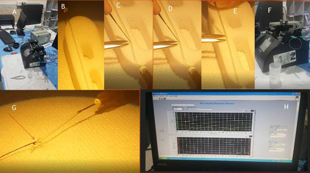
Fig 1: A. Experimental set up with pump system, B. AADI tube, C,D&E. making slit of different
sizes. F. Pump system and tubing, G. Fluid flowing through slit, H. Pressure monitor
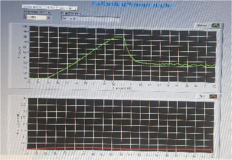
Fig 2: showing pressure recording on the viewing system, arrow represented opening pressure.
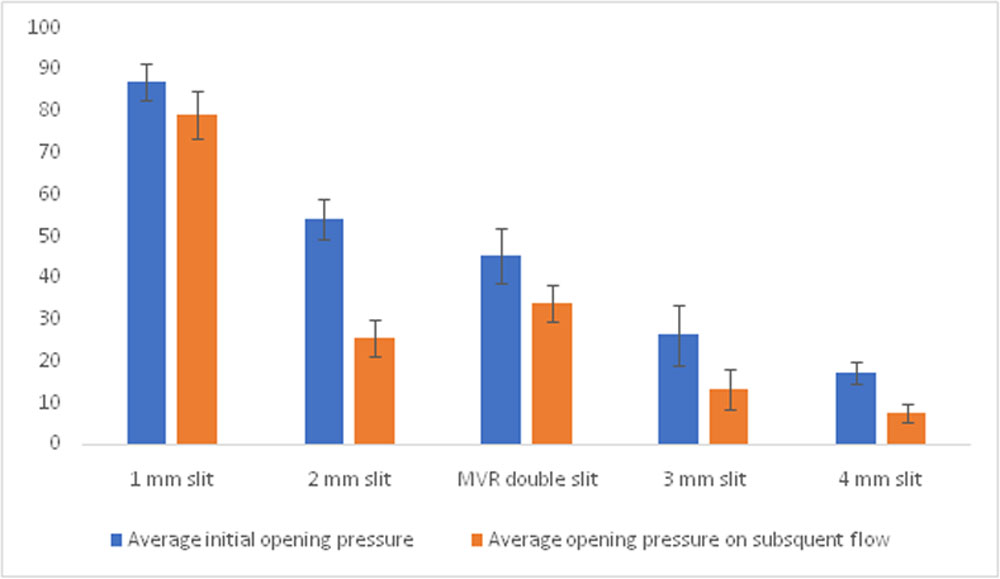
Table 3: Bar diagram showing opening pressure of slits of various size.
| Average initial opening pressure
Mean ± SD in mm Hg |
Opening pressure on subsequent flow Mean ± SD in mm Hg | P value | |
| 5-0 Needle puncture | Not open | Not open | – |
| 1 mm slit | 87.16±4.33 | 79±5.66 | 0.020 |
| MVR double slit | 45.5±6.53 | 34±4.40 | 0.0001 |
| 2mm slit | 54.16±4.91 | 25.66±4.53 | 0.0002 |
| 3mm slit | 26.33±7.13 | 13.33±4.71 | 0.027 |
| 4 mm Hg | 17.26±2.76 | 7.66±2.25 | 0.0006 |
Table 1: showing initial opening pressure and opening pressure on subsequent flow. P<0.05is significant
Leave a Comment