

Dr.Jatin Ashar,
Evaluation of topical human epithelial growth factor for treatment of epithelial defect
Dr. Jatin Ashar
Mumbai Eye Care
What is human epidermal growth factor?
- Small molecular polypeptide existing extensively in human bodies
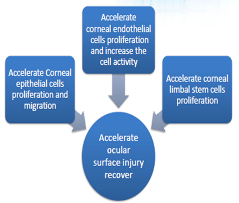
- Promotes migration, proliferation and differentiation of various kinds of epithelial cells, endothelial cells and fibroblasts taking part in the process of wound healing
Highlights of Recombinant Human Epidermal Growth Factor Eye drops
- Promote the regeneration of corneal epithelial cells
- Reduce the healing time of damaged cornea
- Safety is well proven by over 10-year clinical application
- More than 4 million vials are sold in China every year
- Being registered in Turkey
- Complete phase I, II, III clinical trial
Dosage Form:
- Colorless and clear liquid
Specifications:
- 20,000IU(40μg)/2ml/vial, 30,000IU(60μg)/3ml/vial, 40,000(80μg) /4ml/vial
Administration and Dosage:
- For eye dropping, 2-3 drops each time, four times a day.
Storage:
- Stored at 4-25℃.
Packaging:
- Polyethylene plastic bottle, 1vial/box
Shelf Life:
- 24 months
Aim
- To evaluate the efficacy and safety topical preparation of Human epithelial growth factor drops in management of epithelial defect due to various etiologies
Method
Dosing and administration:
- r hEGF eye drops are recommended to be instilled 2-3 drops 4 times a day, during 4 weeks of study period
- Each vial contains r hEGF 40000 IU / 80μg/4ml
Important instruction:
- During the study period of 4 weeks artificial tears are not prescribed unless and until physician feels the need to prescribe the same (to be evaluated at each time point)
Storage of r hEGF eye drops:
- 4-25℃
Method
- 20 patients/ 37 eyes with epithelial defect due to post Photorefractive keratectomy (17), infective keratitis (1), post cornea transplant (2) received human epithelial growth factor drops along with other standard treatment
Primary Outcome Measure:
- Mean Change in size of corneal epithelial defect (in mm) seen under slit lamp bimicroscope using fluorescein stain
- Time point 1: At week 0 (Baseline)
- Time point 2: At week 2 from baseline
- Time point 3: At week 4 from baseline
Secondary Outcome Measure:
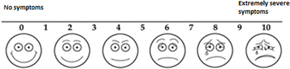
- Ocular discomfort evaluated using Visual Analog scale: Time point 1,2 and 3
- Incidence and nature of adverse events: Time point 2 and 3
- Global evaluation based on clinical examination: Time point 2 and 3
Criteria for evaluation
- Measurement of corneal epithelial defects:
- Corneal epithelial defects are measured with slit lamp biomicroscopic examination under 10 x magnification with fluorescein staining.
Ocular Discomfort score:
- Patient will rate symptoms on a scale of 0 to 10
Global evaluation based on clinical examination
- Rate improvement in patient
- No improvement from baseline
- 10% improvement from baseline
- 25% improvement from baseline
- 75% improvement from baseline
- 90% improvement from baseline
Results
- Mean epithelial defect size after PRK was 8.2mm day 1 & decreased to 1.4 mm day 2 & 0 day 3
- This was faster recover than historical controls
- Pain on day 1 -8,on day 2 -0
- Post transplant the epithelial defect resolved after 8 days
- In infective keratitis it resolved in 15 days
Conclusion
- Human epithelial growth factor drops can help hasten the recovery of corneal epithelial
Leave a Comment