

Prof.Ajay Dudani,Dr.Anjali Israni
Abstract
Leathery brown cataract with its non-complaint nature has always been a major challenge for a phaco surgeon. Plenty of problems like difficult capsulorrhexis, cracking the nucleus and a weak posterior capsule can give a tough time to the surgeon. Even a slight pressure change during cracking or chopping can lead to a nucleus drop. In this surgical technique we are addressing one of the problems of a brown cataract phacoemulsification which is “cracking”. We describe an efficient, repeatable technique of “piece-meal” removal of hard nucleus which gives stable and predictable outcomes even in inexperienced hands.
Introduction
As the lens ages, it increases in weight and thickness. As new layers of cortical fibers are formed concentrically, the lens nucleus undergoes compression and hardening (nuclear sclerosis). Crystallins (lens proteins) are changed by chemical modification and aggregation into high-molecular-weight proteins1.
In the hard and even in the very hard1 nuclei (or the rock-hard lens that is composed of almost all densely compacted, sclerotic endonucleus with very little surrounding epinucleus2) phacoemulsification requires a technique that allows this crystalline to be broken in order to be emulsified. There are several techniques used for this purpose however, high intensity of phaco power and much time is needed to achieve the emulsification of the hard nuclei. Since high phaco power is necessary, this poses risks to the cornea and all anterior chamber tissues causing severe inflammation and loss of endothelial cells. Descmet’s Membrane is the basement membrane of the corneal endothelium, which is essential for the maintenance of corneal transparency. DMD after cataract extraction was first described by Scheie as a vision-threatening complication3. Intraoperative risk factors include the use of blunt instruments, inadvertent insertion of instruments between the corneal stroma and DM, inappropriate incisions, tight main incision, damage of DM during irrigation/aspiration, IOL or phaco probe insertion, and surgeon inexperience.4 The greatest challenge in phacoemulsifying these nuclei is breaking them down because it is difficult to separate the hardened and compressed cortical fiber layers.
The present study is the description of a technique in which the cataract is emulsified without the need of using a great amount of phaco power for an extended amount of time.
Type of study: Surgical technique
Surgical technique
Preoperatively, 1% tropicamide and 10% phenylephrine eye drops are administered every 10 to 20 minutes until a good mydriasis is obtained.
In the cases of very hard cataracts, a peribulbar injection of 2% lidocaine without adrenaline is given with a 25/7-gauge needle. In hard cataracts, topical anesthesia with 0.5% proparacaine is given 4 times at an interval of 10 minutes before beginning of surgery.
Clear cornea temporal incision is performed with a 2.8 mm diamond blade. In most surgeries, 0.1% tripan blue is used to make it easier to visualize the curvilinear capsulorrhexis. The diameter varies from 5 to 5.5 mm. Dispersive viscoelastic is applied in all cases. The hydrodissection is confirmed by rotating the nucleus to certify that it is totally free inside the capsular bag. Phacoemulsification begins with the phaco tip positioned with the bevel upward in the central area of the lens, and a trench is made using 80 % power and 40 mmHg suction on a Venturi machine, so that it penetrates 2/3 inside. Then the nucleus rotated 180degree and a chop is performed getting a good hold on the half sculpted edge with settings 80% power linear and 150 mmHg vaccum.
The two halves are formed which have the posterior lens fibres joined
[ Fig 1]
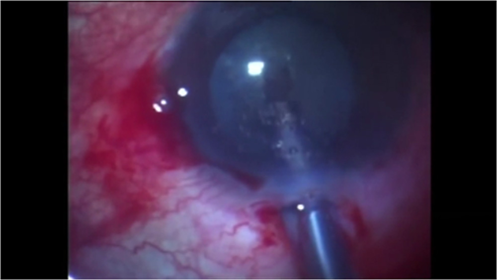
Figure 1: Sculpt having 2/3rd depth
Due to the hardness of the nucleus, in many cases it is not possible to totally break down the nucleus, because the posterior crystalline lens fibers or hard epinucleus are intertwined. Because of that the posterior plate is almost impossible to crack.
So, after the trench is made, in chop mode, we try to break the nucleus into small sectors while leaving the posterior plate intact which in the end resembles a corolla of a flower.
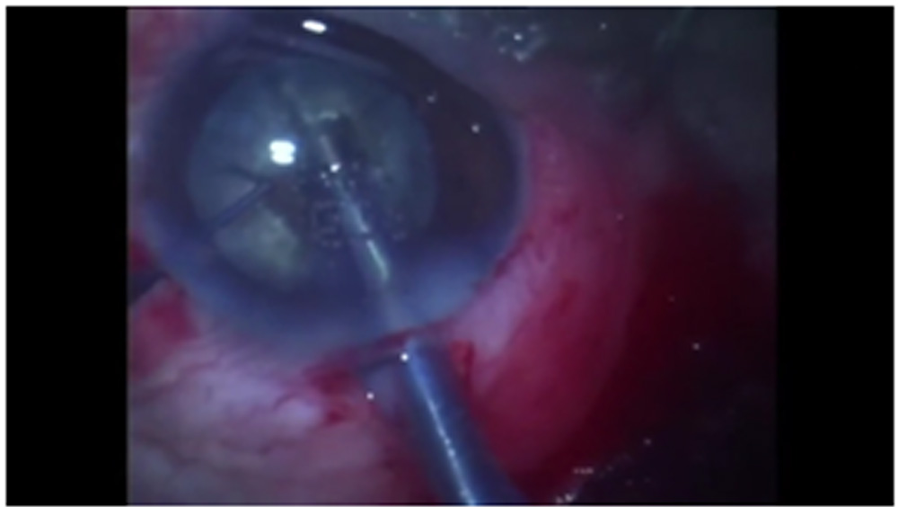
[Fig 2.]
Figure 2: corrolla of flower with stigma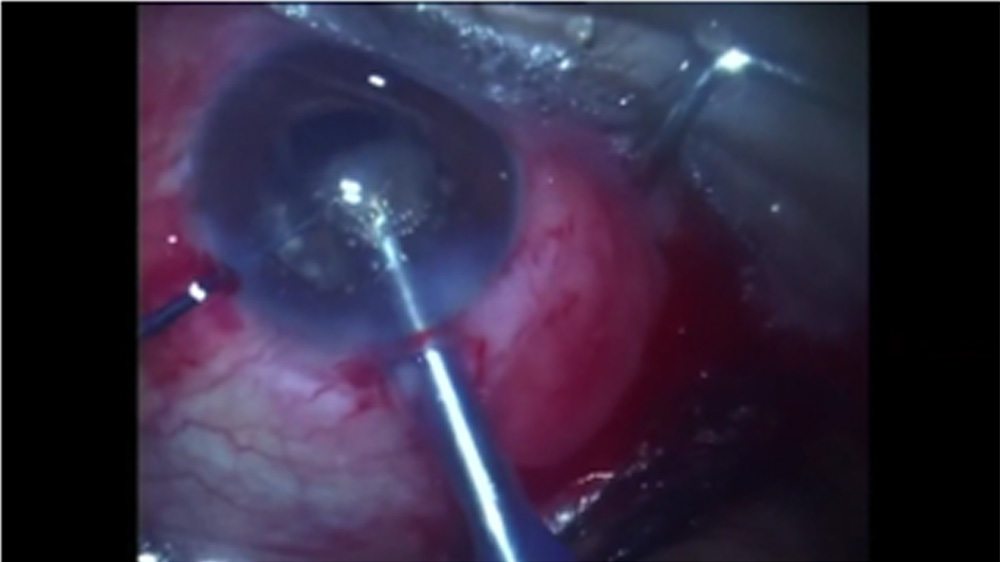
Then, with a vaccum settings at 320/min, each of the “petal” is “plucked” from the posterior nuclear plate, till all the petals are removed, leaving behind a small “stigma” of posterior nuclear plate which can then be easily emulsified. [Fig 3] [Fig 4]
Figure 3: De-flowering of the nucleus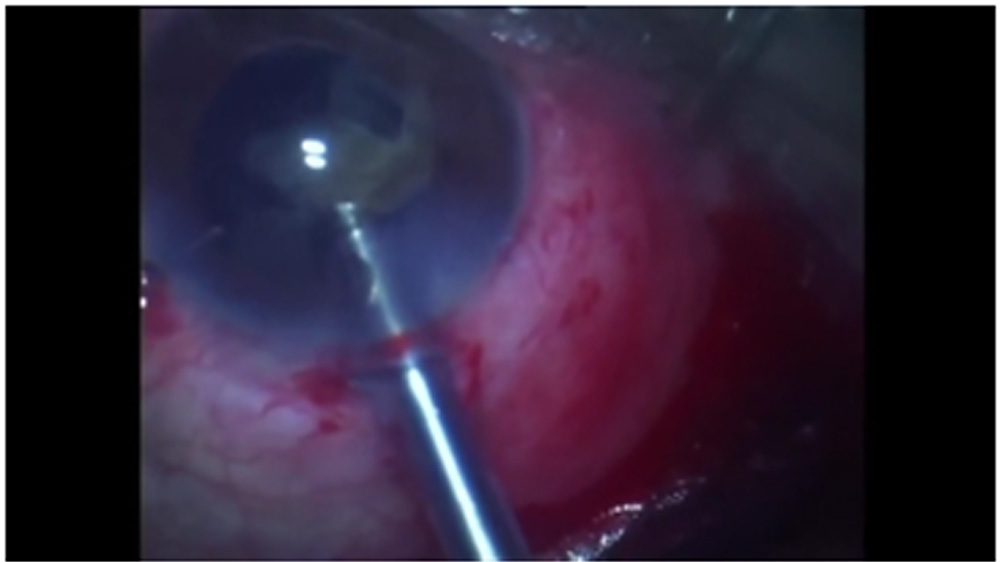
Figure 4: emulsification of the posterior nuclear plate
Discussion
The posterior chamber is now considered the preferred location for hard nucleus phacoemulsification.1 In cases of hard nuclei that are emulsified as a whole in the anterior chamber, in the plane of the iris, or in the posterior chamber, much of ultrasound is necessary and consequently significant endothelial trauma occurs.
Other techniques which are used currently, such as divide and conquer described by Gimbel5, phaco chop made popular by Koch6, and others7, need a longer time of phaco power to sculpt the nuclei and to obtain pieces. In the chop technique, described by Nagahara8 and others7, total breakdown of the nucleus and epinucleus is often not achieved in some hard and very hard cataracts.Prasad et al’s terminal chop technique using a specially developed chopper 9,10 was used to create two halves. However, with absence of epinuclear plate in hard cataracts, it too carries a risk of posterior capsular rupture
Our technique of “piece-meal” removal of hard cataract provides easy removal of all parts of the leathery brown nucleus without putting pressure on zonules and the posterior capsule. This technique is easy to learn, effective and provides safe and repeatable results.
During the development of this technique, we faced few posterior capsular tears due to the pressure we were putting to crack the posterior nuclear plate. Also there were few cases of keratitis which were easily managed with topical medications. However, once this technique was fully developed and understood, we had very less of such complications.
Conclusion
- Phacoemulsification of the very dense cataract presents the surgeon with a series of specific and difficult challenges. Successful management of these cases requires a thoughtful, step-by-step approach that anticipates these challenges and allows the surgeon to surmount them.
References
- American Academy of Ophthalmology. Lens and cataract. San Fransisco, CA, American Academy of Ophthalmology,1998. [ Basic and Clinical Science Course, Section 11]. [ Links]
- Dillman DM. Endolenticular phacoemulsification. Int Ophthalmol Clin. 1994;34(2):91-101.
- Scheie HG. Stripping of Descemet’s membrane in cataract extraction. Arch Ophthalmol. 1965;73:311–4. [PubMed]
- Benatti CA, Tsao JZ, Afshari NA. Descemet membrane detachment during cataract surgery: Etiology and management. Curr Opin Ophthalmol. 2017;28:35–41. [PubMed]
- Gimbel HV. Divide and conquer nucleofractis phacoemulsification: development and variations.. J Cataract Refract Surg. 1991;17(3):281-91.
- Koch PS. Mastering phacoemulsification: A simplified manual of strategies for the spring. crack, and sop and chop technique. Thorofar, NJ: Slack, 1994.
- Arshinoff SA. Phaco slice and separate. J Cataract Refract Surg. 1999;25(4):474-8.
- Nagahara K. Phaco chop [videotape]. Presented at the American Society of Cataract and Refractive Surgery fim festival, Seattle, 9-12 May 1993.
- Chang DF. Ten strategies for the rock-hard nucleus.Eye Net 2005.https://www.aao.org/publications/eyenet/200503/pearls.cfm
- Prasad R, Badhani A,Badhani G. Terminal chop: New technique for full thickness nuclear segmentation in mature hard cataract.Indian J Ophthalmol. 2017 Dec; 65(12): 1415–1418.
Leave a Comment