

Dr.Thirumalesh M B,Dr.Rwituja Thomas,Dr.Sugaranjini G
Introduction
Almost three decades ago, Kelly and Wendel attempted the use of modern vitrectomy techniques, such as internal limiting membrane (ILM) peeling, for the closure of idiopathic macular holes1. In 1997, Goldbaum and Brooks attempted macular hole surgery with silicone oil tamponade to avoid post-operative prone positioning and found that the majority of holes sealed with a single tamponade2. The same year, Tornambe et al investigated whether postoperative facedown positioning is necessary for macular hole closure and found that the success rate after single surgery was 79% without positioning, using C3F8 tamponade3. SD-OCT has now evolved to have a major role in preoperative planning of macular hole surgery and post-operative prognostication. However, obtaining a discernable OCT image of the macula post-operatively, under gas, is a challenge. Since the advent of SS-OCT, image acquisition time has drastically reduced. Our paper assesses the role of SS-OCT in the guiding of post-operative positioning of patients treated for large macular holes using a modified version of the inverse-flap technique.
Methods
Patients and Examination
A retrospective chart review of 10 eyes of 9 patients was conducted at Narayana Nethralaya Eye Hospital, Bangalore, India over a period of 12 months. One of the patients had a traumatic macular hole and all underwent vitrectomy by a single surgeon. Pre- and post-operative (after 12-16 hours of prone positioning) swept source optical coherence tomography (SS-OCT) images were analyzed. All patients had a complete ophthalmic examination before and after surgery and best corrected visual acuity was checked using a Snellen’s acuity chart.
Table 1 – Demographic details and OCT parameters of patients
| No. | Age/Gender | Eye | Pre-op BCVA | Preop. OCT – Hole base diameter | Preop. OCT- Hole height | Macular Hole Index (MHI) | Vitreous substitute | Type of Closure
(Day 1) |
Post-op BCVA |
| 1. | 60/F | LE | 6/36 | 1020 | 419 | 0.41 | SF6 | TYPE 1 | 6/24 |
| 2. | 76/M | RE | 6/36 | 1373 | 429 | 0.31 | SF6 | TYPE 1 | 6/24 |
| 3. | 22/M | RE | 4/60 | 1202 | 306 | 0.25 | Silicone oil | TYPE 1 | 6/7.5 |
| 4. | 75/M | RE | 4/60 | 908 | 329 | 0.36 | SF6 | TYPE 1 | 6/36 |
| 5. | 61/M | RE | 6/24 | 1247 | 398 | 0.32 | SF6 | TYPE 1 | 6/18 |
| 6. | 61/M | LE | 6/36 | 1021 | 420 | 0.41 | SF6 | TYPE 1 | 6/24 |
| 7. | 67/F | LE | 1/60 | 1633 | 431 | 0.26 | SF6 | TYPE 1 | 6/60 |
| 8 | 62/M | RE | 6/18 | 1022 | 543 | 0.53 | SF6 | TYPE 1 | 6/9 |
| 9. | 70/M | RE | 6/36 | 1043 | 382 | 0.37 | SF6 | TYPE 1 | 6/18 |
| 10. | 52/F | LE | 4/60 | 856 | 334 | 0.39 | SF6 | TYPE 1 | 6/36 |
Treatment
All surgeries were performed using a 23-gauge pars plana vitrectomy. Brilliant blue dye was used to stain the internal limiting membrane (ILM). ILM peeling by “pinch and grasp” technique wasdone for all patients, and a modified inverse flap technique was used. A multi-layered flap technique was used to close the hole, with peeling starting a the periphery. Multiple flaps were raised and folded across the hole with the edge of the flap intact. This was done with the knowledge that ILM once peeled, has a tendency to scroll and contracts centripetally once folded, thus shrinking the hole. Also, the peeled layers of ILM tend to act as a barrier for fluid. Fluid-air exchange and exchange of Air to 20% SF6 was performed. Patients were advised prone position post-operatively for 12-16 hours until the first post-operative day follow-up visit. One patient underwent silicone oil injection as the patient was scheduled for air travel back to his state.
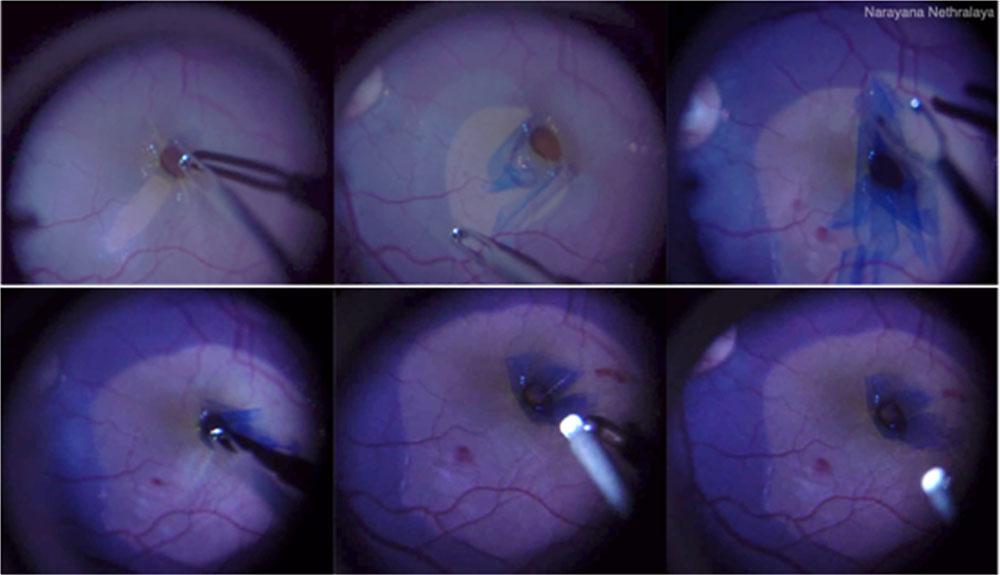
Figure 1 – Intraoperative images of multi-layered inverse flap technique
Swept-Source Optical Coherence Tomography Imaging
OCT imaging was done prior to and after surgery in all operated eyes using SS-OCT (Topcon Deep Range Imaging OCT Triton). This machine utilizes a 1050-nm wavelength light source with a testing rate of 100,000 A-scans per second and an axial resolution of 8 μm. Patients can fixate on the target, thus reducing involuntary eye movements. Also, due to its eye-tracking capabilities and faster scan acquisition time, motion artifacts are minimal5.Post-operative imaging (under gas) was done on Day 1 to confirm closure of the hole. Measurements were done a by single observer (who is a technician). Macular hole index (MHI) was calculated using the ratio the height of the hole(H) by the basal hole diameter (B)6.When complete anatomical closure was obtained it was labeled as Type 1 closure. Imaging was repeated at 6 months to confirm hole closure by imaging under fluid.
Results
In this study, we included 10 eyes of 9 subjects. The mean age was 60.6±15.42 years. 3 were females and 6 were males. The demographic and imaging details are shown in Table 1.The mean basal hole diameter was 1132.5 ±235.33 mm and mean hole height was 399.1±67.96 mm. Average macular hole index (MHI) was found to be0.36 (all <0.5). Mean preoperative baseline BCVA was 0.96± 0.38 (Snellen’s equivalent 20/120) and mean postoperative BCVA was 0.57 ± 0.27. Post-operative visual improvement was significantly (p=0.003) better on the basis of match-pair analysis. There were no cases that had any complications intra- or post-operatively.
All patients achieved Type 1 closure on the first post-operative day as per SS-OCT imaging and positioning was discontinued once the scan was performed. All patients maintained normal foveal contour for a minimum of 6 months post-operatively.
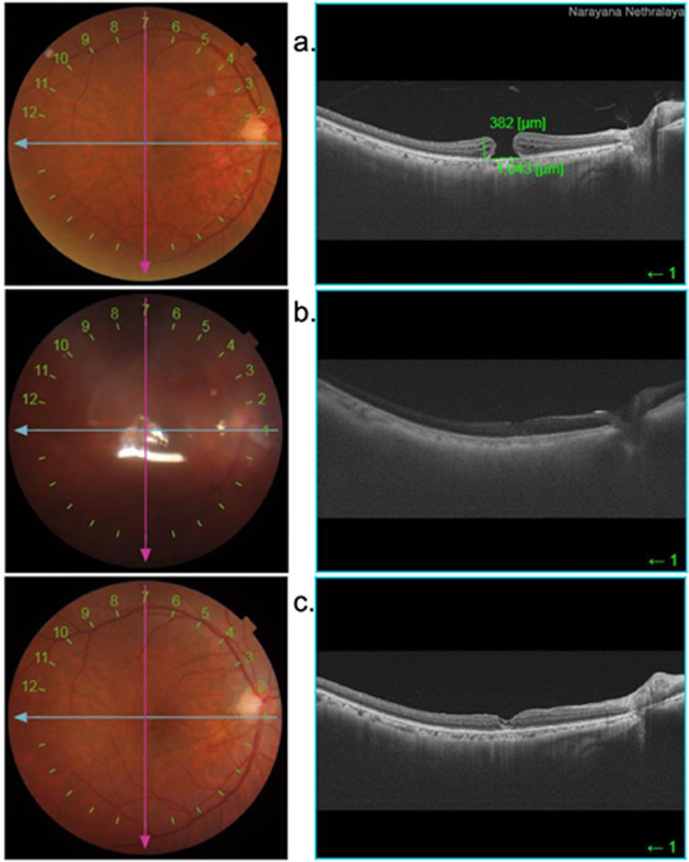
Figure 2 – (a) Preoperative SS-OCT, (b)1st post-operative day, (c) 6 month visit of the same patient
Discussion
The primary objective of this study was to determine the efficacy of the modified inverse flap technique in the closure of large macular holes as well as to determine the duration of postoperative positioning needed for hole closure, with definitive evidence of the same. The average macular hole index of all patients being less than 0.5 entailed labeling of the cases as those with poor prognosis. Imaging of gas-filled eyes has been noted to be inaccurate using SD-OCT. We also noticed however, that patients with a better preoperative visual acuity had a better post-operative visual recovery as well.
In a study by Ahn et al, it was noted that SS-OCT fared better as compared to SD-OCT in gas-filled eyes, for the purpose of visualizing the macula7. We attribute this to the fact that that SS-OCT images 12mmx12mm sections and hence covers the entire macula, using the disc as the reference point in line-scan. Also, as the SS-OCT has a faster speed, fixation can be achieved by telling the patient to look straight ahead or by giving the fixation target to the other eye. Thus it can be concluded that this modality of imaging would lead to superior decision-making on further management during the early post-operative period, specifically regarding that of positioning7.
We used gas tamponadewith SF6 as opposed to the longer acting C3F8. These eyes on follow-ups of 1 month and beyond had a normal foveal contour and remained closed, confirming the findings under gas.
All eyes had anatomical closure by day 1 as confirmed by SS-OCT and hence prone positioning was discontinued. Other studies have shown that closure of large holes under gas can take 1 day to 1 month, following ILM peeling8.
Visual improvement is maximal after over 6 months of hole closure after ILM peeling, as noted by multiple authors9,10,11.
Michalewska et al have also reported that the inverted-flap technique had a better visual prognosis, with 2 lines of improvement and a U-shaped closure11.
On the basis of our data, we can conclude that prone positioning can be discontinued if the hole is found to be closed on day 1 on SS-OCT. Using a larger cohort, including patients with various other risk factors might be of value to assess the full range of benefits and possible pitfalls of single day positioning.
References
- Kelly N, Wendel R. Vitreous Surgery for Idiopathic Macular Holes. Retina. 1991;11(4):447.
- Goldbaum M, McCuen B, Hanneken A, Burgess S, Chen H. Silicone oil tamponade to seal macular holes without position restrictions. Ophthalmology. 1998;105(11):2140-2148.
- Tornambe P, Poliner L, Grote K. Macular hole surgery without face-down positioning. Retina. 1997;17(3):179-185.
- Goldberg R, Waheed N, Duker J. Optical coherence tomography in the preoperative and postoperative management of macular hole and epiretinal membrane. British Journal of Ophthalmology. 2014;98(Suppl 2):ii20-ii23.
- Choma M, Sarunic M, Yang C, Izatt J. Sensitivity advantage of swept source and Fourier domain optical coherence tomography. Optics Express. 2003;11(18):2183.
- Kusuhara S, TeraokaEscaño M, Fujii S, Nakanishi Y, Tamura Y, Nagai A et al. Prediction of postoperative visual outcome based on hole configuration by optical coherence tomography in eyes with idiopathic macular holes. American Journal of Ophthalmology. 2004;138(5):709-716.
- Ahn S, Park S, Lee B. VISUALIZATION OF THE MACULA IN GAS-FILLED EYES. Retina. 2018;38(3):480-489.
- Chow D, Chaudhary K. Optical coherence tomography–based positioning regimen for macular hole surgery. Retina. 2015;35(5):899-907.
- Kase S, Saito W, Mori S, Saito M, Ando R, Dong Z et al. Clinical and histological evaluation of large macular hole surgery using the inverted internal limiting membrane flap technique. Clinical Ophthalmology. 2016;Volume 11:9-14.
- Kuriyama S, Hayashi H, Jingami Y, Kuramoto N, Akita J, Matsumoto M. Efficacy of Inverted Internal Limiting Membrane Flap Technique for the Treatment of Macular Hole in High Myopia. American Journal of Ophthalmology. 2013;156(1):125-131
- Michalewska Z, Michalewski J, Cisiecki S, Adelman R, Nawrocki J. Correlation between foveal structure and visual outcome following macular hole surgery: a spectral optical coherence tomography study. Graefe’s archive for clinical and experimental ophthalmology. 2008;246(6):823–830
Leave a Comment