

Dr. Marian Anne Jacob, P13465, Dr.Mekhla Naik
Introduction
Metastatic orbital lesions have been estimated to accountfor 1–13% of all orbital tumorsreported1-8. Theprevalence of orbital metastasis in cancer patients isestimated to range from 2 to 4.7%2-4.The clinical presentation varies from mild ptosis to severe proptosis depending on the primary tumour site and various other factors. Often the diagnosis is missedin spite of various recent advances in the diagnosis leading to delay in the definitive treatment.
Ophthalmologists have a definite role in the early diagnosis of metastasis providing valuable information that can guide to specific therapy in various cases. In this scenario, we did a study to evaluate the clinical features, management and outcome in a series of patients diagnosed with orbital metastasis.
Materials and Methods:
A retrospectiveinterventional case series of 5 patients who presented to a tertiary center in South India were included. The history,clinical features, imaging findings, histopathology and the treatment outcome were analysed. All metastatic tumourinvolving the orbit were included. Secondary orbital tumour arising from the adnexa and surrounding structures were excluded.
Results:
There were three males (60%)andtwo females(40%). The age group ranged from 43 years to 89 year with a mean of 61 yrs. All tumours were unilateral. Four patients had involvement of right orbit whereas one had left orbit involvement. The duration of symptoms ranged from 2weeks to 24months with a mean of 9.3months .Lid oedema was the presenting symptom in three patients and dropping was present in two cases .Pain was there in 4patients(80%).The most common signs were proptosis(n=3), strabismus(n=1) and visual loss(n=2) and ptosis(n=3).Breast carcinoma (40%),prostatic cancer (20%) ,lymphoma (20%) and unknown (20%)were the primary sites.
| Case | Age
(Yrs) |
Sex | Side | Durn of symptoms
(months) |
Primary site | Duration between primary and mets | Treatment | Follow up |
| 1 | 43 | F | R | 12 | Breast | 21yrs | RT/CT/HT | 3 yrs |
| 2 | 61 | F | R | 8 | Breast | 3yrs | RT/CT/HT | Expired at 8months |
| 3 | 48 | M | L | 0.5 | Lymphoma | 0.5yrs | RT | Expired at 6months |
| 4. | 89 | M | R | 2 | Prostate | 10yrs | RT | 1yr |
| 5. | 64 | M | R | 24 | Unknown | NA | SURGERY/RT | 1yr |
RT -Radiotherapy; CT-Chemotherapy;HT-Hormone Therapy
Computed Tomography scan showed extraconal lesion in 4 cases .One patient showed both intraconal and extraconal involvement.One patient underwent MRI to confirm muscle involvement.Regarding the location of lesion, predominantly superior orbit was involved in 2 patients followed by superomedial(n=1)and medial orbit (n=1)in others.None of the patients showed any bony involvement.
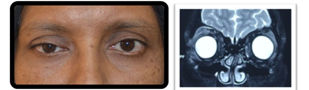
Fig 1A. Patient 1 – a case of Carcinoma breast treated 21yrs back presented with mild ptosis. Fig 1B-MRI showing ill defined mass along supero temporal quadrant(arrow)
The time duration between primary tumour presentation and orbital involvement ranged from 6months to 21 yrs .One patient was treated was metastasis of unknown primary.
All patients had open biopsy to confirm thediagnosis.Once diagnosis is conformed patient was referred to Oncologistand further investigations were done in coordination with Oncologist. Serological studies, molecular biology ,immunohistochemistryincludinghormonereceptors were used in relevant cases.
Treatment was multimodal. 4 patients(80%) underwent external beam radiotherapy(EBRT) as primary treatment. The total dose varied from 20 to 40 Gy delivered in 10-20 sessions over 3-5 weeks. Chemotherapy and hormone therapy were combined for metastatic breast cancer. One patient underwent debulking as primary procedure followed by radiation.(Table-1)
Survival ranged from 3 months to 24 months after the diagnosis .After detecting orbital metastasis three patients were diagnosed with metastasis on other sites on Positron Emission tomography(PET) imaging. Of these three patients, two patientscase no.2 and 3died at 8 and 6 months respectively.Three patients were alive with a mean follow up of 1.6 yrs(1-3yrs)
Discussion
The orbit is an unusual site for metastasis. Several authorshavereported their experience with metastatic orbital tumours1-15. The largest series was reported by Shields et alin which they described 100 patients18. Our case series comprised of 5 patients and this is the first series reported from India .
The clinical presentationof metastatic orbital cancer is well known. In our series the duration of symptoms varied from weeks to years. Inacute presentation it canmimic orbital cellulitis or inflammatory pseudotumour but the clinical history will give the clue towards metastatictumour.When patient presents with long standing ptosis or proptosis or when the tumour is history is not known, the diagnosis can be delayed . Proper history and imaging can help in detecting earlydisease .Shields at al18 reported proptosis and limited ocular motility in majority of cases(57%)wheres in our series proptosis and ptosis (60%)were the most common signs . Pain was the predominating symptom(80%) in our series whereas in other series double vision predominates. The clinical presentation varies with the type of tumour. The metastasisfrom carcinoid mimics orbital cellulitis which was the most commontumour in Shieldsseries .
Orbital imaging gives an an important diagnostic clue towards metastasis. We have done CT scan in all cases and MRI in one case. Some authors prefer magnetic resonance imaging(MRI)superior to CT scan in detecting metastasis.CT scan is valuable in detecting bony lesions which can give a diagnostic clue especially in prostate metastasis where sclerotic lesions present.The pattern of orbital involvement in imaging also helps in detectingthe primary site. The orbitalmetastasis from breast cancer tends to be diffuse andirregular, often growing along the rectus muscles andfascial planes18. In contrast, orbital metastasis from carcinoidtumor, renal cell carcinoma, and melanoma tends tobe more circumscribed, at least in the early stages. In our series supertemporal quadrant involvement in metastatic carcinoma breast mimicked chonic dacryoadenitis leading to delayed diagnosis but on close examination the tumour was infiltratinglevator muscle which was more evident in MRI rather than CT scan .Judicial interpretation and selection of imaging modality is very important in early diagnosis .
The final diagnosis is made by open biopsy and histopathological examination of the tissue.Inour series all tumours were anterior and we could do biopsy by transcutaneous anterior orbitotomy. In one patient we did anterior exenteration as the eye was painful blind eye and the tumour was infiltrating the ocular coats also. If the tumour is wellcircumscribed and amenable to complete removal, it can be done without causing any additional damage to the vitalstructures.In posterior orbital tumours, if the tumour is very deep and in presence of a known primary ,Fine Needle AspirationCytology(FNAC)under ultrasound guidance will help. FNAC is also useful as a minimally invasive procedure in situations where the lesion is easily accessibleand the patient is too frail or unwell for an openbiopsy.Valenzuela et al ,has used fine needle biopsy successfully in 18%of their cases19.
Management of orbital metastasis varies in each case. The main aim is to maximise the quality of life and to restore or stabilise the vision. The quality of life ,effects of treatment and the life expectancy should be considered in choosing the treatment option.The most common modality used is external beam radiotherapy17 . The recommended dose is 20 to 40 Gy delivered in multiple fractions over a period of 2-3 weeks .It is beneficial in reducing the proptosis ,exposure keratopathy and restoring vision in somecases. Side effects have to be kept in mind and usage of corneal shield is alsorecommended .Chemotherapy19-20 is useful in chemosensitive tumours like metatasis from small cell carcinoma lung and neuroblastoma.Hormonetherapy useful in carcinoma breast and prostate .Radical surgery will not offer any advantage in terms of long term survival . It is useful in reducing the pain, diplopiaandproptosis .In our series 4 patients underwent radiotherapy as first line treatment and one patient underwent radical surgery followed by irradiation.
Generally, orbital metastasisis associated with poor prognosis in terms of survival. Most reported series are small and only limited date regarding the same in the literature16. In shields series metastatic carcinoid has the longest survival followed by carcinoma breast. In our series those patients with isolated metastasis had a better survival than the patients with multiple metastasis However the series is small to reach a definite conclusion.
Conclusion
Orbital metastasis is mostly a disease of adulthood and usually unilateral.The diagnosis is based on clinical features,imaging and histopathological examination. Management involves radiation, chemotherapy and hormone therapy based on the primary tumour Radical surgery has only limited role .Eventhough local tumour control is good, the overall survival is limited .Early diagnosis and treatment is crucial in detecting orbital metastasis to achieve life salvage .
References
1.Albert DM, Rubenstein RA, Scheie HG. Tumor metastasis to the eye : incidence in 213 adult patients with generalized malignancy.Am J Ophthalmol1967;63:727–32.
2. Bloch RS, Gartner S. The incidence of ocular metastatic carcinoma. Arch Ophthalmol1971;85:673–5.
3. Hutchinson DS, Smith TR. Ocular and orbital metastatic carcinoma. Ann Ophthalmol1979;11:869–73.
4. Bullock JD, Yanes B. Metastatic tumors of the orbit. Ann Ophthalmol1980;12:1392–4.
5. Castro PA, Albert DM, Wang WJ, et al. Tumors metastatic to the eye and adnexa. Int Ophthalmol Clin 1982;22:189–223.
6. Freedman MI, Folk JC. Metastatic tumors to the eye and orbit: patient survival and clinical characteristics. Ophthalmol1987;105:1215–9.
7. Shields CL, Shields JA, Peggs M. Tumors metastatic to the orbit. OphthalPlast and Reconstr Surg 1988;4:73–80.
8. Goldberg RA, Rootman J, Cline RA. Tumors metastatic to the orbit: a changing picture. SurvOphthalmol1990;35:1–24.
9. Tijl J, Koorneef L, Eijpe A, et al. Metastatic tumors to the orbit: management and prognosis. Graefes Arch Clin Exp Ophthalmol1992;230:527–30.
10. Shields CL, Shields JA. Metastatic tumors to the orbit Int Ophthalmol Clin 1993;33:189–202.
11. Gunalp I, Gunduz K. Metastatic orbital tumors. Jpn J Ophthalmol1995;39:65–70.
12. Char DH, Miller T, Kroll S. Orbital metastasis: diagnosis and course. Br J Ophthalmol1997;81:386–90. Perri P, Brienza D, Mazzeo V, et al. Orbital metastases: echographic findings our experience. Ophthalmologica1998;212(suppl 1):82–4.
13.Ahmad SM, Esmaeli B. Metastatic tumors of the orbit and ocular adnexa. CurrOpinOphthalmol2007;18(5):405–413.
14.Font RL, Ferry AP. Carcinoma metastatic to the eye and orbit III. A clinicopathologic study of 28 cases metastatic to the orbit. Cancer 1976;38(3):1326–1335.
15.Forrest AW. Intraorbital tumors. Arch Ophthal1949;41(2):198–232.
16.Freedman MI, Folk JC. Metastatic tumors to the eye and orbit. Patient survival and clinical characteristics. Arch Ophthalmol 1987;105(9):1215–1219.
17.Glassburn JR, Klionsky M, Brady LW. Radiation therapy for metastatic disease involving the orbit. Am J Clin Oncol 1984;7(2):145–148.
18.Shields JA, Shields CL, Brotman HK, Carvalho C, Perez N, Eagle RC, Jr. Cancer metastatic to the orbit: the 2000 RobertM.Curts Lecture. OphthalPlastReconstr Surg 2001;17(5):346–354
19.Valenzuela AA, Archibald CW, Fleming B,et al. Orbital metastasis: clinical features, management and outcome.Orbit. 2009;28(2-3):153-9.
20.Holland D, Maune S, Kovács G, Behrendt S. Metastatic tumors of the orbit: a retrospective study.Orbit. 2003 Mar;22(1):15-24
Leave a Comment