

Dr.Divya Patel,Dr.Tanuja Kate,Dr.Urvija Choudhary,Dr.H.K.Karmalkar
Introduction :
Ocular blood flow may be affected in some ocular and systemic diseases.Glaucoma is a chronic progressive optic neuropathy characterized by retinal ganglion cell death and associated visual field loss.1It is usually associated with raised intraocular pressure(IOP),but IOP alone is not sufficient to damage optic nerve.Other mechanism like vascular factor is also implicated in causation of glaucomatous optic nerve damage2.The vascular theory describing the mechanism of glaucoma considers the optic neuropathy as a consequence of insufficient blood perfusion due to either increased intraocular pressure or other risk factors which may lead to a reduction in the ocular blood flow 3.Inability of the perfusion system to adapt to tissue blood flow requirements or changes in perfusion pressure may lead to chronically low or unstable ocular perfusion 4,
which in turn may cause ischemia, oxidative stress or both, possibly leading to glaucomatous damage to the optic nerve head. It seems that vascular factors are important in the development and progression of Primary open angle glaucoma (POAG).Systemic hypertension is one of the systemic entity which causes blood vessels sclerosis and narrowing and hence can affect blood flow to optic disc.There are various techniques available for ocular blood flow assessment out of which color Doppler imaging (CDI) has particular advantages in that it is noninvasive, is not affected by poor ocular media, requires no contrast or radiation, and has been used in ophthalmology for 20 years.5,6CDI uses the principle of Doppler shift in frequency in hertz to measure blood flow velocity in cm/sec.The usual parameters studied in CDI are Peak systolic velocity(PSV),End diastolic velocity(EDV) and Resistivity index(RI).
A higher value of RI represents greater peripheral vascular resistance, which generally implies deleterious significance.The purpose of this study was to evaluate the effect of optic nerve head blood circulation using CDI in POAG and systemic hypertension.
Material and Methods–
This was a prospective study which included16 subjects divided in two groups equally.Group 1 included POAG without systemic hypertension and Group 2 included systemic hypertension without POAG.The study was carried out at Rajas eye and Retina research centre,Indore(M.P.) from December 2017 to may 2018.The radiologist who performed CDI was blinded regarding the diagnosis.Patients with evidence of ocular inflammation, previous intraocular surgery ,any other types of glaucoma and other systemic diseases were excluded.
Complete ocular examination was performed.The diagnosis of POAG was based on the presence of glaucomatous optic nerve head damage on slit lamp (90 D lens), characteristic glaucomatous visual field defect (humphry automated perimetry), raised IOP >21 mmHg (GoldmannApplanation Tonometer), open and normal anterior chamber angles on gonioscopy (Goldmann 3-mirror lens).Systemic hypertension was confirmed by physician reference and opinion.
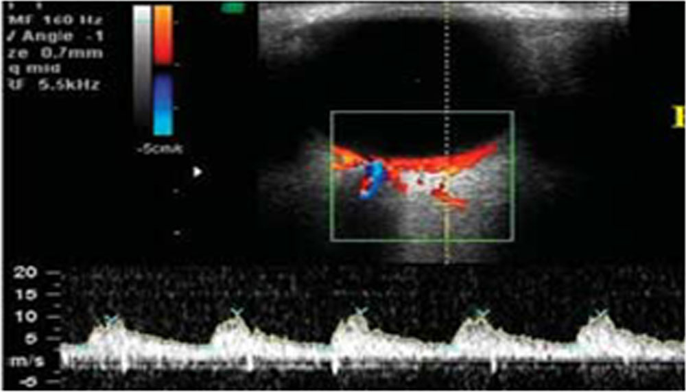
CDI was performed with a probe of 5MHz. The parameters measured in each eye were Peak systolic Velocity (V max), End Diastolic Velocity (V min) and Resistivity index (RI) of Ophthalmic artery(OA)[Fig-1] ,Central retinal artery(CRA )[Fig-2]and Short posterior ciliary artery(SPCA)[fig-3].RI was calculated using the formula RI= V max–V min/V max.The following figures are showing normal specific graph for different arteries.
Fig 1:Ophthalmic artery
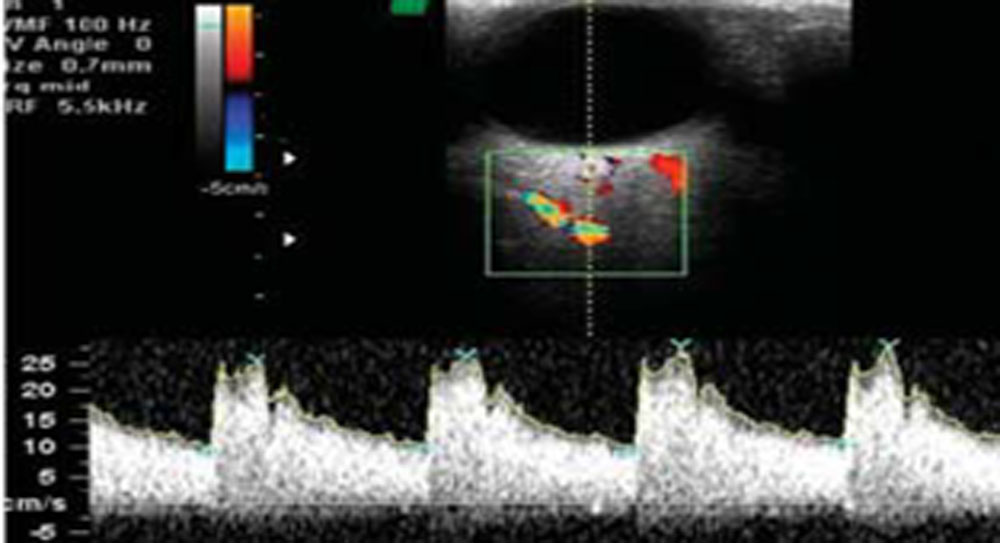
Fig.2:Central retinal artery
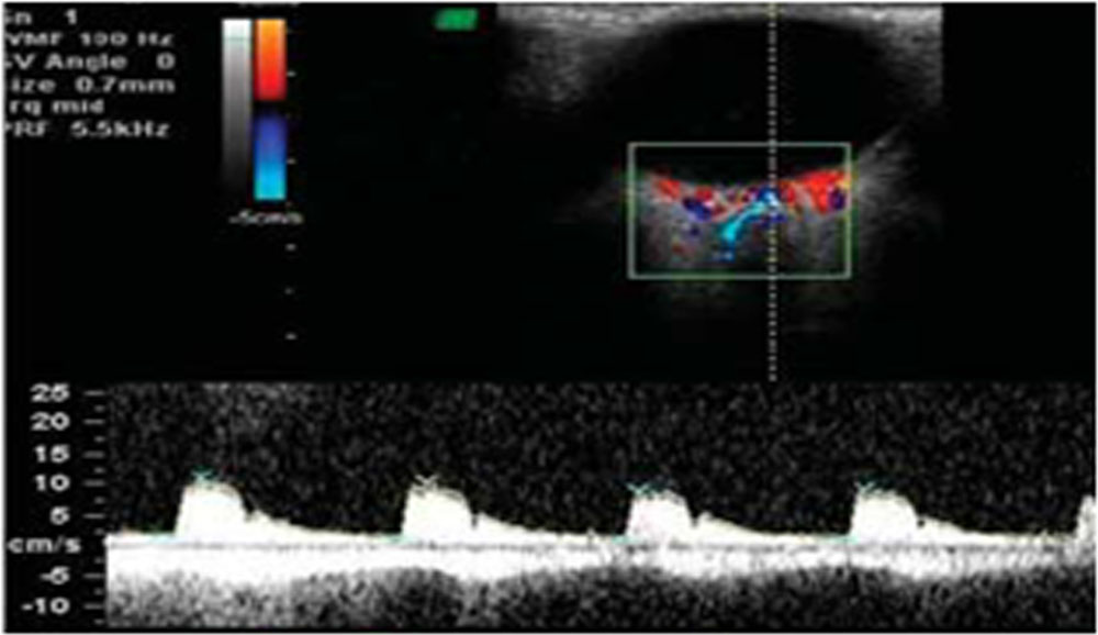
Fig.3:Short posterior ciliary artery
Results-The study sample consisted of 16 subjects,8 in each group.There were total 13 males and 3 females. The mean age of group 1 was 65year(Range 54-70years) and group 2 was 59.75 year(Range 55-70years).
Table 1:CDI parameters in OA(Mean value and Standard deviation)
| PSV | EDV | RI | |
| GROUP 1 | 23.50(SD-11.98) | 5.41(SD-2.30) | 0.76(SD-0.06) |
| GROUP 2 | 32.26(SD-7.97) | 7.27(SD-1.91) | 0.75(SD-0.06) |
| PVALUE | 0.02 | 0.16 | 0.87 |
Table 1 is showing that PSV was significantly decreased in POAG group as compared systemic hypertension group.Also,EDV was decreased and RI increased in POAG group compared with systemic hypertension group, but it was not statistically significant.
Table 2: CDI parameters in CRA( Mean value and Standard deviation)
| PSV | EDV | RI | |
| GROUP 1 | 8.60(SD-3.35) | 2.48(SD-0.04) | 0.70(SD-0.07) |
| GROUP 2 | 12.20(SD-4.16) | 3.59(SD-1.0) | 0.68(SD-0.07) |
| P VALUE | 0.05 | 0.04 | 0.51 |
Table 2 is showing that PSV and EDV were significantly decreased in POAG group compared with Systemic hypertension group.RI was increased in POAG group compared with systemic hypertension group but this difference was not statistically significant.
Table. 3: CDI parameters in SPCA(Mean value)
| PSV | EDV | RI | |
| GROUP 1 | 8.61(SD-2.58) | 2.33(SD-0.68) | 0.72(SD-0.12) |
| GROUP 2 | 11.55(SD-3.19) | 3.95(SD-1.75) | 0.65(SD-0.09) |
| P VALUE | 0.08 | 0.03 | 0.44 |
Table 3 is showing EDV was significantly decreased in group1(POAG) compared with group 2(Systemic hypertension).PSV was decreased and RI increased in group 1(POAG) compared with group2 (systemic hypertension) but this difference was not statistically significant.
Statistical analysis: Comparison of two groups was done by mann-whitney nonparametric test and significant P values (0.05) in OA -PSV,CRA-PSV, CRA-EDV and SPCA-EDV were found.
Discussion:POAG has long been recognized as the major cause for legal blindness worldwide.7Vascular etiology in glaucoma inspite of normal IOP has been implicated for glaucomatous damage.The OA,CRA and SPCA were important vessels providing evidence of the influence of vascular factor in pathogenesis of glaucomatous optic neuropathy.Systemic hypertension causing vascular sclerosis results in narrowing and altered blood flow,which in turn can affect the optic nerve head blood circulation.The volume of blood flow can not be measured by Doppler but velocity of blood flow and resistivity index are good enough indicators for perfusion of Optic nerve head.
Significantly slower mean blood flow velocities (e.g., PSV and EDV) were recorded in the OA and CRA of POAG patients in ourstudy.This finding was similar to a previous report by Rojanapongpun et al., who reported slower mean PSV and EDV of the OA in comparison with that in the control group in the same Asian population.8Mokbel et al.,9 studied ocular color Doppler imaging of the CRA in Egyptian patients with POAG and found slower EDV in the POAG group as compared with the control group subjects in the same population.
Numerous studies have reported reduced PSV and EDV and increased RI in the OA, CRA and SPCA in eyes of POAG 10,11.Although Between the ocular perfusion pressure and open angle glaucoma, it was proven that there is a certain association, due to the fact that intraocular pressure is part of ocular perfusion pressure12. We had conducted this study with the hypothesis that atherosclerosis in systemic hypertension could cause narrowing of vessels and decreased blood flow to optic nerve head,which could be a risk factor for POAG.But we could not find any significant association between systemic hypertension and altered blood flow by color dopper in our study.We could not find any studies regarding effect of systemic hypertension on ocular blood flow.Our study supported the vascular theory for POAG but we did not find any effect of systemic hypertension on optic nerve blood flow.
Conclusion:Although the sample size in the present study was very small,but what we could find was that colour Doppler Imagingshowed significant optic nerve head perfusion abnormality in patients with POAGwhich strongly suggests vascular theory for POAG but systemic hypertension cohort did not show any effect on ocular blood flow.
No financial or competing interests
References-
1.Moore D, Harris A, Wudunn D, Kheradiya N, Siesky B. Dysfunctional regulation of ocular blood flow: A risk factor for glaucoma? ClinOphthalmol 2008;2:849-61
2.Yanagi M, Kawasaki R, Wang JJ, Wong TY, Crowston J, Kiuchi Y. Vascular risk factors in glaucoma: a review. Clin. Experiment ophthalmol. 2011;39:252–8. [PubMed]
3.Flammer J, Orgul S, Costa VP, Orzalesi N, Krieglstein GK, Serra LM, et al. The impact of ocular blood flow in glaucoma.ProgRetin Eye Res. 2002;21(4):359-93
4.Leske MC, Heijl A, Hyman L, Bengtsson B, Dong L, et al. (2007) Predictors of long-term progression in the early manifest glaucoma trial. Ophthalmology 114: 1965–1972.
5.Lieb WE, Cohen SM, Merton DA, Shields JA, Mitchell DG, et al. (1991) Color Doppler imaging of the eye and orbit. Technique and normal vascular anatomy. Arch Ophthalmol 109: 527–531.
6.Kaiser HJ, Schotzau A, Flammer J (1996) Blood-flow velocities in the extraocular vessels in normal volunteers. Am J Ophthalmol 122: 364–370.
7.Quigley HA, Broman AT (2006) The number of people with glaucoma worldwide in 2010 and 2020. Br J Ophthalmol 90: 262–267.
8.Rojanapongpun P, Drance SM, Morrison BJ. Ophthalmic artery flow velocity in glaucomatous and normal subjects. Br J Ophthalmol. 1993;77:25–9
9.Mokbel TH, Shahin MM, El-Said EM, Abd EL-Ghaffar WM. Potential Diagnostic value of fluorescein angiography and color dopplerimaginh in primary open angle glaucoma. Eur J Ophthalmol. 2009;19:957–62.
10.Garhofer G, Fuchsjager-Mayrl G, Vass C, Pemp B, Hommer A, et al. (2010) Retrobulbar blood flow velocities in open angle glaucoma and their association with mean arterial blood pressure. Invest Ophthalmol Vis Sci 51: 6652–6657.
11.Cellini M, Possati GL, Caramazza N, Caramazza R (1996) Colour Doppler analysis of the choroidal circulation in chronic open-angle glaucoma. Ophthalmologica 210: 200–202.
12.Wishal D. Ramdas, Roger C.W.Wolfs, Albert Hofman, Paulus T.V.M. de Jong, Johannes R. Vingerling, Nomdo M. Jansonius. Ocular Perfusion Pressure and the Incidence of Glaucoma: Real Effect or Artifact?: The Rotterdam Study. Invest Ophthalmol Vis Sci. 2011;52:6875–6881. [PubMed]
Leave a Comment