

Dr.Komal Khetwani, K18458, Dr.Prasanna Aradhye, Dr.Rushikesh Naigaonkar, Dr.Abhishek Desai
Abstract
PURPOSE:
To assess the effectiveness of a combined procedure (pars plana vitrectomy with temporary keratoprosthesis, vitreoretinal surgery, and penetrating keratoplasty) in patients with endophthalmitis and to determine which factors influence corneal transplant success rates.
METHODS:
Records for 54 severely involved eyes of 54 patients who underwent penetrating keratoplasty in combination with vitreoretinal surgery between 2010 to 2017, with a follow-up of at least 6 months, were evaluated retrospectively. Analysis was focused on ocular history, functional and anatomic anterior and posterior segment outcome, and complications.
RESULTS:
Males were 42.6% and females were 57.4%.right eye was involved in majority of cases 64.8% and left eye 35%.the major causative factor was post penetrating trauma 21 eyes (38.8%).post operative 17 eyes(31.4%).corneal ulcer leading to endophthalmitis 13 eyes(24%) and endogenous endophthalmitis 3 eyes (5.5%).BCVA was improved in 24 eyes(44.4%).,remained stable in 14 eyes(25.9%),worsened in16 eyes(29.6%). Reasons of poor visual outcome were graft failure in 9 eyes(16%),phthisis in 7 eyes(13%),secondary glaucoma in 19 eyes(35%) manageable with antiglaucoma medications and surgical intervention.
CONCLUSION:
Combined procedure is often complicated by ciliary body malfunction and secondary transplant failure. Although the functional outcome of a combined procedure is limited by primary and secondary tissue destruction, preserving ambulatory vision is possible and thus improves the quality of life, at least in patients with single remaining eyes.
Keywords: Vitreoretinal surgery,temporary keratoprosthesis,penetrating keratoplasty,endophthalmitis.
INTRODUCTION
Endophthalmitis is one of the most feared complications in modern ophthalmic surgery and is discussed on all surgical consent forms for intraocular surgery.The combination of vitreoretinal surgery with penetrating keratoplasty(PKP) is a useful method for treating patients with vitreoretinal disorders complicated by corneal opacification.Endophthalmitis ,especially after trauma, is one of the biggest challenges for ophthalmologic surgeons,opacification and oedema of cornea,inflammatory material in the anterior chamber,nontransparent vitreous and fragile,necrotic retina makes the surgery extremely difficult..
Studies evaluating combined penetrating keratoplasty(PKP) and pars plana vitrectomy(PPV) for infectious etiologies are limited.Majority of these eyes need evisceration/ enucleation for eliminating the infection and many of them as a primary treatment.1-4 Management with combined surgery is helpful in such eyes associated with corneal opacity/infection, which causes insufficient visualization of the fundus. Previous studies have reported favourable outcomes of combined PKP and PPV using a temporary keratoprosthesis5-10But most of these studies have included non infectious causes of combined anterior and posterior segment diseases. Dae Sung Lee et al in their retrospective case series of 11 eyes reported the outcomes of patients who underwent combined PKP and PPV. Six of their 11 patients had infectious endophthalmitis with either microbial keratitis or corneal edema obscuring posterior segment visualization. All 6 eyes had poor final visual outcome with only one eye maintaining clear graft and one eye progressing to phthisis bulbi, but in 4 of the 6 cases the globe could be salvaged.7 In combined surgery.long -term follow up of is important because these eyes often sustain severe damages and complications can arise at a later date.
In this study we describe how effective is the combined procedure penetrating keratoplasty with vitrectomy using temporary keratoprosthesis focussing only on endophthalmitis cases of different causes or mechanisms and analyze their anatomic and functional outcomes. This device provided excellent visualisation of posterior and peripheral intraocular structures and maintained a closed system during surgery.
SUBJECTS AND METHODS
We reviewed the medical records of 54 eyes of 54 patients who had undergone combined PKP and vitrectomy using temporary keratoprosthesis in our hospital between 2010-2017.This study was approved byInstitutional review board,followed the declaration of Helsinki.The patients with follow up of atleast 6 months were included.Inclusion criteria were patients with age more than 18 years with diagnosed endophthalmitis, with poor corneal clarity so as to impede intraoperative posterior segment visualization.Diagnosis of endophthalmitis was clinical, supplemented with ultrasound B-scan findings confirming vitreous exudates. Patients with age less than or equal to 18 years and a follow up of less than 6 months were excluded from the study. Patients with no perception of light underwent primary evisceration and were also excluded.The following data were collected:demographic data,including age and sex,which of the two eyes were involved,preoperative ocular characteristics(corneal and retinal),inciting factor,preoperative and postoperative BCVA ,VA was divided into 3 categories depending on whether acuity improved,remained stable,or worsened,lens status,status of vitreous and retina(based on bscan ultrasound),surgical method,Intraoperative details, lens or IOL extraction, intravitreal antibiotic injection, silicone oil injection or any other additional procedure were noted. The microbiological profile of vitreous sample and corneal button were documented. Postoperative complications, such as rejection ,infection,glaucoma,phthisis and the resolution or the recurrence of posterior segment infection were noted.Successful surgical outcome was defined as maintenance of clear graft,anatomic attachment of retina,and controlled IOP5.Ambulatory vision was defined as 20/200-20/800 or counting finger.Poor visual outcome defined as postoperative visual acuity less than ambulatory vision(hand motions or less).
Anatomic and functional outcomes were defined prior to review. Since all patients presented with beyond Snellen acuities [i.e., count fingers (CF)/ Log MAR 1.7-2.0, hand movements (HM)/ Log MAR 2.1-2.4, perception of light (PL) or no perception of light (NPL)], visual results were obtained from the difference in VA pre-treatment and post-treatment and categorised into three groups: (1) improved, (2) no change, and (3) deteriorated. An improvement was defined as either a gain of one measured step or more (e.g., from HM to CF, or CF to 20/100). A deterioration was defined as loss of one measured step or more (e.g., from CF to PL). Anatomic success was defined as elimination of infection with restoration of tectonic integrity of the globe. Retinal anatomic success was defined as resolution of vitreous exudates with attached retina. Corneal success was defined on the basis of a clear and compact graft, and any instances of graft rejection or failure were noted.Graft failure was defined as the occurrence of epithelial or stromal edema and irreversible loss of corneal opacity under slit lamp examination..graft rejection was defined as the sudden loss of the corneal clarity,with the presence of a rejection line or keratic precipitates on the graft.
Anatomic failure was defined when the eye progressed to phthisis bulbi or needed evisceration for uncontrolled infection.endophthalmitis cases in which intervention was done within 24 hrs of presentation are termed as early intervention and after 24 hrs are late intervention.
Eyes with combined ocular hypotonia and corneal edema defined eyes at risk for phthisis bulbi.Analysis was focused on functional and anatomic anterior and posterior segment outcome, and complications.
SURGICAL PROCEDURE AND POSTOPERATIVE CARE.
Written informed consent was taken. After adequate anaesthesia was obtained with a Van Lint facial block and a peribulbar injection, a lid speculum was inserted. A 23- gauze infusion port was inserted through the pars plana. Two more sclerotomies were made at the pars plana and temporarily closed with plugs. The affected portion of the cornea was excised with a corneal trephine of diameter 7.25 or 8.25mm depending on the size of the pathology. A Eckardttemporary keratoprosthesis(TKP), was sutured onto the recipient cornea with six interrupted 7-0 vicryl sutures.Standard three-port pars plana vitrectomy was then performed. Cataract was removed by pars plana lensectomy or extracapsular cataract extraction was done before applying TKP; intraocular lens was removed if it was infected or was decentred and distorted the view of the posterior segment. Depending on the retinal pathology, vitreoretinal surgery was performed. Than a TKP was later removed and replaced with donor corneal button with diameter 0.5mm larger than the host trephination. The donor cornea was sutured onto the recipient cornea with 16-24 interrupted 10-0 nylon sutures. During PKP, intraocular pressurewas adjusted by regulating the flow through the infusion port. All corneal suture knots were buried. Intraocular pressure was slowly brought to normal. Intraocular antibiotics were injected based on the preliminary microbiology report (Gram’s and Potassium hydroxide staining of intraoperative vitreous tap). Intravitreal vancomycin 1mg/0.1ml and ceftazidime 2.25mg/0.1ml were injected if staining demonstrated bacterial organism or no organism. Intravitreal injection of voriconazole 100micrograms/0.1ml was given if staining showed fungal organism. Silicon oil was injected at this stage if indicated. After closure of the sclerotomy sites moxifloxacin was injected subconjunctivally. Vitreous aspirate and corneal button were plated on blood agar, chocolate and Sabouraud dextrose agar for bacterial and fungal cultures.
Postoperatively, patients were prescribed oral antibiotics (ofloxacin, 400 mg twice daily), topical antibiotics ( moxifloxacin 0.5% ,cefazolin sodium 5% and tobramycin sulfate 1.3%, 2 hourly), and topical steroids (prednisolone acetate 1%, 4 hourly) in cases of bacterial endophthalmitis. Topical natamycin (5%, 1 hourly) and oral ketoconazole (200 mg, 12 hourly) were given for fungal keratitis (topical steroids were withheld in fungal keratitis). Antibiotics were changed based on culture-sensitivity results. Patients were followed up regularly on case to case basis till the infection resolved, then monthly for 3 months, 2 monthly for 1 year. Any additional procedure or intravitreal injection needed to control the infection was documented. In case of presumed or confirmed bacterial endophthalmitis, intravitreal injections of vancomycin 1mg/0.1ml and ceftazidime 2.25mg/0.1mlwere injected and were repeated every 48 hours till a clinical resolution of the infection was noted. In case of a presumed or confirmed fungal endophthalmitis, intravitreal injection of voriconazole 100micrograms/0.1ml was given with repeat injections being given depending onclinical resolution.
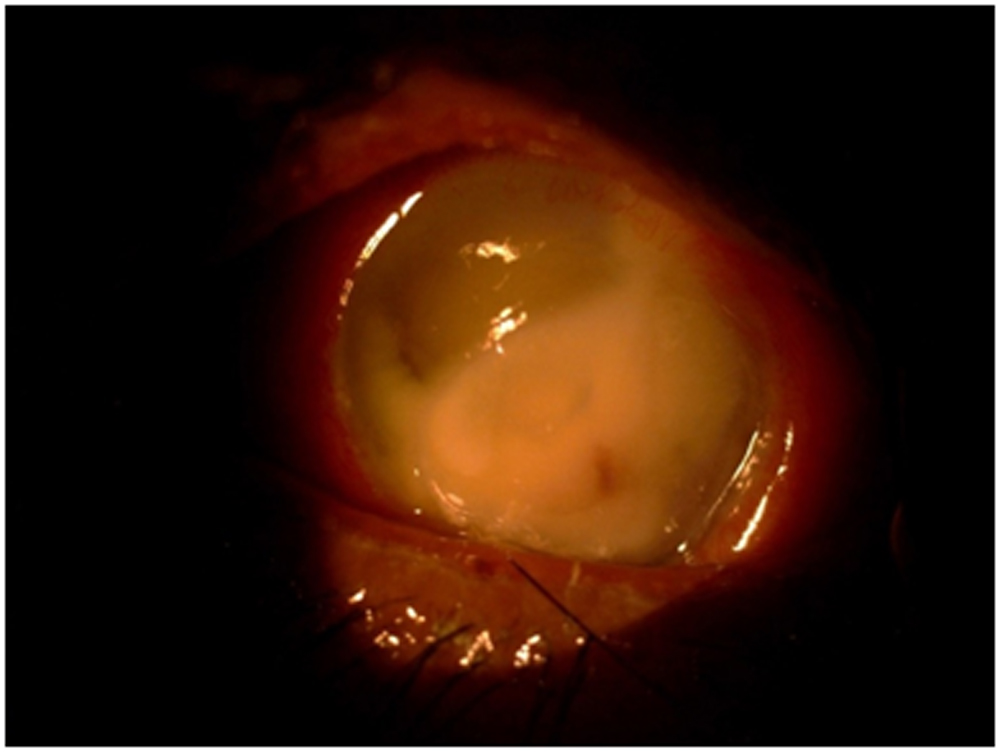
Figure 1(case example,preoperative)-A 54 year old female with aspergillus corneal infection in her right eye for 20 days.
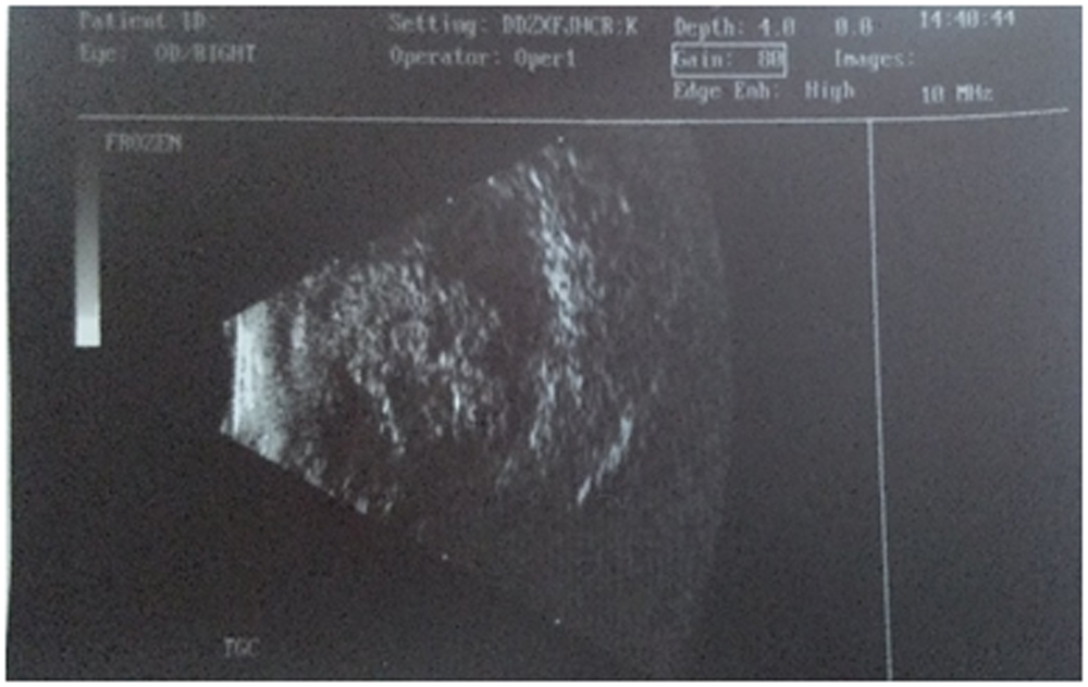
Figure 2-USG Bscan of same patient shows dense vitreous exudates.
STATISTICAL ANALYSIS
SPSS software version 20.0 was used for statistical analysis. For description, quantitative variables have been expressed as mean ± SD, and qualitative variables have been expressed as a percentage. The chi square tests were performed for categorical variables. All the tests were 2-tailed, and P<0.05 was considered significant.
RESULTS
CombinedPKP with vitrectomy for infectious endophthalmitis was performed in 54 eyes of 54 patientsduring the defined study period.
Table 1: Baseline characteristics of the study population.
| Variables | Values |
| Age,mean(range) | 50+/-13.14(23-78)years |
| Male/female | 23(42.6%) / 31(57.4%) |
| Right eye/left eye | 35 (64.8%) / 19 (35%) |
| Follow up period | 6 months |
| Vision at presentation | No of eyes(%) |
| PL | 30 (55.55%) |
| HM (Log MAR 2.4) | 16 (29.6%) |
| CF (Log MAR 2.0) | 5 (9.25%) |
| 20/650 | 3 (5.55%) |
| Inciting event | No of eyes(%) |
| Trauma | 21 (38.8%) |
| Postoperative | 17 (31.4%) |
| Postcataract surgery | 11 (64.7%) |
| Post trabeculectomy (bleb associated) | 4 (23.5%) |
| Penetrating keratoplasty | 1 (5.88%) |
| Vitreo-retinal surgery | 1(5.88%) |
| Corneal ulcer leading to endoph | 13(24%) |
| Endogenous endophthalmitis | 3 (5.55%) |
Abbreviations: counting fingers (CF).hand movements(HM),perception of light(PL)
The most common inciting agent for endophthalmitis was post penetrating trauma(38.8%). All 21eyes of trauma patients had the history of primary repair of corneal and scleral lacerations.the median time between the trauma and surgery using TKP was 6 days(range,1-50 days).All eyes had corneal abscess or corneal infiltration precluding adequate visualisation of the vitreous and retina.B-scan performed on presentation was suggestive of endophthalmitis.lens examination revealed presence of crystalline lens in 14 eyes( 25.9%) ,pseudophakia in 29 eyes (53.7%) and aphakia in 11 eyes (20.3%).Nine out of 54 patients were monocular and had a vision less than 20/400(Log MAR 1.3) in the fellow eye.
Pars plana vitrectomy was carried out using TKP in all 54 eyes.corneal graft size varied from 7.5-11mm.IOL explantation was done in 20 eyes, crystalline lens extraction in 10 eyes.4 eyes had detached retina in which one eye has intraoperative detachment,in them silicon oil insertion was done as a primary procedure.
Microbiological culture positivity was seen in 50 eyes( 92.6%).gram negative bacterial growth was observed in 16 eyes(29.6%) ,gram positive in 18 eyes(33.3%) ,fungal in 13 eyes(24%) mixed gram negative and fungal in 2 eyes(3.7%),mixed gram positive and fungal in 1 eye(1.85%),there was no growth documented in 4 eyes(7.40%).
Table 2:Details of microbiological analysis of vitreous sample/corneal button.
| Organisms | Isolates(%) |
| gram positive organisms | |
| staphylococcus aureus | 6(11.1) |
| staphylococcus epidermidis | 8(14.8) |
| Streptococcus | 3(5.55) |
| Bacillus | 1(1.85) |
| mixed growth | 1(1.85) |
| gram negative organisms | |
| pseudomonas aeruginosa | 13(24) |
| Moraxella | 2(3.7) |
| Klebsiella | 1(1.85) |
| Mixed | 2(3.7) |
| fungus | |
| Fusarium | 9(16.6) |
| Aspergillus | 2(3.7) |
| dematicious fungi | 2(3.7) |
| no growth | 4(7.4) |
At 6 month follow up the BCVA improved in 24 eyes(44.4%),remained stable in 14 eyes(25.9%),worsened in 16 eyes(29.6%).final BCVA was PL in 11 eyes(20.37%) ,HM in 25 eyes(46.3%),CF in 2 eyes(3.7%).20/650 in 2 eyes(3.7%).20/500 in 5 eyes(9.25%),20/400 in one eye(1.85%),20/250 in one eye (1.85%),no PL in 7 eyes(12.9%
Table 3- Post operative characteristics of study population
| Variables | No of eyes(%) |
| Post operative visual acuity | |
| Improved | 24(44.4%) |
| Stable | 14(25.9%) |
| Worsened | 16(29.6%) |
| Graft outcome | |
| Clear | 20(37%) |
| Hazy | 25(46.3%) |
| Failure | 9 (16%) |
| Retinal success | |
| Achieved | 45(83%) |
| Not achieved | 9(16.6%) |
| Secondary glaucoma | 19(35%) |
| Final fate | |
| Doing well | 38(70.3%) |
| Phthisis | 7(13%) |
| Evisceration | 9(16.6%) |
4 : Comparison of anatomical (retina) and functional outcome in relation to microbiological growth:-
| Status | retinal anatomical success | functional outcome(BCVA) | |||
| attached retina | detached retina | improved | stable | Worsen | |
| gram positive | 18 | 0 | 14 | 2 | 2 |
| gram negative | 12 | 4 | 5 | 2 | 9 |
| mixed infection | 10 | 3 | 4 | 7 | 2 |
| fungal growth | 2 | 1 | 0 | 0 | 3 |
| no growth | 3 | 1 | 1 | 3 | 0 |
| Total | 45 | 9 | 24 | 14 | 16 |
| P value | 0.232 (not significant) | 0.000 (significant) | |||
Abbreviations:BCVA-best corrected visual acuity
Table 5: Comparison of corneal graft outcome in relation to microbiological growth:-
| Status | corneal graft outcome | ||
| clear | failure | Haze | |
| gram positive | 13 | 1 | 5 |
| gram negative | 3 | 3 | 9 |
| mixed infection | 4 | 3 | 6 |
| fungal growth | 0 | 2 | 1 |
| no growth | 0 | 0 | 4 |
| Total | 20 | 9 | 25 |
| P value | 0.007(significant) | ||
Table 6-Comparison of intraocular pressure in relation to microbiological growth:-
| Status | Intraocular pressure | ||
| Increased | hypotony | normal | |
| gram positive | 7 | 1 | 10 |
| gram negative | 3 | 9 | 4 |
| mixed infection | 6 | 3 | 4 |
| fungal growth | 1 | 1 | 1 |
| no growth | 2 | 1 | 1 |
| Total | 19 | 15 | 20 |
| P value | 0.126(not significant) | ||
P value at 95% confidence interval
Gram negative bacterial infection has shown worse anatomical outcome in our study in contrast to the recent study published by Abhishek Dave et al in which globe survival was worse when fungus was isolated as the causative agent.11
Corneal graft was clear in 20 eyes(37%) at 6 months,hazy in 25 eyes(46.3%), frank failure seen in 9 eyes(16%).the causes of graft failure were recurrence of infection in 7 eyes(12.9%),graft rejection in 1 eye(1.85%) and primary graft failure in 1 eye(1.85%).graft acceptance was significantly more in gram positive infections (P=0.007).
Table 7-Comparison of the final outcome in relation with the intervention.
| Intervention | Outcome | P value | ||
| DW | Evisceration | Phthisis | ||
| 0.102 | ||||
| Early | 27 | 3 | 4 | (Not-Significant) |
| Late | 11 | 6 | 3 | |
Abbreviations: doing well(DW).early- intervention within 24 hrs of presentation ,late-after 24 hrs of presentation.P value at 95% confidence interval
There is no significant difference noted between the timing of presentation of endophthalmitis with final outcome.(P=0.102).
Significant P value=0.000 was shown between microbiological profile and visual acuity outcome as more number of gram positive bacterial infection was associated with better functional outcome.Retinal anatomical success was achieved in 45 eyes(83%) at final follow up.The infection failed to clear in 16 eyes(29.6%) inspite of repeated surgical attempts or intravitreal antibiotics..9 of these eyes were eviscerated and rest progressed to phthisis bulbi.9 eyes developed retinal detachment in which 7 eyes developed phthisis bulbi eventually.
At the final follow up visit 49 eyes were aphakic and 5 eyes were pseudophakic.raised IOP was seen in 19 eyes(35%).which were controlled with topical antiglaucoma medications and surgical intervention.there was no significant correlation seen between microbiological profile and IOP changes.( P value=0.126).
Intravitreal injection of antibiotics was done in 42 eyes .repeat vitrectomy was needed in 11 eyes ,repeat combined surgery was done in 1 eye. Of the 12 eyes needing repeat surgery, only 2 eyes achieved anatomical success, 7 eyes underwent evisceration and 3 eyes had a closed funnel detachment with retinal fibrosis and eventually progressed to phthisis. Of the 9 patients who had poor vision in the fellow eye, all 9 eyes could be salvaged and 7 of those had BCVA above CF at 1metre.
No serious complications with combined surgery were noted. None of the eyes lost light perception immediately after surgical procedures..no intraoperative failure occurred.
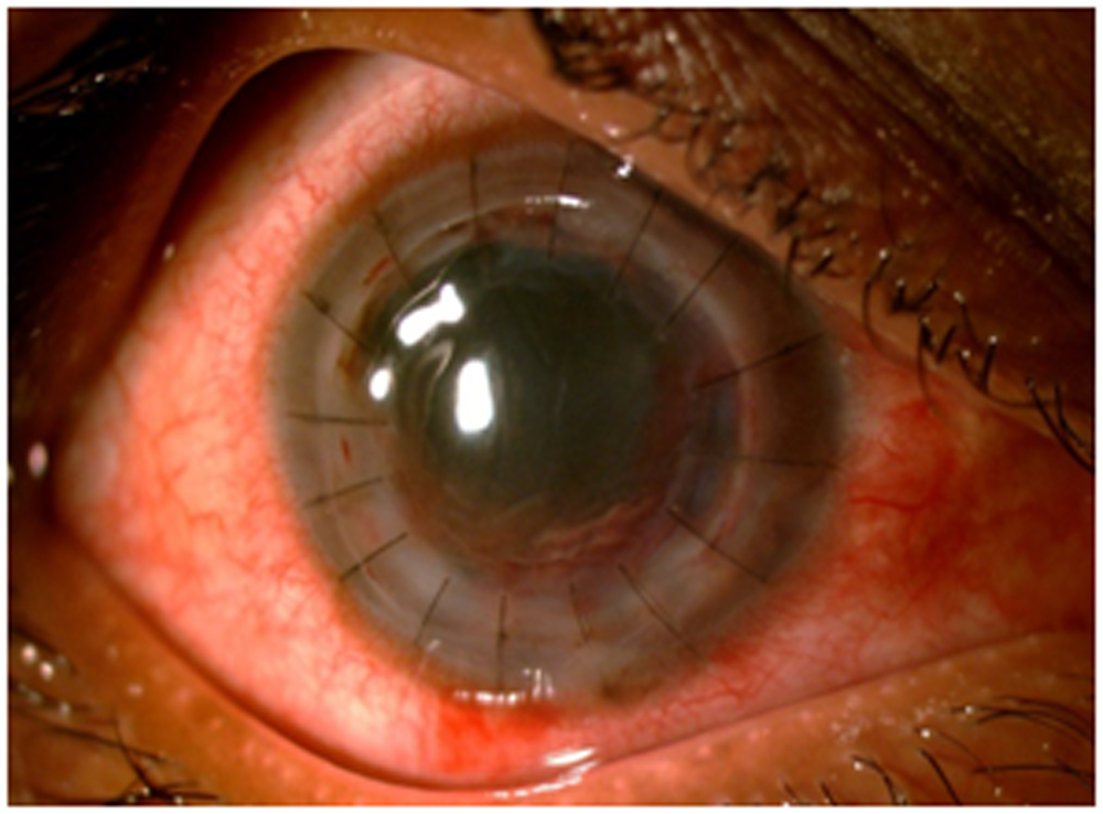
Figure 3-Postoperative 3 weeks of same patient as mentioned before.
Discussion
The main aim of combined surgery for treatment of infective endophthalmitis with corneal infection/opacity is the eradication or reduction of infective load and preservation of structural integrity of the eye. Visual rehabilitation is often difficult and of secondary consideration. In our study we evaluated the results of 54 eyes which underwent combined PKP with PPV with a follow-up of atleast 6 months. 38 eyes (70%) could be salvaged and 24(44.4%) eyes noted an improvement in visual acuity.
One thing worth mentioning was that 9 patients were monocular. All 9 eyes were salvaged and regained useful visual function.
Intraoperative factors play an important role in surgical outcome. If hypotony occurs during the opensky situation, this may result in suprachoroidal haemorrhage which can be avoided by adjusting the flow through the infusion port if the eye is filled with balanced salt solution.Another precaution during surgery is to inject viscoelastics into the anterior chamber angle just prior to suturing the donor cornea to ensure that there is no peripheral anterior synaechia, which can result in chronic angle closure glaucoma.
Secondary glaucoma is mostly d/t extensive peripheral synechiae and can result from trabecular meshwork damage, obstruction by inflammatory cells or neovascularization and may lead to graft failure and blind eye.we noted incidence of secondary glaucoma of 35% compared to that reported by sharma et al and sukhija and jain et al who mentioned 25% and 22% respectively.although presence of secondary glaucoma is not significant statistically,and is controlled with either medical or surgical management. Consequently, one should be aware of the potential risk of elevated intraocular pressure and instigate aggressive antiglaucoma treatment if necessary.12-14
Corneal ulcers presenting at our institute are severe ,already perforated or impending to perforate often leading to endophthalmitis.that is why 24% of combined procedureswere done for corneal ulcers associated enoph.sedghipour et al and sukhija and jain reported even greater proportion of perforated ulcers,76-88%12-15.
Postoperatively,TPK graft has a high incidence of severe AC reaction,shallow AC,synechiae,high intraocular pressure.sony et al mentioned a range of postoperative AC shallowing as 9%-31% in various studies.13In our study 21% patients needed AC reformation.some cases require cataract extraction deepening of ACand prevention of synechiae.There is high chance of graft failure on repeated intervention.graft failure in our cases were mainly due to recurrence of infection could be due to residual infection in the recipient rim or not doing complete vitrectomy as many corneal ulcers presenting in our institute were advanced with extensive invasion of anterior segment structures.
Graft clarity with resolution of posterior segment infection also depends on follow up time.In our study with average follow up period of 6 months.graft clarity was 37%.the majority of patients 52% were from far rural areas and did not regularly follow up,and this had an implication in the graft survival.long -term study on these patients is underway.
Landers et al reported the first use TKP for posterior segment disease with associated corneal opacification in 1981.Since then several studies have reported outcomes of combined PKP with vitreoretinal surgeries using TKP. We preferred using the Eckardts TKP in comparison to landers as it provides a excellent view of the posterior and peripheral retina.16-17 A TKP maintains a closed globe system with normal IOP to allow easy performance of vitreo-retinal procedures without major complications. We used the TKP in all 54 eyes.this might be the reason of good outcome in our study.
Full visual rehabilitation such as cataract surgery,regraftand correction of astigmatism was not studied in our study.
Combined surgery allows rehabilitation or preservation of eyes with severe ocular pathology..functional outcomes of this combined procedures are limited by primary and secondary tissue damage in this series ,however,preservation of vision was possible in most eyes despite severe endophthalmitis..in some eyes with guarded visual prognosisrates of retinal attachment and corneal graftclarity were high,but longer period of follow up are needed to establish the long term success of such outcomes.a large series of patients will better identify factors associated with success and failure.
Our study does have some limitations. First it is a retrospective study with a relatively small sample size. But seeing the unusual occurrence of such complex pathologies and relative absence of literature on combined surgeries for infectious endophthalmitis we could do relatively large series.Second, all our cases had a compromised corneal clarity to begin with, we were unable to document and diagnose any coexisting retinal pathologies apart from retinal detachment which would significantly affect the final visual outcome in our cases.Third is the short follow up as in combined surgery.long -term follow up of is important because these eyes often sustain severe damages and complications can arise at a later date.
To conclude, Combined procedure is an effective way of managing endophthalmitis with poor corneal clarity. Although the functional outcome of a combined procedures is limited by primary and secondary tissue destruction and is often complicated by ciliary body malfunction and secondary transplant failure,preserving ambulatory vision is possible and thus improves the quality of life, at least in patients with single remaining eyes.The primary aim of surgery is restoration of the structural integrity of the eye.In our studyGram negative infection carries a poorer prognosis. Aggressive approach with early surgical intervention, close in-patient postoperative follow-up, intensive topical and systemic treatment and frequent injection of intraocular antibiotics helps in achieving a favourable outcome.
Financial support and sponsorship
Nil
Conflicts of interest
There are no conflicts of interest.
References
1.Wong TY, Chee SP. The epidemiology of acute endophthalmitis after cataract surgery in an Asian population. Ophthalmology 2004;111:699- 70-705.
2.O’Neill EC, Yeoh J, Fabinyi DC et al. Risk factors, microbial profiles and prognosis of microbial keratitis-associated endophthalmitis in high-risk eyes. Graefes Arch Clin Exp Ophthalmol 2014;252:1457-62.
3. Scott IU, Flynn HWJr, FeuerW, Pflugfelder SC et al. Endophthalmitis associated with microbial keratitis. Ophthalmology 1996;103:1864-70.
4. Bourcier T, Thomas F, Borderie V, Chaumeil C, Laroche L. Bacterial keratitis: predisposing factors, clinical and microbiological review of 300 cases. Br J Ophthalmol 2003;87:834-8.
5.Dong X,Wang W, Xie L, Chiu AM. L ong -term outcome of combined penetrating keratoplasty and vitreoretinal surgery using temporary keratoprosthesis. Eye (Lond)2006;20(1):59-63.successful surgical outcome
6.Tanaka M, Ando M, Kitagawa H et al. Penetrating keratoplasty surgery combined with vitrectomy after failing previous corneal surgery. Retina 2003;23:41-7.
7. Lee DS, Heo JW, Choi HJ, Kim MK, Wee WR, Oh JY. Combined corneal allotransplantation and vitreoretinal surgery using an Eckardt temporary keratoprosthesis: analysis for factors determining corneal allograft survival. Clin Ophthalmol 2014;25:449-54.
8.Roters S, Hamzei P, Szurman P et al. Combined penetrating keratoplasty and vitreoretinal surgery with silicone oil: a 1-year follow-up. Graefes Arch Clin Exp Ophthalmol 2003;241:24-33.
9. Roters S, Szurman P, Hermes S, Thumann G, Bartz-Schmidt KU, Kirchhof B. Outcome of combined penetrating keratoplasty with vitreoretinal surgery for management of severe ocular injuries. Retina 2003;23:48–56.
10. Garcia-Valenzuela E, Blair NP, Shapiro MJ, Gieser JP et al. Outcome of vitreoretinal surgery and penetrating keratoplasty using temporary keratoprosthesis. Retina 1999;19:424–429.
11.Abhishek Dave MD,Manisha Acharaya MS,.outcome of combined keratoplasty and pars plans vitrectomy for endophthalmitis with compromised corneal clarity.clinical and experimental oph 2018.
12.Sukhija J,Jain AK.Outcome of therapeutic penetrating keratoplsty in infectious keratitis.Ophthalmic Surg Lasers Imaging.2005;36(4):303-309.
13.Sony P,Sharma N,Vajpayee RB,Ray M.Therapeutic keratoplasty for infectious keratitis;a review of the literature.CLAO J.2002;28(3):111-118…ac shallowing
14.Sharma N,Jain M,Sehra SV, et al.outcomes of therapeutic penetrating keratiplasty from a tertiary eye care centre in northern india.
15.Sedghipour MR,Sorkhabi R,Shenasi A,Dehghan H.Outcome of penetrating keratoplasty in corneal ulcer: a single-centre experience.Clin Ophthalmol.2011;5:1265-1268.
16.Landers MB, Foulks GN, Landers DM et al. Temporary keratoprosthesis for the use during pars plana vitrectomy. Am J Ophthalmol 1981;91:615-619
17.Eckardt C. A new temporary keratoprothesis for pars plana vitrectomy. Retina 1987;7:34-7.
18.Gallemore RP, Bokosky JE. Penetrating keratoplasty with vitreoretinal surgery using the Eckardt temporary keratoprosthesis: modified technique allowing use of large corneal graft. Cornea 1995; 14: 33–38.
Leave a Comment