

Prof. Kamaljeet Singh, Dr.KSHAMA DWIVEDI, Dr. SATYA PRAKASH SINGH, Dr. Pranav Saluja
Introduction:
Macular edema is the main cause of visual impairment in diabetic patients. According to Early Treatment diabetic Retinopathy Study (ETDRS) laser photocoagulation is beneficial for clinically significant diabetic macular edema but for diffuse macular edema the results of laser photocoagulation are unsatisfactory.
Various alternative methods in the form of intravitreal injections has been clinically tried and proven to be better than laser photocoagulation for treatment of diabetic macular edema. They range from Triamcinolone Acetonide , Anti-VEGFs and Dexamethasone implants which had been extensively studied in previous studies and are now being used effectively for reducing central macular thickness and improving vision in diabetic macular edema.
Triamcinolone Acetonide use dates back to 2001 when Jost B Jonas used 15- 20 mg of crystalline triamcinolone acetonide for complicated proliferative diabetic retinopathy associated with central retinal traction detachment1. Raised IOP, cataract formation, sterile endophthalmitis were some of the common complications noted with intravitreal injections. Various studies followed with varying doses of triamcinolone acetonide evaluating the efficacy along with invcidence and severity of side effects. Until recently 0.1ml(4mg) of the drug was being considered the best in terms of efficacy.
In our study we aim to highlight the importance of triamcinolone acetonide in a low dose of 2mg (0.05ml of preservative free triamcinolone acetonide) with similar or better efficacy as compared to anti-VEGFs but with reduced side effects.
The primary outcome of the study was anatomic decrease in central macular thickness and secondary outcome was assessment of improvement of visual acuity.
Inclusion criteria
- Age >18 years
- Diffuse diabetic macular edema (as diagnosed on fundus flourescein angiography)
- Central macular thickness >250 microns
- No prior ocular laser within 12 weeks
- No prior ocular surgery within 6 months of intravitreal injection
- Systemic parameters –HbA1c <9.5% and BP < 150/90 mmHg.
Exclusion criteria
- Aphakic/ pseudophakic eyes
- Other ocular co-morbidities
- OCT showing vitreomacular traction / subretinal fibrosis
- Glaucoma / steroid responsive patients
- Any patient who does not fit in inclusion criteria.
Vision was converted to ETDRS after recording in Snellen’s.
Baseline characteristics
| Features | Group 1 (antivegf) | Group 2 (IVTA) |
| Mean Age (in years) | 64.5 | 66 |
| Sex ratio (M:F) | 1:1 | 2:3 |
| Rural:urban | 1:9 | 3:7 |
| Socioeconomic status
(U:M:L income group) |
U.M.L.(8:2:0) | U.M.L. (0:5:19) |
| Mean HbA1c (%) | 8.2 | 8.6 |
| Mean Visual acuity (in logMAR ) | 0.48 | 0.43 |
| Mean IOP (in mmHg) | 17.3 | 17 |
| Mean Central macular thickness (in microns) | 420.50 | 412.00 |
Materials and Methods
In our study 244 eyes of 133 patients of diffuse macular edema were enrolled during a 4 month period from January 2016 to April 2016. Out of these 7 patients (13eyes) opted for anti-VEGF treatment (group 1 IVA) and 126 patients (231 eyes) were put in triamcinolone acetonide group (group 2 IVTA). 2 patients (3 eyes) in group1 and 5 patients (8eyes) in group 2 lost to followup. 1 patient of group 2 developed cataract and was excluded from study. Thus, in a total of 125 patients 5/125 (10 eyes i.e 4% of patients) opted for ranibizumab and remaining 120 (222 eyes i.e 96% of patients) due to cost effectiveness opted for triamcinolone acetonide. Out of 120 patients 25 (20.8%) belonged to poor socioeconomic status and 79% from middle class. In all the patients visual acuity (ETDRS), IOP (Goldmann applanation tonometry) and central macular thickness(Cirrus HD OCT) were recorded at baseline.
The central macular thickness significant enough for giving intravitreal injections was taken as >= 300 microns (range 300 to 600 microns) . Mean IOP was 17.3mmHg and 17mmHg in group 1 and group 2 respectively (range 10 -21mmHg). All patients received intravitreal injections by 30 gauge needle through infero temporal pars plana route in OT under topical anaesthesia. 5/125 patients received intravitreal ranibizumab and remaining 120 received 0.05ml (2mg) of intravitreal triamcinolone acetonide (40mg/ml of preservative free triamcinolone). Strict sterile protocols were followed in all cases. Immediately after injection antibiotic eye drops were instilled and 250mg of oral acetazolamide was given to all patients.
The patients were followed up at 1st week, 6 weeks, 3 months, 6 months and then followed 6 monthly for 2 years. In each follow up visit visual acuity (in logMAR), IOP and central macular thickness were recorded. “Improvement” was defined as >= 20% reduction in macular thickness (or normalization of macular thickness even if there is <20% reduction) and “resolution” was defined as normalization of the macular thickness (i.e., <260 microns) .
The need for further injections, was thus evaluated. All patients in group 1 received repeat dose at around 6 weeks and in group 2 received repeat injections at nearly 6 months.. Safety outcomes focused primarily on the risk of elevation of IOP to >= 24mmHg at any point of time during the entire 2 year followup period. Any other complications like hypotony (IOP <6mmHg), sudden marked diminision of vision, sudden rise in IOP, cataract formation, sterile endophthalmitis and retinal detachment were also recorded.
Between Jan 16-Apr 16 (4 Months periods)
244 eyes of 133 patients were enrolled in the study.
| IVA
(group 1 ) |
IVTA
(group 2) |
|
|
|
| 13 eyes (7 patients) | 231 eyes (126 patients) |
|
|
|
| 3 eyes (2 patients) lost to follow up | 8 eyes (5 patients) lost to follow up
& 1 eye of 1 patient developed cataract |
|
|
|
| Final 10 eyes of 5 patients | Final 222 eyes of 120 patients |
Observation:
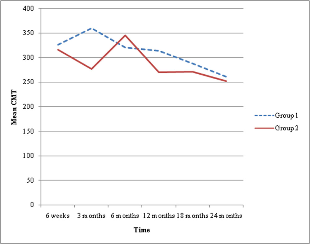
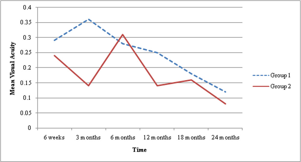
We followed 10 eyes of DME who received anti-VEGF and 222 eyes of DME received IVTA . In both the groups there was reduction in CMT and improvement in visual acuity. In group 1, after an initial reduction at 6 weeks, there was slight trend towards increase in central macular thickness, hence repeat injection was given leading to almost anatomical resolution of macular edema which correlated well with improvement in visual acuity.
In group 2, the maximum decrease in edema was slightly delayed at about 3rd month with a mean reduction of 135.2 micron. It gradually faded at near about 6 months and required repeat injections for maintaining the reduction in edema. The average number of injections required per person over a period of 2 years was 7.8 in group 1 and 3.2 in group 2.
Although the mean reduction in CMT and improvement in visual acuity was almost similar in both the groups .
Only 2 patients of group 2 showed increase in IOP >/= 24mm Hg requiring IOP lowering medications. None of the patient required surgery for glaucoma. Only 1 patient developed significant cataract in group 2 and was excluded from statistical analysis. None of the other patients showed significant progression of lenticular changes.No cases of sterile endophthalmitis, hypotony, or retinal detachment were reported in the entire 2 year followup .
CMT (In microns)
| S.No. | Time | Group 1
mean CMT |
Group 1mean
Reduction from baseline |
Group 2
mean CMT |
Group 2 mean
Reduction from baseline |
| 1. | Baseline | 420.5 | 412.0 | ||
| 2. | 6 weeks | 326.7 | 94.8 | 316.4 | 95.8 |
| 3. | 3 months | 360.2 | 67.5 | 276.8 | 135.2 |
| 4. | 6 months | 321.2 | 100.1 | 345.7 | 66.3 |
| 5. | 12 months | 314 | 107.5 | 270.2 | 131.8 |
| 6. | 18 months | 288.4 | 132.4 | 270.9 | 141.1 |
| 7. | 24 months | 261.6 | 159.7 | 251.9 | 156.4 |
VISUAL ACUITY (in LogMAR)
| S.No. | Time | Group 1
mean visual acuity |
Group 1 mean improvement in visual acuity | Group 2
mean visual acuity |
Group 2 mean improvement in visual acuity |
| 1. | Baseline | 0.48 | 0.43 | ||
| 2. | 6 weeks | 0.29 | 0.18 | 0.24 | 0.37 |
| 3. | 3 months | 0.36 | 0.11 | 0.14 | 0.29 |
| 4. | 6 months | 0.28 | 0.22 | 0.31 | 0.12 |
| 5. | 12 months | 0.25 | 0.23 | 0.14 | 0.24 |
| 6. | 18 months | 0.18 | 0.30 | 0.16 | 0.15 |
| 7. | 24 months | 0.12 | 0.36 | 0.08 | 0.35 |
| Group 1 | Group 2 | |
| No. of injections | 7.8 (range 4-10) | 3.2 (range 1-6) |
| Cost of injections | +++ | + |
Complications
| Group 1 | Group 2 | |
| Glaucoma | 0/10 (0%) | 2/10 (1.6%) |
| Endophthalmitis | 0/10 (0%) | 0/120 (0%) |
| Cataract progression | 0/10 (0%) | 0/120 (0%) |
Discussion:
Corticosteroids have been the drug of choice for controlling the inflammation systemically. Systemic administration of corticosteroids has a poor ocular penetration. The direct intraocular injection of steroid was first of all administered by Robert Machemer et al in case of Proliferative treoretinopathy. Machemer et al propogated the use of intravitreal injection of crystalline cortisone to suppress intraocular proliferation of tissues2. On his suggestion Jost B. Jonas and Jochen K. Hayler et al in 2001 used 15-20mg of crystalline triamcinolone in complicated proliferative diabetic retinopoathy with central retinal traction detachment after pars plana vitrectomy.
Triamcinolone acetonide in a low dose of 2mg was 1st used in 2001 by Richard J. Antcliff and David J. Spalton et al for refractive uveitic cystoids macular edema3. They reported complete anatomic and marked functional recovery in most of their patients within 1 week confirming the potential benefits of triamcinolone in macular edema even in such low doses.
The half life of drug injected intravitreally is longer than the one given systematically. Beer et al studied this and reported that in a non vitrectomised eye intravitreal injection of 4 mg Triamcinolone persists for three month4. And so does its therapeutic effects.
Intravitreal triamcinolone acetonide has increasingly been used recently as treatment for other intraocular edematous and neovascular diseases, such as central retinal vein occlusion, branch retinal vein occlusion, neovascular glaucoma with or without cataract surgery, chronic uveitis, persistent pseudophakic cystoid macular edema, exudative age related macular dgeneration, proliferative diabetic retinopathy & diffuse diabetic macular edema5-9.
It is useful in treatment of patients with diffuse diabetic macular edema refractory to laser and even anti-VEGFs. Using a dosage of about 20 mg , the increase visual acuity is especially pronounced at 3-6 months of injection and observable up till 9 month. With a 4mg dose, the effect was maintained upto 6 months. In both these doses of triamcinolone, the increase in IOP is seen in a significant number of patients. In the end of follow- up, visual acuity & central macular thickness returned to baseline
David Hauser and Amir Bukelman in 2008 compared the efficacy of intravitreal triamcinolone acetonide in 1mg, 2mg and 4mg doses in diffuse diabetic macular edema10. They reported improvement in central macular thickness in all three doses which was maintained to upto 3 months with 1mg dose and upto 6 months with 2mg dose. The results of 4mg dose were significantly worse than the other two. Also, a significant increase in IOP to >10mmHg was reported with 4mg triamcinolone use as compared to 1mg and 2mg.
With the development of intravitreal anti-VEGFs the use of intravitreal triamcinolone declined and many comparative studies reported the superiority of anti-VEGFs over triamcinolone. To the best of our knowledge, through previous studies, the efficacy of triamcinolone acetonide, along with its side effect profiles in lower doses of 4mg and 2mg, are not only similar but are even better than anti-VEGFs. Considering the cost effectivity and therapeutic efficacy of triamcinolone, particularly in developing countries like ours where mass is either from middle class or from low socio-economic status, we strongly advocate the use of triamcinolone as first line intravitreal therapy.
In our study the mean reduction in central macular thickness and mean improvement in visual acuity in both groups were almost similar when considered in a longer follow-up. The need for repeat injections of anti-VEGFs was at shorter durations of around 6 weeks as compared to triamcinolone which was repeated at around 6 months to achieve better results. This added to the overall cost of treatment in group 1 as compared to group 2
In our study maximum improvement in visual acuity was seen at 6 months which correlated with the previous studies. By 12 months the effect started weaning off, and we gave repeat injections at nearly one year which again improved visual acuity & central macular thickness by 18 months. Effect gradually weaned off towards 24 months, but nevertheless it was better than the presenting condition.
Since the dose of IVTA was lesser than in previous studies, the significant increase in IOP was found in only 2 patients which did not require long medical treatment. None of the patients required trabeculectomy in our study group, even at follow-up of two years.
Conclusion:
In conclusion, intravitreal injection of triamcinolone is better than intravitreal injection of anti-VEGF. Firstly, lesser number of injections are required which also avoids all the injection related complications and stress related to both doctor and patients.
Secondly, repetitive cost of anti-VEGF injections is prohibitive for its use, especially for the patients of middle class and poor socioeconomic status. Thirdly, injection of 2mg of triamcinolone is safe in terms of development of complications. Fourthly, the vision improvement and reduction of CMT is comparable in both the group. Further studies are required to confirm the findings.
References:
- Jost B. Jonas, Jochen K. Hayler, Antje Sofker, Songhomitra Panda-Jonas. Intravitreal injection of crystalline cortisone as adjunctive treatment of proliferative diabetic retinopathy. Am J Ophthalmol 2001;131:468-471.
- Machemer R., Sugita G, Tano Y. Treatment of intraocular proliferations with steroids. Trans AM Ophthalmol Soc 1979; 77: 171-180.
- Richard J. Antcliff, David J. Spalton, Miles R. Stanford, Elizabeth M. Graham, Timothy J. Ffytche et al. Intravitreal triamcinolone for uveitic macular edema: An optical coherence tomography study. Ophthalmol 2001;108:765-772.
- Beer PM, Bakri SJ, Singh RJ, LiuW, Peters GB, Miller M. intraocular concentration and pharmacokinetics of triamcinolone acetonide after a single intravitreal injection. Ophthalmology 2003;110(4):681-686.
- Mandi D. Conway, Christina Canakis, Charalampos Livir-Rallatos, Gholam A. Peyman. Intravitreal triamcinolone acetonide for refractory chronic pseudophakic cystoids macular edema. J Cataract Refract Surg 2003;29:27-33.
- Sushmita Kaushik, Vishali Gupta, Amod Gupta, Mangat Ram Dogra, Ramandeep Singh. Intractable glaucoma following intravitreal triamcinolone in central retinal vein occlusion. Ophthalmol 2004;137:758-760.
- JB Jonas I Akkoyun, B Kamppeter, I Kreissig, RF Degenring. Branch retinal vein occlusion treated by intravitreal triamcinolone acetonide. Eye 2005;19:65-71.
- Maca SM, Abela-Formanek C, Kiss CG, Sacu SG, Benesch T, Barisani-Asenbauer T. Intravitreal triamcinolonefor persistent cystoid macular edema in eyes with quiescent uveitis. Exp. Ophthalmol 2009 May;37(4):389-396.
- Koch R, Schmidt M, Gebauer S, Busse H Uhlig CE. Intravitreal treatment in patients with exudative age related macular degeneration and visual acuity <=0.05. BMC Ophthalmol 2015Oct21;15:138.
- David Hauser, Amir Bukelman, Russell Pokroy, Haia Katz, Ariela Len et al.Intravitreal triamcinolone for diabetic macular edema. Retina 2008;28:825-830.
Leave a Comment