

Dr. Soumya N, S21446, Dr.Kavitha Toopalli, Dr. Modini Pandharpurar
Abstract :
Context:
Ocular surface squamous neoplasia (OSSN), first described by Lee and Hirst in 19951, encompasses a spectrum of benign, pre- malignant and malignant epithelial lesions of the conjunctiva and cornea ranging from simple dysplasia to invasive squamous cell carcinoma. OSSN has been reported to present in HIV positive patients as a higher-grade malignancy with a higher incidence of invasion and recurrence.
Aims:
To study the clinical behaviour and pathological characteristics of OSSN in HIV positive patients as compared to HIV negative patients.
Settings and Design:–
It is a retrospective study conducted over a period of three years from2015 to 2018.
Methods and Material:
All the cases diagnosed as ocular surface squamous neoplasia (OSSN)on histopathological examination were included in the study. Pre-operative HIV status of the patients was noted. Lesions were assessed for their clinical presentation, histopathological differentiation and extent of local invasion. Tumour proliferation was analysed using ki-67.
Statistical analysis used:
Proportions, odds ratio, chi square test, p – value
Results:
A total of 77 cases of OSSN were encountered of which13(16.9%) were HIV positive and 64(83.1%) were non-HIV cases. In HIV positive cases mean age at presentation was 41.6 years and mean size of the lesion was 1.5cms. On histopathology, invasive tumours were seen in 6/13(46.1%) cases as compared to 13/64, (20.3%) in non-HIV cases (p<0.05).
Conclusions:
OSSN in HIV positive patients tends to present as a larger and aggressive high-grade tumour in relatively younger age groups.
Key-words:
HIV, ocular surface squamous neoplasia, Ki-67
Key Messages :
A high index of clinical suspicion of HIV is important in Ocular surface squamous neoplasia (OSSN) patients and an HIV test should be considered in all the patients, especially in younger age groups, wherein, it may be the only apparent manifestation of HIV.
Ocular surface squamous neoplasia(OSSN) is a broad term, first described by Lee and Hirst in 1995, that encompasses a spectrum of benign, pre- malignant and malignant epithelial lesions of the conjunctiva and cornea ranging from simple dysplasia to invasive squamous cell carcinoma. [1]Morphologically there are three types of lesions, the Gelatinous type (leukoplakic or papilliform), Nodular type and the Diffuse variant. Ocular surface squamous neoplasia (OSSN) has been reported to be an aggressive tumour in HIV positive patients presenting as a larger lesion and higher-grade malignancy with a higher incidence of invasion and recurrence. Ki67, a nuclear proteinexpressed in G1, S, G2 and M phases of cell cycle, is a marker of cell proliferation and was first described in 1983.It is considered a prognostic factor of many types of malignancies. High percentage reflects worse prognosis with higher chances of recurrence. [2]
Subjects and Methods:
It is a retrospective study conducted over a period of three years from2015 to2018. All the cases diagnosed as ocular surface squamous neoplasia (OSSN) on histopathological examination were included in the study. Clinical data and pre-operative HIV status of the cases was collected from the medical records. Lesions were assessed for their clinical presentation, invasion into surrounding structures and intraocular spread. All the histopathological slides of OSSN were reviewed and slides with good staining showing definite features of OSSN were included in the study. Tumours were assessed for their differentiation and invasiveness into underlying tissues. Tumour proliferation using immunohistochemical (IHC) staining for ki-67 was analysed in a total of only 13 out of 77 histologic specimens, due to poor microwave antigen retrieval from most of the tissues. Out of these, six specimenswere of HIV positive cases and seven were from HIV negative cases. IHC was done on 3µm thin paraffin sections using pre-diluted antibodies, MIB -1, an antibody to Ki-67 antigen in the nuclei of neoplastic cells.
Statistical analysis
In order to compare the frequency of various clinical and histopathological features between the HIV-positive and HIV-negative groups, proportions (expressed as percentages) and odds ratios were calculated. p – value was calculated using chi square test. Results of ki67 index were expressed as average percentage of positive cells to the total number of cells in at least 10 fields at ×40 magnification. Grading was done as follows: low proliferation of 1+(1% to 10%); borderline 2+ (10% to 20%); and high proliferation of 3+ (> 20%). [2-3]
Results:
A total of 77 cases of ocular surface squamous neoplasia (OSSN) were retrieved.Excisional biopsy along with cryotherapy was performed in 72(93.5%) cases, enucleation was done in 4(5.2%) cases and exenteration was done in 1(1.3%) case. Of these patients, 13(16.8%) cases were HIV positive and 64(83.2%) cases were HIV negative. Age at presentation in HIV positive cases ranged from 35-55 years (mean 41.6) as compared to non-HIV patients (range 40-83, mean54.8). Male predominance was seen in both HIV positive 7/13(53.8%) and negative cases 50/64(78.1%). Proportions of various clinical featuresand histopathological features of OSSN in both HIV positive and HIV negative groups are shown in Table 1. Onimmunohistochemical (IHC) examination, grading of tumour proliferation using the immunohistochemical marker Ki-67 in both HIV positive and HIV negative groups is shown in table 2.
Discussion:
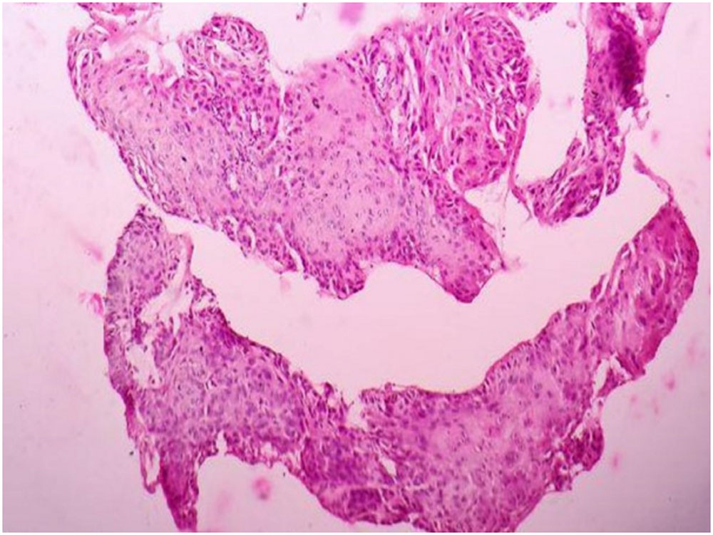
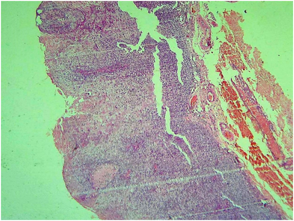
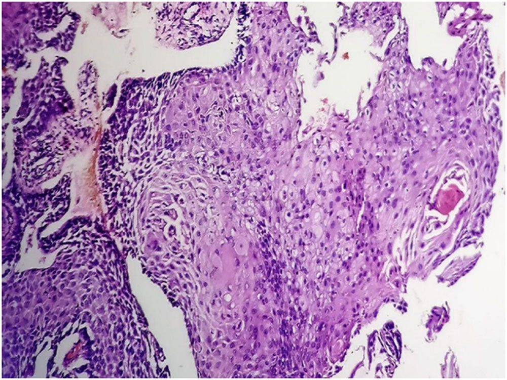
As per the India HIV estimation 2015 report, adult (15-49 years) HIV prevalence in India was estimated at 0.26% in 2015.In the present study,18.5% (n=13) of the cases were HIV positive. Thus, in Ocular surface squamous neoplasia (OSSN) patients, a high index of clinical suspicion of HIV is important and the current recommendation is to conduct HIV screening in all cases of OSSN to rule out undetected HIV infection. [3-5] OSSN may be the primary and only apparent manifestation of HIV in these patients, especially in younger age groups, thus, it may be a possible marker for HIV infection. [6] The most commonlesion was a gelatinous/leukoplakic limbal lesion on the temporal side of the right eye in both HIV positive and negative cases. The size of the lesion ranged from 0.5 cm – 3cms (mean- 1.5cms) in HIV positive cases and 0.2cms – 4.5cms (mean- 0.5cms) in HIV negative cases. There was only one case in the HIV negative group wherein the lesion measured 4.5cms and showed extension into the orbit for which exenteration was done. Whereas in rest of the cases, the size ranged from 0.2 -1cm. In the HIV negative group, corneal involvement was observed in four cases for which enucleation was done and tumour extension into the orbit was seen in one case for which exenteration was done. In the HIV positive group, one case showed invasion into the sclera with calcifications and enucleation was done for this case. There was no significant difference between HIV positive and HIV negative groups when corneoscleral involvement or orbital extension of the tumour was considered. This may be due to the inherent nature of the tumour which is a slow growing, low-grade malignancy. However, it may act aggressively and result in recurrences and invasion in some immunocompromised patients. [7] On histopathology, the HIV negative cases showed a wide spectrum of differentiation ranging from mild dysplasia to invasive squamous cell carcinoma. Whereas, tumours in HIV positive patients were of a higher-grade ranging from carcinoma in situ to invasive squamous cell carcinoma. (Figure 1). Invasive tumours were significantly higher in frequency in the HIV positive group 6/13(46 %) than in the HIV negative group 13/64(20.3 %) (p value <0.05).
However, the most common histological diagnosis in both HIV positive 7/13(53.8%) and HIV negative groups 33/64(51.5%) was carcinoma in situ. The proportion of cases showing high and borderline tumour proliferation (+2/+3) with ki67 staining were significantly higher in HIV positive patients (5/6;83%) than the HIV negative patients (2/7;28.5%) (p < 0.05).(table 2 and figure 2).Various studies described the prognostic factors for OSSN recurrence as greater lesion size, tumour invasion, non-radical excision, absence of cryotherapy and an increased level of positive expression of the biomarker, Ki-67. [7-9]Due to a high Ki 67 index in HIV positive cases, there might be high chances of tumour recurrence and thus, radical excision with cryotherapy and regular follow up to detect any early recurrence or early detection of lesion in the fellow eye is necessary.CD4 lymphocyte count monitoring is also recommended in all OSSN patients affected by HIV. [10] Immunosuppression resulting from HIV infection is suspected to play a role in the etiopathogenesis of OSSN. In few studies, at the time of OSSN detection, a CD4 lymphocyte count <200 cells/mm3 was observed in 85%–100% patients [12,13,14] However, no significant association was observed between CD4 count and detection of OSSN in HIV positive cases. [11,12,15] The effect of highly active antiretroviral therapy on OSSN is also controversial. However, there is a need to explore and practice a multi-modality approach in the treatment of OSSN in HIV patients starting with a high index of suspicion of HIV infection in young adults, early detection of lesions in already known seropositive cases, use of alternative methods like the use of topical antimetabolites alone or in combination with surgery in early OSSN and a regular close follow up to address the possible high recurrence rates. As there is not much information available about various treatment modalities or their outcomes in OSSN patients with HIV infection, there is a need to frame standard treatment guidelines for OSSN in HIV positive patients. Effective evidence-based interventions are needed to allow early diagnosis and treatment, as well as prevention of the disease.
References :
- Lee GA, Hirst LW. Ocular surface squamous neoplasia. SurvOphthalmol. 1995; 39:429–450.
- Roychowdhury, M. Ki67. PathologyOutlines.com website. https://www.pathologyoutlines.com/topic/stainski67.html.
- Mahomed A, Chetty R. Human immunodeficiency virus infection, Bcl-2, p53 protein, and Ki-67 analysis in ocular surface squamous neoplasia. Arch Ophthalmol. 2002;120(5):554–558.
- Robert J nutt, John L Clements, William H Dean.Ocular surface squamous neoplasia in HIV positive and HIV-negative patients and response to 5-fluorouracil in Angola. Clinical Ophthalmology Dovepress 2014:8 2435–2440
- Nagaiah G, Stotler C, Orem J, Mwanda WO, Remick SC. Ocular surface squamous neoplasia in patients with HIV infection in sub-Saharan Africa. CurrOpin Oncol. 2010; 22:437–442.
- Carreira H, Coutinho F, Carrilho C, Lunet N. HIV and HPV infections and ocular surface squamous neoplasia: systematic review and meta-analysis. Br J Cancer. 2013;109(7):1981–1988.
- Karp CL, Scott IU, Chang TS, Pflugfelder SC. Conjunctival intraepithelial neoplasia: a possible marker for human immunodeficiency virus infection? Arch Ophthalmol. 1996;114(3):257–261.
- Kiire CA, Srinivasan S, Karp CL.Ocular surface squamous neoplasia.Int Ophthalmol Clin. 2010 Summer; 50(3):35-46.
- McKelvie PA, Daniell M, McNab A, Loughnan M, Santamaria JD. Squamous cell carcinoma of the conjunctiva: a series of 26 cases. Br J Ophthalmol. 2002; 86:168–173.
- Tunc M, Char DH, Crawford B, Miller T. Intraepithelial and invasive squamous cell carcinoma of the conjunctiva: analysis of 60 cases. Br J Ophthalmol. 1999; 83:98–103.
- Rathi SG, Ganguly Kapoor A, Kaliki S.Ocular surface squamous neoplasia in HIV-infected patients: current perspectives. HIV/AIDS – Research and Palliative Care.2018; 10:33—45.
- Makupa II, Swai B, Makupa WU, White VA, Lewallen S. Clinical factors associated with malignancy and HIV status in patients with ocular surface squamous neoplasia at Kilimanjaro Christian Medical Centre, Tanzania. Br J Ophthalmol. 2012;96(4):482–484.
- Waddell KM, Lewallen S, Lucas SB, Atenyi-Agaba C, Herrington CS, Liomba G. Carcinoma of the conjunctiva and HIV infection in Uganda and Malawi. Br J Ophthalmol. 1996;80(6):503–508.
- Pradeep TG, Gangasagara SB, Subbaramaiah GB, Suresh MB, Gangashettappa N, Durgappa R. Prevalence of undiagnosed HIV infection in patients with ocular surface squamous neoplasia in a tertiary centre in Karnataka, south India. Cornea. 2012;31(11):1282–1284.
- Gichuhi S, Sagoo MS, Weiss HA, Burton MJ. Epidemiology of ocular surface squamous neoplasia in Africa. Trop Med Int Health. 2013;18(12):1424–1443. [PMC free article] [PubMed]
- World Health Organization HIV/AIDS. 2017. [Accessed October 12, 2017]. Available from: https://www.who.int/mediacentre/factsheets/fs360/en.
Tables and figures
Table 1
Summary of clinical and histological features of OSSN with absolute numbers (proportions) and odds ratios.
| Clinical features | Number of patients (proportions) | Odds ratio | |
| Hiv positive | Hiv negative | ||
| Age range and mean | 35-55 years (mean -41.6) | 40-83 years (mean -54.8) | NA |
| Sex :females | 6 | 14 | 3.2 |
| Sex : males | 7 | 50 | 0.3 |
| Leukoplakia | 9 | 56 | 0.3 |
| Nodular | 3 | 3 | 6.0 |
| Pigmentation | 1 | 3 | 1.6 |
| Cystic lesion | 0 | 2 | NA |
| Laterality : Nasal | 5 | 24 | 1.0 |
| Temporal | 8 | 40 | 0.9 |
| Right | 7 | 50 | 0.3 |
| Left | 6 | 14 | 3.0 |
| Corneo scleral involvement | 4 | 1 | 30 |
| Orbital extention | 1 | 0 | NA |
| Range and Mean size of the lesion (largest dimension) | 0.5 cm – 3cms (1.5cms) | 0.2cms 4.5cms (0.5cms) | NA |
| Non invasive :
Dysplasia ( mild to severe ) |
0 | 18 | NA |
| Carcinoma in situ | 7 | 33 | 1.0 |
| Invasive:
Well differentiated squamous cell carcinoma |
2 | 8 | 1.2 |
| Moderately differentiated squamous cell carcinoma | 4 | 5 | 5.9 |
| Poorly differentiated squamous cell carcinoma | 0 | 0 | NA |
Table 2
Summary of Ki 67 index in OSSN with absolute numbers (proportions)
| Tumour proliferation – Ki67 grading
|
Total | |||
| % | Low
(<10%) |
Boderline
(10-20%) |
High
(>20%) |
|
| Grade | 1+ | 2+ | 3+ | |
| HIV positive | 1 | 3 | 2 | 6 |
| HIV negative | 5 | 1 | 1 | 7 |
| Total | 6 | 4 | 3 | 13 |
Figure 1
a – H&E 10X Severe dysplasia in a case of OSSN
b – H&E 4X Carcinoma in situ in a case of OSSN
c – H &E 10X Well differentiated squamous cell carcinoma in a case of OSSN
d – H&E 10X. Moderately differentiated squamous cell carcinoma in a case of OSSN
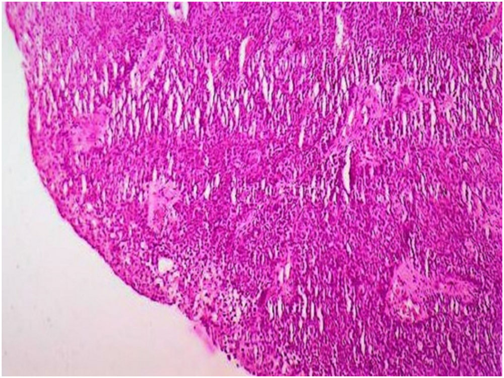
Figure 2
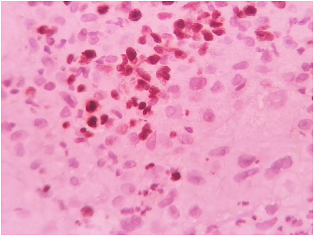
a – IHC 40x showing nuclear staining of ki-67
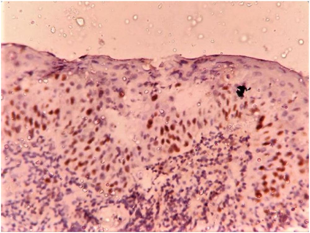
b – IHC 10X showing nuclear positivity of ki-67 < 10%, grade 1+
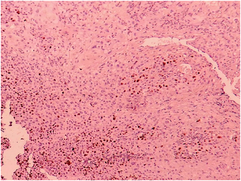
c – IHC 10X showing nuclear positivity of ki-67 15%, grade 2+
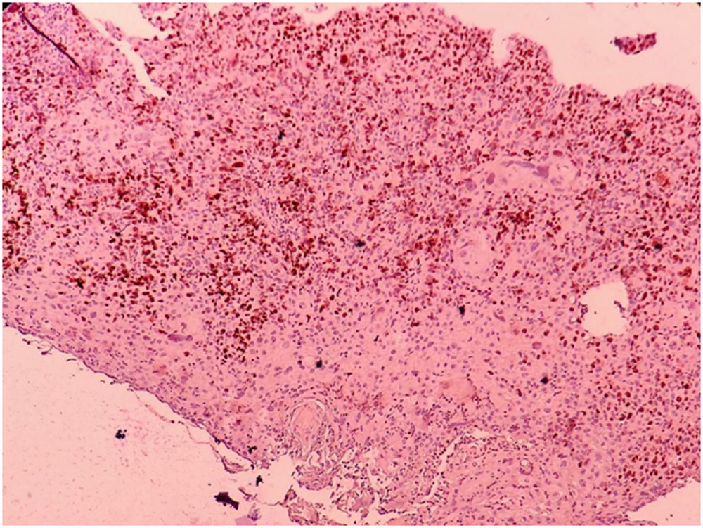
d – IHC 10x showing nuclear positivity of ki-67 >20%, grade 3+
Leave a Comment