

Dr.Devendra aheshwari, Dr.SWATHI KANDURI,Dr.Neelam Pawar,Dr.Rama Krishnan R
Abstract
Purpose:To determine the safety and efficacy of Mitomycin C (MMC) injection versussponge duringtrabeculectomy.
Methods: It’s a prospective analysis of patients who underwent Trabeculectomywith Mitomycin C&followed for 1year, divided in 2 groups: group1- injection (n=21), group2 – sponge (n=21).Sameconcentration of Mitomycin C was used for both groups. Inclusion criteria were Trabeculectomies withMitomycin C for Intraocular pressure control in eyes with glaucoma (primary + secondary) with a follow up of 1 year.
Results:Mean preoperativeintraocular pressure in group1 was 29.00±11.92mmhg& group 2 was 25.87±11.09 mm hg which reduced to 12.19±4.03 &15.56±10.72mmhg at final visit with P value of 0.0002 &0.001 respectively. Mean preoperative number of anti-glaucoma medications was 2.4±0.87 in group 1 & 2.3±0.96 in group 2 which reduced to 0.38± 0.5 &0.91±0.85 with P value of 0.001 &0.0003 respectively. Overall, success rate was 90.5% &87% in group1&group 2at final visit.All complications were encountered in sponge group. 11.1 % developed Choroidal detachment ,Malignant glaucoma which underwent medical management.33.3% hadencapsulated bleb which received bleb needling . 44.4% underwentArgon laser suturelysis postoperatively. There is a significant difference between baseline and final visual acuity in Sponge group (p-value – 0.024) compared to injection group.
Conclusion: MMC injection may be as safe and as effective as conventional sponge application with comparable estimated complete treatment success.
Key Words:Trabeculectomy,Mitomycin C
Full Text
Introduction
The introduction of Mitomycin C (MMC) as an adjunct to trabeculectomy was a major advance in the ability to improve the intraocular pressure (IOP)loweringefficacy of the procedure.[1] MMC is an antineoplastic antibiotic agent isolated from the fermentation filtrate of Streptomyces caespitosus, has been shown to suppress fibroblastic activity.It acts as a deoxyribonucleic acid cross-linker, which inhibits fibroblast proliferation. It is used widely in medicine as a chemotherapeutic agent to treat a variety of cancers. Its use and application in ophthalmology is common practice because of its modulatory effects on woundhealing.[2]
Current applications of MMC include glaucoma surgery,pterygiumsurgery, corneal refractive surgery,cicatricialeye disease, conjunctivalneoplasia, and allergic eye disease.[3]. For more than two decades, MMC has been routinelyused during trabeculectomyto reduce postoperative episcleral fibrosis and bleb failure due to scarring by the wound healing process.[4]The use of MMC in trabeculectomy is indicated in patients who are young, African-American, or have had previous surgery, and has been shown to increase fibroblast density and compact connective tissue over time.[5] Studies have shown that the use of MMC improves outcomes in glaucoma filtration surgery with good long –term IOP control.[6-8]
The time tested route of administration of Mitomycin C is via a sponge soaked in it.[9]This sponge is applied to the subconjunctivalspace. Both the concentration of the drug used and the duration of exposure can be altered, depending on the risk of failure. A subconjunctivalinjection of Mitomycin C instead of these sponges is recently being studied as an alternative.[10] Initial result suggest that this new method of application of Mitomycin Cis associated with superior surgical outcomes and no increase in complication.
Our current study is a randomized, prospective, open label, interventional one aimed at comparing these two modalities ofMitomycin C administration intrabeculectomy surgery in Indian population. The purpose of this study was to determine the safety and efficacy of intraoperative injection of MMC against conventional sponge – applied MMC duringtrabeculectomy.It iswith this background that this study was undertaken.
Materials and Methods
Study Design
This study was a prospective, comparative case series designed from a consecutive series of Trabeculectomies with MMC performed in Aravind eye hospital, Tirunelveli, Tamilnadu. Inclusion criteria were trabeculectomieswith MMC for IOP controlin eyes withglaucoma (primary + secondary) with afollowupof 1 year. The study group (injection group)included all trabeculectomies that met the above inclusion criteria and were performed consecutively betweenJanuary2016 and January 2017 (n = 21). The control group (sponge group) was selected fromtrabeculectomy proceduresperformed by the same surgeon thatmet the inclusion criteria and was matchedfor baseline IOP and Visual acquity(VA) (n = 21). Exclusions were patients undergoing any other glaucomaprocedure such as tube-shunt procedures,nonpenetrating glaucoma surgery, combination surgery (i.e., phacoemulsification +trabeculectomy),use of an antimetabolite, such as 5-FU and any patients with glaucoma(i.e., uveitic, neovascular, traumatic glaucoma). This study was approved by the Institutional Review Board/ Ethics Committee of Aravind eye hospital.
TrabeculectomyOutcome
Data were collected preoperatively and postoperatively at 2weeks,1 month,3 months, 6months and1 year after surgery.Demographic data and burden of postoperative care (number of visits within 3 months) were recorded. Baseline IOP and VA were calculated using the averageof measurements from the two most recent visits prior to surgery.Goldmannapplanation IOP, best-corrected VA, number of glaucoma medications, the need for postoperative interventions, and postoperative complications wererecorded at each examination. Specifically, postoperative data on complications including bleb leak,hypotony(defined as IOP<6 mm Hg), shallow AC (defined asiris/cornea touch beyond the mid – iris centrally),infection, corneal edema/haze, and cataract formation were collected.
Operation Procedure
All trabeculectomies were performed at a single institutionby a single surgeon (Dr .DevendraMaheshwari).To prepare the MMC injection, the surgeon used a 20-μg preparation starting with MMC 0.4 mg/mL, diluting0.1 mL of MMC (40 μg) in0.1 mL oflidocaine (1:1, total volume of 0.2 mL). Half of that solution (0.1 mL of MMC:lidocaine[20 μg]) was used for injection. Topical anesthesia was instilled. Snip peritomy was performed with alimbalincision.A blunt 30-gauage cannula was introduced 7 to 8 mmfrom the limbus. The MMC preparation was injected posterior to the anticipated flap location subconjunctivally.In order to avoid egress to the surface, theincision was kept small and the conjunctival entry was compressed with a surgical sponge to prevent any MMCfrom escaping.
The blunt cannula was withdrawn, and the solution was further spread over a larger surface area using a surgical sponge. The conjunctivalperitomywas then completed. Wet-field bipolar cautery was performed for hemostasis with copious irrigation using salinesolution. Thetrabeculectomy was completed in the standard fashion by delineating a 6 × 4mm scleralflap. A 15 noblade wasthen used to dissect the partial thickness scleral flap. Aparacentesiswas performed using a 1-mm side port blade in the temporal cornea. A sclerostomywas created with a Kelly’spunch. A peripheraliridectomy was created with a DeWeckerscissors. The scleral flap was repositioned inplace using three 10-0 nylon sutures using releasable suturetechnique. Once flow was determined to be adequate, with the anterior chamber remaining well maintained,conjunctival closure proceeded using a running 8-0 vicrylsuture.At the end of the case, the conjunctivalincision was checked for lack of leakage.
The conventional sponge-applied technique was used in the control group. On two separate semicircular surgical sponges (7-mm corneal light shield cut in half), aMMC solution of 0.4 mg/mL was used and then inserted subconjunctivallyat the surgical site. The sponges were applied for 2 minutes and removed, and then the area was copiously irrigated with salinesolution beforethe caseproceeded in the usual fashion as described earlier.
Statistical Analysis
Descriptive variables were presented with Frequency (Percentage) or Mean (Standard deviation). Chi square test was used to find out the association between categorical variables. Student’s t-test or Mann-Whitney U test was used to find out the significant difference of continuous variables between the study procedure (MMC and Sponge). Pre and Post comparisons were done using Wilcoxon sign rank test. To find out the relationship between Intra Ocular Pressure with Bleb characteristics and Blebvascularities, Spearman rank ordercorrelation was used. To find out the cumulativeprobability of success,Kaplan-Meier survival analysis was performed. P value less than 0.05wasconsidered as statistically significant and the statistical analysis was performed using statistical software STATA 14.1 (Texas, USA).
Surgical outcome Definition
Complete success – Postoperative IOP ≤18 mmHg but more than 6 mmHg without addition of anti-glaucoma medication or other interventions.Qualified success s – IOP ≤18 mm Hg but more than 6 mmHg with additional anti-glaucoma medication.Failure – IOP>18 mmHg with additional anti-glaucoma medication.
Results
In total, 43 eyes were included: 21intraoperative injection and 21 sponge – applied MMC.
IntraocularPressure (IOP):
There is a significant difference between baseline and final IOP in Sponge (p-value – 0.001) & Injection (p-value – 0.0002)(Table 1).
NumberofAntiglaucoma Medications (AGM):
Number of AGM was significantly reduced inthe injection group (p-value = 0.0001) and in the sponge group (p value = 0.0003) from the baseline. The p-value (0.021 < 0.05) shows there is a significant difference between Injection and Sponge at the end of 1 year(TABLE2).
SuccessCriteria
Overall, successrate (complete + qualified) was 90.5% in the MMC injection group and 87.0% in the MMC sponge group at postoperative year 1.
Kaplan MeierSurvival Analysis
This plotshows the cumulative probability of success against time. From the graph, the predicted probability of success at month1, month3,month6 and month12 in Injection group is 100.0%, 100.0%, 85.7%& 42.9% and in sponge group is 100.0%, 88.9%, 81.5% & 61.1%. The Log rank test forequality of survivor function P-value (0.917) shows that there is no significant difference between the curve of injection and sponge group
(FIG1).
Postoperative Complicationsand Procedures:All complications were encountered in sponge group. 11.1 % developed Choroidal detachment, Malignant glaucoma which underwent medicalmanagement.33.3% had encapsulated bleb which received bleb needling . 44.4% underwentArgon laser suturelysis postoperatively(TABLE 4).
BestCorrected Visual Acuity:There is a significant difference between baseline and final visual acuity in Sponge group (p-value – 0.024) &there is no significant difference in Injection group (p-value – 0.470 >0.05).
Discussion
Treatment of patients having glaucoma requires both clinical skill and keen judgement. Consultants treating these patients have to decide on the timing of surgery, the type of surgical procedure and the details of the procedure. In order to increase the success rate of the surgical procedure, an augmentation with an antimetabolite is usually done. The most commonly used antimetabolite for an augmented glaucoma filtration surgery is Mitomycin C. Out of the many modes of administration of Mitomycin C, sponge soaked method is the most common. Administration ofMitomycin C as asubconjunctival injection is a newly developed method for augmentation of a glaucoma filtration surgery. This study was done to compare these two methods of augmentationof Trabeculectomy withMitomycinC.
Our study shows that the efficacy of injection of MMC is comparable to sponge application, with less need forvisits and 5-FU intervention. Overall complete treatmentsuccess in the MMC injection group at 1 year was 90.5% compared to sponge(87%) which is consistent with a prior comparative study reporting 1-year outcomes of MMC injection in trabeculectomy versussponge .[11]
Intraoperative injection of MMC in trabeculectomy has several advantages over conventional sponge application.A large MMC treatment area produces more diffuse and elevated blebs.[12]Large-area MMC application also seems to increase long-term success without increasing the complication rates in trabeculectomies.[13,14]Direct and diffuse application of MMC by injection maypromote less scarring and vascularization of the bleb.[15] In order to achieve the same surface area of exposure with sponges, i.e., achieved with injection, the surgeon must use multiple sponges, all of which must be carefully collected thereafter. The injection method therefore, eliminates the risk of retained sponges.
Another advantage of using injection vssponge application of MMC is the predictable dose of delivery. In sponge application, the surface area of cut pieces of surgicalsponges is very variable. A study found that the quantities of MMC contained in sponges prepared for glaucoma surgery differed for a given surgeon and between surgeons. The estimated actual dose delivered in a sponge soaked with MMC 0.2 mg/mL varied between 1.9 and 17.3 μg.16[16] With this unpredictable sponge dosing, surgeons runthe risk of overdosing MMC. Irrigation is often used after deliveryof MMC; however, it appears to only have an effect at reducing MMC concentrations in the superficial scleral layers, with no effect on MMC concentrations in the deepscleral and subsclerallayers.[17] Another advantage of thesubtenoninjection technique is that the tumescenttenonis more easily dissected and provides anaccessible handle for manipulation and thereby reducesconjunctivaldamage.
Although IOP is frequently considered as the primary outcome measure of success in glaucoma surgery, the success oftrabeculectomy actually relies on a functioning bleb. In contrast to postoperative measurement of IOP that may sometimes be misleading, bleb morphology could be a surrogate of bleb functionality and IOP.[18]In the current study, we usedMoorfieldsbleb gradingsystem to evaluate and compare bleb morphology in our study groups.
The bleb characteristics were studied for each visit in each group. In general a diffuse bleb with normalvascularization is associated with greater survival. A thickenedvascularizedbleb is associated with failure while a thin cystic avascular bleb is associated with increase in risk of bleb infections and bleb leaks.[19]In our study, theblebs were graded by the MoorfieldsBleb Grading System. Thus more diffuse blebs are seen with injectableMitomycin C as well as withsponge soaked Mitomycin C in contrast tothe findings of Hung et al, who found more diffuse blebs by using subconjunctivalinjections of Mitomycin C for trabeculectomy over a 12 month follow up period.[20]
MMC bleb-related complications, namely thin-walled and cystic blebs, hypotony,Blebleakage,blebitis and endophthalmitis, others include hypotonymaculopathy, corneal epithelial toxicity etc. compromise the outcome of surgery.[21,22]Hypotony and itssequelae may be related to intraoculartoxicity of MMC.[23]
Routinely, glaucoma surgeons apply titrated doses of MMC-soaked sponges under the scleral flap for 1–3 min to modulate wound healing and prevent fibrosis.[24]There are concerns regarding the sponge method of MMCdelivery including a physical barrier to treat a larger area, conjunctivaldamage during sponge manipulation, risk of sponge fragmentation and retention and the need for some extra minutes. The pharmacologicalaction of MMC is limited to itsarea of exposure and larger treatment areas are believed to produce more diffuse blebs and reduce the risk of cystic bleb formation.[25,26]Occasionally, sponge application can also create a whitish MMC “burn” often due to overdosing of MMC. The avascular, thin bleb produced is at increased risk of early and late bleb leaks as well as of infection.Premature scar formation around an insufficiently treated area could lead to an encapsulated bleb, the ‘ring of steel’ phenomenon and bleb failure.[27]
Our study shows that the injection of MMC is safe, with nil postoperative complications compared to conventional sponge in contrast to apriornoncomparativestudy of MMC injection wherethe most frequent early postoperative complicationsencounteredwere hypotony, hyphema, and serous choroidaldetachments.[28]
Lee et alfirst reported the outcome of 108 consecutivetrabeculectomiesand phacotrabeculectomies who received a variable concentration ofintratenonMMC, 0.2–0.5 mg/mL at the time of surgery. Although they considered their outcomes ‘favourable’ in comparison with conventional method, almost a quarter of patients developed cystic bleb, defined as area of marked conjunctivalthinning andavascularity. The higher incidence ofcystic bleb in their case series compared with current study could be attributed to several factors such as higher concentration (0.02% in 68% of cases) of MMC, 5 min duration of tissue contact before washing and ‘milking out’ MMC and longer follow-up.[28]
In our study, there was no difference in IOP and number of AGM between injection and sponge application. This is consistent with a single report on intra-Tenon injection of MMC during trabeculectomythat showed the injection group had a similar result and also had lower mean IOP and need for fewer glaucomamedications.[15]
Limitations of this study includes its small sample size, Surgeonsfactor, relatively short follow up period (limited to 1 year).While IOP tends tostabilize 6 months postoperatively,[29]many complications of MMC bleb occur even years after operation.[30]Studies with longer follow-up period are needed to evaluate long-term side effects ofsubconjunctivalMMC.Further study in a prospective, long-term, larger cohort is necessary to further assess the efficacy and safety of this modality.
Inconclusion, injection of MMC may be as safe and as effective as conventional sponge application of MMC with comparable estimated complete treatment success with relatively lower complication rates. Surgeons may consider intraoperative injection of MMC in appropriate patient cohorts given comparable safety and efficacy and several advantages over traditional sponge application.
References
1.LeeJJ, Park KH, YounDH. The effect of lowand highdose adjunctive mitomycin C intrabeculectomy. Korean J Ophthalmol1996;10:42-7.
2.Singh P, Singh A. Mitomycin-C use in ophthalmology. J Pharm 2013; 3:12-14.
3.MearzaAA, Aslanides IM. Uses andcomplications ofmitomycin C in ophthalmology. ExpertOpinDrug Saf2007; 6:27-32.
4.Hollo G. Wound healing and glaucoma surgery: modulating the scarring process with conventional antimetabolites and new molecules.DevOphthalmol.2012; 50:79-89.
5.NuzziR, Vercelli A, Finazzo C, CraccoC. Conjunctiva andsubconjunctival tissue in primary open-angle glaucoma after long-term topical treatment: an immunohistochemical andultrastructural study.Graefes ArchClinExpOphthalmol. 1995; 233:154-162.
6.Azuara-Blanco A, Bond JB, Wilson RP, MosterMR, Schmidt CM. Encapsulated filtering blebs after trabeculectomywith mitomycin-C. Ophthalmic SurgLasers. 1997; 28:805-809.
7.Wilkins M,IndarA, WormaldR. Intra-operative Mitomycin C for glaucoma surgery. Cochrane DatabaseSyst Rev. 2001; 2:28-97.
8.KyprianouI, Nessim M, Kumar V, O’Neill E. Long-termresults of trabeculectomy with mitomycin C applied underthe scleral flap.IntOphthalmol. 2007; 27:351-355.
9.Robin AL, Ramakrishnan R, Krishnadas R, Smith SD, Katz JD,Selvaraj S, et al. A long-term dose-response study of mitomycin in glaucoma filtration surgery. Arch Ophthalmol. 1997; 115: 969-74.
10.Hung PT. Mitomycin-C inglaucoma filtering surgery. Asia PacJ Ophthalmol. 2000; 2: 21-24.
11.Lee E, Doyle E, Jenkins C.Trabeculectomy surgery augmented with intra-Tenoninjection of Mitomycin C. ActaOphthalmol. 2008;86:866-870.
12.Cordeiro MF, Constable PH, Alexander RA, Bhattacharya SS, Khaw PT. Effect of varying the mitomycin-C treatment area in glaucoma filtration surgery in the rabbit. Invest OphthalmolVis Sci. 1997; 38:1639-1646.
13.Onol M, Aktas Z, Hasanreisoglu B. Enhancement ofthe success rate in trabeculectomy: Large-area mitomycin-C application.ClinExpOphthalmol. 2008; 36:316-322.
14.KarmelM.RethinkingMitomycin C; 2014. Accessed 2014 Jun 4. Available from: https://www.aao.org/publications/eyenet/200503/feature.cfm.
15.Lim MC. A comparison of trabeculectomysurgery outcomes with mitomycin-C applied by intra-Tenon injection versus sponge method. American Glaucoma Society 23rd AnnualMeeting; 2013, SanFrancisco, CA.
16.MehelE, Weber M, Stork L,Pechereau A. A novel method for controlling the quantity ofmitomycin-C applied during filtering surgery for glaucoma. J OculPharmacolTher. 1998; 14:491-496.
17.GeorgopoulosM, Vass C, Vatanparast Z. Impact of irrigation in a new model for in vitro diffusion of mitomycin-C after episcleralapplication. Curr Eye Res. 2002; 25:221-225.
18.Vesti E. Filtering blebs: follow-up of trabeculectomy. Ophthalmic Surg. 1993; 24:249–55.
19.ParkSC, RitchR. Resurrecting thefailing filtering bleb. In: Shaarawy TM, Dada T, Bhartiya S. ISGS Textbook of Glaucoma surgery. JaypeeBrothers Medical Publishers; 2014. P. 114-5.
20.Hung PT, Lin LLK, Hsieh JW, Wang TH. Preoperative mitomycin-C subconjunctival injection and glaucoma filtering surgery. J Ocular Pharmacol. 1995; 11: 233-41.
21.JampelHD, Solus JF, Tracey PA, et al. Outcomes andbleb related complications oftrabeculectomy. Ophthalmology.2012; 119:712–22.
22.Al Habash A, AljasimLA, Owaidhah O, et al. A review of the efficacy of Mitomycin C in glaucoma filtration surgery. ClinOphthalmol. 2015; 9:1945–51.
23.MietzH, Diestelhorst M, Rump AF,TheisohnM, Klaus W, KrieglsteinGK.Ocular concentrations ofmitomycinC using different delivery devices. Ophthalmologica. 1998; 212:37-42.
24.KhawPT. Advances in glaucoma surgery: evolution of antimetabolite adjunctive therapy. J Glaucoma. 2001; 10:S81–4.
25.Lee E, Doyle E, Jenkins C. Trabeculectomy surgery augmented with intra-Tenoninjection of mitomycinC. ActaOphthalmol. 2008;86:866–70.
26.Jones E, Clarke J, Khaw PT. Recent advances intrabeculectomytechnique. CurrOpinOphthalmol. 2005; 16:107–13.
27.KhawPT, Chang LPY. Modulating scarringand new surgical techniques in glaucoma surgery. In:Duker D, Yanoff M, eds. Ophthalmology: a practicaltextbook. London:Churchill Livingston, 2003:1596–603.
28.Lee E, Doyle E, Jenkins C.Trabeculectomy surgeryaugmented with intra-Tenoninjection ofmitomycin C. ActaOphthalmol 2008;86:866-870.
29.Sihota R, AngmoD, Chandra A, et al. Evaluating the long-term efficacy of short-duration 0.1 mg/ml and 0.2 mg/ml MMC in primary trabeculectomyfor primary adult glaucoma. GraefesArchClinExpOphthalmol. 2015; 253:1153–9.
30.BindlishR, Condon GP, Schlosser JD, etal. Efficacy andsafety of mitomycin-C inPrimarytrabeculectomy: five-year follow-up. Ophthalmology.2002; 109:1336–41.
Table 1: Intraocular Pressure
| Visits | Sponge soaked | Injection | P – value | ||||
| Mean (SD) |
Median | Range | Mean (SD) | Median | Range | ||
| Pre-op | 25.87 (11.09) |
24 | 12 – 46 | 29.00 (11.92) |
27 | 12 – 56 | 0.417 |
| 2 week | 16.74 (8.81) |
14 | 4 – 37 | 12.00 (6.12) |
10 | 4 – 30 | 0.047 |
| 1 month | 14.61 (7.85) |
14 | 4 – 36 | 11.95 (5.93) |
12 | 4 – 26 | 0.283 |
| 3 month | 12.52 (5.52) |
12 | 4 – 24 | 11.00 (3.61) |
10 | 6 – 23 | 0.319 |
| 6 month | 12.87 (7.54) |
10 | 2 – 40 | 12.30 (3.70) |
12 | 6 – 20 | 0.599 |
| 12month | 15.56 (10.72) |
12 | 8 – 62 | 12.19 (4.03) |
12 | 6 – 25 | 0.134 |
| P – value | 0.001 | 0.0002 | – | ||||
Pre-op – Preoperative, SD – Standard Deviation
Table 2: Number of Antiglaucoma Medications
| AGM | n | Injection | n | Sponge | P – value |
| Mean(SD) | Mean(SD) | ||||
| Baseline | 21 | 2.4(0.87) | 21 | 2.3(0.96) | 0.620* |
| At 12 month | 21 | 0.38(0.5) | 21 | 0.91(0.85) | 0.021* |
| P – value | 0.0001** | 0.0003** | – | ||
AGM – Antiglaucoma Medications, SD – Standard Deviation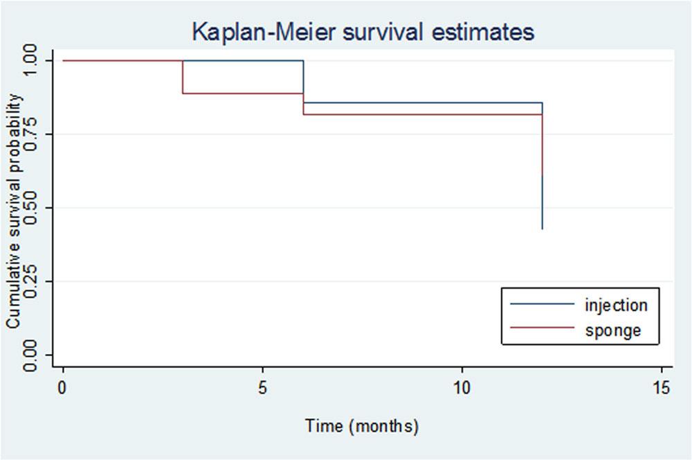
Table 3: Cumulative Probability of Success
| Visits | Survival probability | |
| Injection | Sponge | |
| Month 1 | 100.0% | 100.0% |
| Month 3 | 100.0% | 88.9% |
| Month 6 | 85.7% | 81.5% |
| Month 12 | 42.9% | 61.1% |
Table 4: Postoperative Complications and Procedures
| Postop complication | N |
| Malignant glaucoma | 1(11.1) |
| Bleb Needling | 3(33.3) |
| ALS | 4(44.4) |
| CD | 1(11.1) |
| Total | 9 |
ALS – Argon laser Suturelysis, CD – Choroidal detachment, N- Number
Leave a Comment