

Dr.Moreker Sunil Ratilal, M09512, Dr.Harshavardhan Ghorpade
Introduction: – Analysis of incidence, prevalence and incidence of misdiagnosed cases of sebaceous gland carcinoma in western region of the Indian Subcontinent with long term follow up of misdiagnosed and advanced meibomian gland carcinoma is missing in literature.
Sebaceous carcinoma is reported to be rare in the western population and is a potentially aggressive tumor but information about incidence, prevalence, risk factors, and prognosis of this disease has been traditionally based on isolated case reports of 70% of sebaceous carcinomas which are usually located in the head and neck region while sebaceous carcinomas are primarily periorbital and arise frommeibomian and Zeis glands and arethe fourth most common tumors of the eyelid which generally metastasize and spread to regional lymph nodes
Sebaceous carcinoma of the eyelid may be mistaken for inflammatory lesions, such as chalazion or blepharoconjunctivitis, resulting in a delayed diagnosis and poorer prognosis. This is the most confusing aspect of sebaceous carcinoma, its ability to mimic and masquerade as other lesions which makes diagnosis to be delayed by 1year to approximately 3 years which to a large extent may increase mortality
Poor prognostic factors include involvement of both upper and lower eyelids concomitantly and involvement of the upper lid, compared with the lower lid as well as lymphatic spread, multicentric disease, poorly differentiated tumors, pagetoid spread, or a highly infiltrative tumor
Given the rarity of sebaceous carcinoma and the relatively limited information available in the literature in India especially Western India we undertook an investigation to look at incidence prevalence and risk factors and prognostic factors
The rate of Sebaceous carcinoma varies according to race and in Caucasians is less than 1–5.5% of eyelid malignancies while in the Chinese and Japanese population it is the second most common eyelid malignancy with the highest reported rates being 39% and 37.5% respectively, whereas in the Indian population and in Singapore reported rates are in range of 31.2% and 10.2% respectively. The reason for higher rates in the Asian population could be genetic in nature
Methods: Epidemiological study of incidence, prevalence, demographic characteristics of misdiagnosed cases & results was done.
Statistical analysis
The continuous data were summarized as mean and standard deviation while discrete (categorical) in numbers (n) and percentage (%). The data were analyzed by independent Student’s t-test and Chi-square (χ2) test. A two-tailed (a = 2) P < 0.05 was considered statistically significant. Statistical analysis was performed using SPSS software (Windows version 16.0 IBM, Chicago).
Results: –Cases were analyzed across 12 hospitals in Mumbai and Navi Mumbai in corporate set up, charitable set up, medical college and standalone eye institutes
We report a rate of 41.2%. The most common was lower lid (56%).
Mean duration of symptoms was 11.4 months (range 3 to 19 months).
The mean size of tumour at diagnosis was 3.1± 0.4 cm.
The mean follow-up was 97 months.
Surgical methods used included wedge excision & primary closure, excision & skin grafting, or tarso-conjunctival flap and lid sharing procedures.
Odds of five-year survival were more if an excision with 10 mm clear margin was done.
Recurrent tumors were sensitive to Mitomycin C
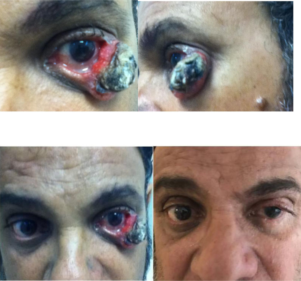
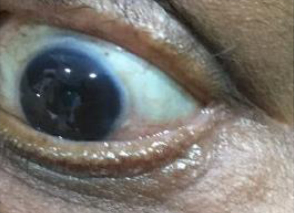
Sebaceous carcinoma masquerading as excoriation of lid, post brow ptosis,in one case was corrected with wide excision yet recurred though it was T 1 but was treated by personalized medicine done with genomic analysis following which three cycles of topical drops of mitomycin C and was completely cured at no recurrences at one year follow up. Mitomycin also worked against the resistant bacteria and the infection too was eradicated and at two years follow up patient had no recurrences and was tumor free and even lymph nodes negative and systemically there was no metastasis
Exoneration was necessary in one case
Successful treatment with lid sharing procedure with five year follow up:-
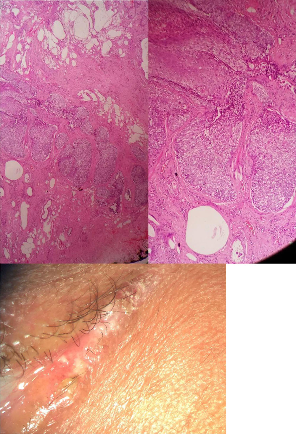
Pagetoid Spread in a case presenting with only excoriation of canthus: –
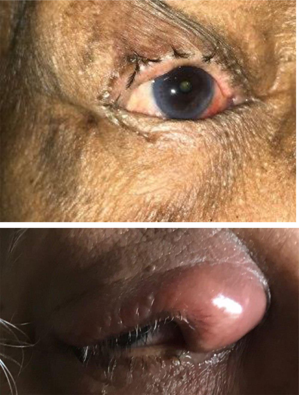
Otherpresentations
Recurrence :- upper and lower lid canthus kissing lesions
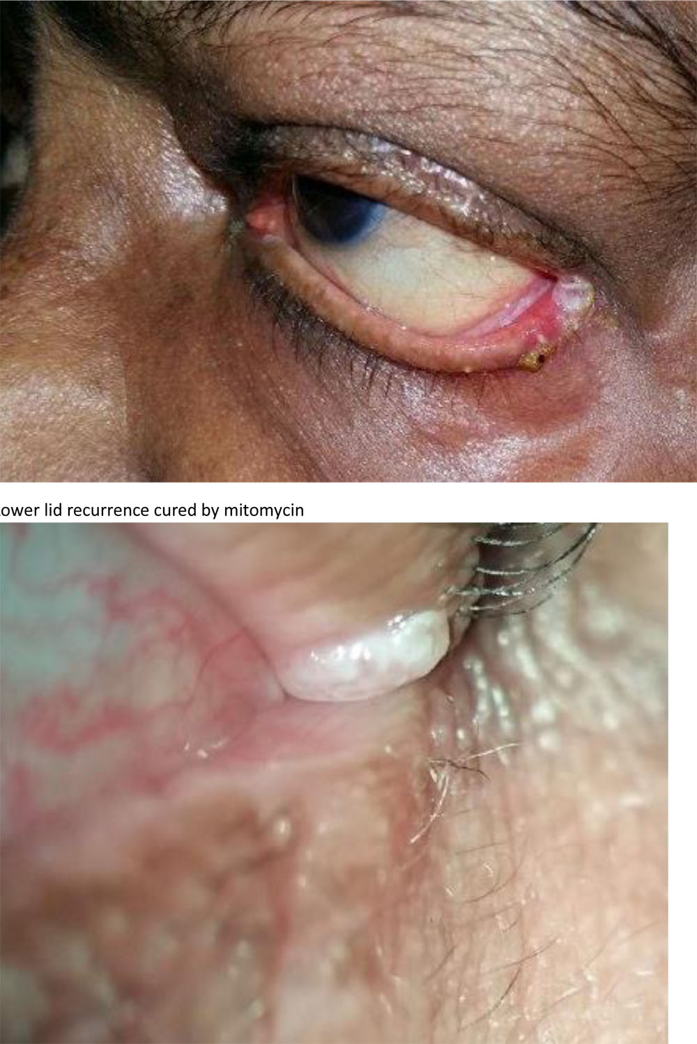
Both lower and upper lid lesions cured by Mitomycin C
Precision medicine can help in cases where one chemotherapeutic agent doesn’t work and one would want to know which agents will be the ones to which it will respond
Discussion:-Kass and Hornblassdescribed an the incidence in Western populations from 0.2% to 0.7% among all eyelid tumors and 1-5.5% among eyelid malignancies. Another analysis of eyelid malignancies other than basal cell or squamous cell carcinoma in Florida,reported the rate of sebaceous carcinoma in Caucasians as 0.5 per (million)10. However, reports of the incidence of sebaceous carcinoma is much higher in China with Ni reporting an incidence of 33% among all eyelid (malignancies). Shields et al recently reported their experience in 60 cases; the upper eyelid was involved in 75% of cases, lower eyelid (22%), caruncle (2%), and bulbar conjunctiva (2% = 1 case) 9. Of all cases 28 (51.9%) were seen on upper eyelid and 24 (44.4%) occurred on the lower eyelid, both lids were involved in 3 (5.5%) cases. Sebaceous gland carcinoma occurred predominantly on upper eyelid and rest of the malignant lesions had predilection for lower lid. We too found a predilection for lower lid. Reported risk factors for sebaceous carcinoma include advanced age Asian/South Asian race, women or history of irradiation to the head and neck in past and a genetic predisposition for Muir-Torre syndrome. The higher incidence we found of both sebaceous carcinoma and Muir Torre syndrome supports this hypothesis and also history of irradiation was seen in three patients.Preauricular and cervical lymph node infiltration is reported to be associated with a 50% to 67% 5-year mortality rate. Periocular and lymph node involved was lower
Conclusion:The incidence of sebaceous carcinoma is more than earlier reported rate of 31% in literature & this could be due to unmasking of masquerade cases in our setting due to high suspicion
References:-
- Jahagirdar SS, Thakre TP, Kale SM, Kulkarni H, Mamtani M. A clinicopathologicalstudy of eyelid malignancies from central India. Indian J Ophthalmol2007;55:109-12
- Hornblass A., Lauer S.A.: Sebaceous carcinoma of the eyelids ophthalmology 2005;111:1641
- Shields JA, Demirci H, Mar BP, et al: Sebaceous carcinoma of the eyelids: personalexperience with 60 cases. Ophthalmology 2004; 111:2151-2157.
- Straatsma, Bradley R. “Meibomian gland tumors.” AMA Archives of Ophthalmology56.1 (1956): 71-93.
- Yeatts RP, Waller RR: Sebaceous carcinoma of the eyelid: pitfalls in diagnosis.OphthalPlastReconstr Surg. 1985; 1(1):35-42.
- Shields JA, Demirci H, Marr BP, et al. Sebaceous carcinoma of the ocular region: a review. SurvOphthalmol. 2005;50:103-122.
- Nelson BR, Hamlet KR, Gillard M, et al. Sebaceous carcinoma. J Am AcadDermatol. 1995;33:1-15.
- Bailet JW, Zimmerman MC, Arnstein DP, Wollman JS, Mickel RA. Sebaceous carcinoma of the head and neck. Case report and literature review. Arch Otolaryngol Head Neck Surg. 1992;118:1245-1249.
- Chao AN, Shields CL, Krema H, et al. Outcome of patients with periocular sebaceous gland carcinoma with and without conjunctival intraepithelial invasion. Ophthalmology. 2001;108:1877-1883.
- Ni C, Guo BK. Pathologic classification of meibomian gland carcinomas of eyelids: clinical and pathologic study of 156 cases. Chin Med J (Engl). 1979;92:671-676.
- Abdi U, Tyagi N, Maheshwari V, et al. Tumours of eyelid: a clinicopathologic study. J Indian Med Assoc. 1996;94:405-409.
- Ni C, Searl SS, Kuo PK, et al. Sebaceous cell carcinomas of the ocular adnexa. IntOphthalmolClin. 1982;22:23-61.
- Rao NA, Hidayet AA, McLean IW, et al. Sebaceous carcinomas of the ocular adnexa: a clinicopathologic study of 104 cases, with 5-year follow-up data. Hum Pathol. 1982;13:113-122.
- Wolfe JT, Yeats RP, Wick MR, et al. Sebaceous carcinoma of the eyelid. Errors in clinical and pathologic diagnosis. Am J SurgPathol. 1984;8:597-606.
- Snow SN, Larson PO, Lucarelli MJ, et al. Sebaceous carcinoma of the eyelids treated by Mohs micrographic surgery: report of 9 cases with review of the literature. Dermatol Surg. 2002;28:623-631.
- Wick MR, Goellner JR, Wolfe JT 3rd, et al. Adnexal carcinomas of the skin. II. Extraocular sebaceous carcinomas. Cancer. 1985;56:1163-1172.
- Kivela T, Asko-Siljavaara S, Pihkala U, et al. Sebaceous carcinoma of the eyelid associated with retinoblastoma. Ophthalmology. 2001;108:1124-1128.
- Stockl FA, Dolmetsch AM, Codere F, et al. Sebaceous carcinoma of the eyelid in an immunocompromised patient with Muir-Torre syndrome. Can J Ophthalmol. 1995;30:324-326.
- Sweebe EC, Cogan DG. Adenocarcinoma of the meibomian gland; a pseudochalazion entity. AMA Arch Ophthalmol. 1959;61:282-290.
- Weedon D. Skin Pathology. 2nd ed. London, United Kingdom: Churchill Livingstone; 2002.
- Rao NA, McLean JW, Zimmerman LE. Sebaceous carcinoma of the eyelid and caruncle: correlation of clinicopathologic features with prognosis. In: Jakobiec FA, ed. Ocular and Adnexal Tumors. Birmingham, United Kingdom: Aesculapuis; 1978:461-476.
- Jensen ML. Extraocular sebaceous carcinoma of the skin with visceral metastases: case report. JCutanPathol. 1990; 17:117-121)
- Muqit MM, Foot B, Walters SJ, et al. Observational prospective cohort study of patients with newly-diagnosed ocular sebaceous carcinoma. Br J Ophthalmol 2013; 97:47
- Shet T, Kelkar G, Juvekar S, Mistry R, Borges A. Masquerade syndrome:sebaceous carcinoma presenting as an unknown primary with pagetoid spread to the nasal cavity. J Laryngol Otol. 2004 Apr;118(4):307-9
- Panda BB, Parija S, Pujahari S, Mallick J. Sebaceous Gland Carcinoma of Eyelid- A Tarnished Masquerade. Journal of Clinical and Diagnostic Research : JCDR. 2016;10(4):ND03-ND05. doi:10.7860/JCDR/2016/16405.7567.
- Bhardwaj M, Sen S, Sharma A, Kashyap S, Chosdol K, Pushker N, Bajaj MS,Bakhshi S. ZEB2/SIP1 as novel prognostic indicator in eyelid sebaceous gland carcinoma. Hum Pathol. 2015 Oct;46(10):1437-42. doi:10.1016/j.humpath.2015.05.026
- Ford J, Thakar S, Thuro B, Esmaeli B. Prognostic Value of the Staging System for Eyelid Tumors in the 7th Edition of the American Joint Committee on CancerStaging Manual. Ophthalmic PlastReconstr Surg. 2017 Sep/Oct;33(5):317-324.
- Shields CL, Naseripour M, Shields JA, Eagle RC Jr. Topical mitomycin-C for pagetoid invasion of the conjunctiva by eyelid sebaceous gland carcinoma.Ophthalmology. 2002 Nov;109(11):2129-33
Leave a Comment