

Dr.Mamta Agarwal, Dr.Jyotirmay Biswas
Abstract
Purpose –
To study the inciting factors, clinical features, diagnosis and treatment outcome in patients with infectious scleritis.
Methods –
Retrospective study between 2012 & 2017 at a tertiary care center in South India.
Results –
23 patients (23 eyes). All except 1 were immunocompetent. Mean age was 50.78 years (±16.35). Mean duration of symptoms was 53.14±35.66 days presenting as redness (100%), pain (86.96%) and decreased vision (39.13%). Risk factors included previous surgery in 6 eyes (26%) (Vitrectomy 4, scleral buckle 1, cataract surgery 1) and injury in 7 eyes (30.4%).Pre treatment mean visual acuity was 1.57 logmar(±1.22). Clinical presentation included 22 anterior and 1 posterior scleritis with multifocal lesions in 10 eyes (43.5%), unifocal in 10 eyes (43.5%) and diffuse in 3 eyes (13%). Associated uveitis was seen in 7 eyes (30.4%), keratitis in 6 eyes (26%) and exudative retinal detachment in 1 eye. The causative organisms included bacteria in 52%, fungus in 39%, herpes virus in 4.3%,and mixed in 4.3%. Bacterial pathogens were Nocardia(3), Mycobacterium tuberculosis(5),Streptococcus viridans,Pseudomonas aeruginosa,Burkholderia and Treponemapallidum each. Fungal pathogens were Aspergillusfumigatus (2), Penicillium,Paecilomyces and Candida albicans each.Average duration of symptoms was longer in fungal (55.22 ±39.93days) than bacterial scleritis (44.73 ±29.36days).
All patients were treated with antimicrobials. Surgical treatment included scleral debridement& patch graft. Average time of healing was longer in fungal (80.87± 53.4 days) than bacterial scleritis (62±46.32 days). Follow up range was 6 weeks to 4.5 years. 21(91.3%) eyes had complete resolution. 1 eye each required enucleation and evisceration. Mean post treatment visual acuity was 1.32 logmar(±1.28). Causes of decreased vision were corneal scar, glaucoma, macular scar and phthisis bulbi. On univariate analysis, poor visual acuity at presentation resulted in a worse visual outcome (P=0.062). The visual outcome was independent of the causative organism (P=0.802) or presence of multifocal abscesses (P=0.073). Surgical intervention did not result in better visual outcome than medical therapy alone (P=0.006).
Conclusion –
Early diagnosis and antimicrobial treatment can result in complete resolution of infectious scleritis but visual prognosis depends on other complications
Introduction
Scleritis is a severe painful inflammation of the sclera that may also involve adjacent ocular structures and present with a wide spectrum of clinical presentations and etiological factors. Scleritis can be idiopathic, autoimmune or infectious. Nearly 90% scleritis is autoimmune associated with systemic inflammatory conditions like rheumatoid arthritis, systemic lupus erythematosus, seronegativespondarthritides, graulomatosis with polyangiitis, relapsing polychondritis, polyarthritis nodosa and others*. Infectious scleritis accounts for 5-10% of all cases of scleritis* where it presents as an ulcerated or non ulcerated scleral nodule resulting from direct or indirect microbial invasion. The main risk factors for infectious scleritis are trauma or surgery most commonly pterygium surgery with beta radiation or mitomycin C, cataract surgery, vitreoretinal surgery, scleral buckle, glaucoma surgery, conjunctival neoplasm excision.
Few cases have also been reported in immunosuppression due to human immunodeficiency virus or chemotherapy*. While bacteria, fungus, viruses and parasites can cause infectious scleritis, Pseudomonas aeruginosa is the most common isolated pathogen in 51-81% of cases.* Other pathogens include Staphylococcus aureus, Streptococcus, Stenotrophomonas, Nocardia, Aspergillus, Fusarium, mycobacteria,andherpes virus.* However, due to similar clinical presentations, diagnosis of infectious scleritis is often missed. Systemic corticosteroid and immunosuppressives resolves autoimmune scleritis but poses a serious threat in infectious scleritis and worsens the visual outcome. The aim of our study was to toanalyse the inciting factors, clinical features, diagnosis and treatment outcome in patients with infectious scleritis in Indian population.
Materials and methods
This is a retrospective review of patients diagnosed with infectious scleritis examined at Uveitis & Cornea services, SankaraNethralaya, Chennai, India from August 2012 to December 2017. The study was conducted in accordance with the principles of Declaration of Helsinki.
All patients underwent a complete ophthalmic examination. Patients demographics and characteristics including age, sex, inciting factor for scleritis, duration of symptoms, past history of treatment, visual acuity pre and post treatment, presenting symptoms and signs, causative organisms, antibiotic susceptibility by Kirby-Bauer disc diffusion method, duration of oral and topical antibiotics, surgical intervention, time to resolution and complications were studied. Associated ocular comorbidities with systemic diseases were also recorded. Diagnosis of infectious scleritis was either presumptive or biopsy proven.
Presumptive diagnosis was based on history of herpes zoster or tuberculosis, scleral inflammation not responding to corticosteroid and immunosuppressives and ancillary investigations. Patients with ulcerative lesions underwent a scleral scraping with a No.15 surgical blade from the base and active edges of the ulcer under topical anesthesia in the out patient clinic and all non ulcerating nodular lesions were scraped in the operating room under peribulbar or topical anesthesia. In the latter cases, the scleral nodule was scraped after dissecting the conjunctiva and deroofing the sclera. Material collected was smeared on glass slides and stained with Grams stain, Potassium hydroxide + Calcofluor white stain, ZeihlNeelson stain using 20% H2SO4or modified ZeihlNeelson stain using 1% H2SO4. Acid fast stainingwas usually done if Grams stain smear was negative and lesions were strongly suspicious of bacteria.
Pus from the lesion was cultured on blood agar, chocolate agar, Sabouraud’s dextrose agar (SDA) and Brain-heart infusion broth. All media were inoculated at 37°C except SDA which was inoculated at 27°C. Microbial cultures were positive if there was confluent growth on one solid medium, or growth of the same organism on 2 or more media, or growth on one solid or liquid medium with the presence of same organism in smears. Antibiotic sensitivity test was also done by Kirby-Bauer disc diffusion method in cases of culture positivity. Treatment was based on smear results and when the smear was negative, empirical treatment was started based on clinical suspicion. The treatment was then changed based on culture, PCR and sensitivity reports.
All statistical analysis was performed using Stata software version.
Results
The study included 23 patients (23 eyes) with infectious scleritis seen between August 2012 and December 2017. All except one patient were immunocompetent. There were 13 men and 10 women. Mean age of presentation was 50.78 ±16.35years (range 21- 80 years). Follow up range was 6 weeks to 4.5 years.
Inciting factors for scleritis were seen in 65.2% (15/23) eyes which included trauma in 7 eyes (30.4%) and surgery in 7 eyes (30.4%) most common being vitrectomy (5 eyes) followed by scleral buckle and cataract surgery in 1 eye each. 1 patient with spontaneous infectious scleritis was HIV+ and 6 patients had diabetes mellitus. Mean duration of symptoms was 53.14±35.66 days. Mean duration of symptoms in scleritis post trauma was 2-8 weeks while post surgery was 3-10 weeks. 11 patients were treated with topical steroids, 5 patients with oral steroid & 1 patients was on azathioprine at the time of presentation.
The most common symptoms were redness in 100% of eyes (n=23), pain in 86.96% of eyes (n=20), decreased vision in 39.13% of eyes (n=9 eyes) and swelling in 21.73% of eyes (n=5 eyes). Themean visual acuity at presentation was 1.57 logmar (±1.22) (range PL – 6/6). 14 (60.86%) patients presented with vision less than 6/60. Clinical presentation included 22 anterior and 1 posterior scleritis with multifocal lesions in 10 eyes (43.5%), unifocal in 10 eyes (43.5%), and diffuse in 3 eyes (13%). Associated uveitis was seen in 7 eyes (30.4%), keratitis in 6 eyes (26%) and exudative retinal detachment with subretinal abscess in 1 eye.
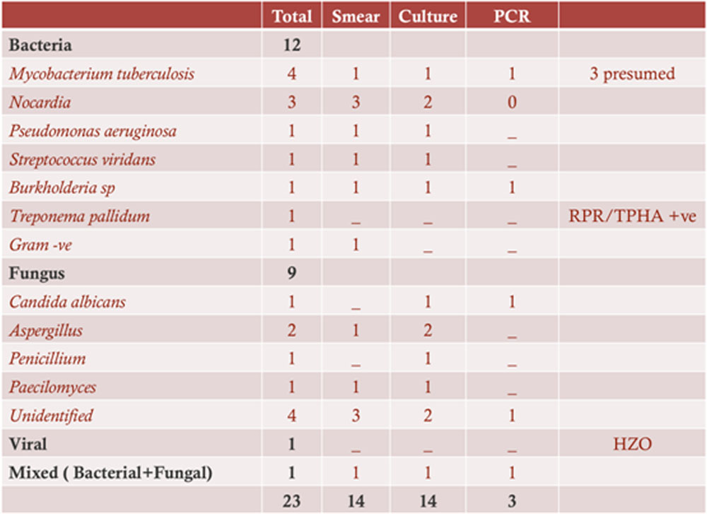
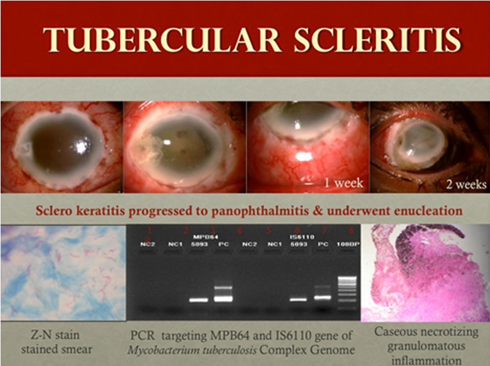
The causative organisms included bacteria in 52% (n=12 eyes), fungus in 39%(n=9 eyes), herpes virus in 4.3%(n=1 eye),and mixed in 4.3% (n=1 eye). Bacterial pathogens isolated on culture were Nocardia species(n = 3), Mycobacterium tuberculosis(n =1),Streptococcus viridans (n=1),Pseudomonas aeruginosa (n=1),Burkholderia (n=1) and Treponemapallidum(n=1). 3 patients were diagnosed as presumed tubercular scleritis based on mantoux test, quantiferon test and HRCT chest.Fungal pathogens isolated on culture were Aspergillusfumigatus (n=2), Penicillium (n=1),Paecilomyces(n=1),and Candida albicans(n=1).2 patients had positive KOH smear with no growth in culture and 2 patients had growth on culture but organisms could not be identified.Average duration of symptoms was longer in fungal (55.22 ±39.93days) than bacterial scleritis (44.73 ±29.36days).
All patients were treated with antimicrobials; topical antimicrobials in 87% (n=20), oral antimicrobials in 96% (n=23) and intravenous antimicrobials in 9% (n=2).Fungal scleritis was treated topically with natamycin 5%, voriconazole 1%, amphotericin 0.25% and systemically with voriconazole 200mg or ketoconazole 200mg or itraconazole 100mg twice daily. Nocardiascleritis was treated with topical fortified amikacin (50mg/ml), fluoroquinolones and oral trimethoprim(160mg) sulphamethoxazole (800mg) (TMX-SMZ) combination. Presumed tubercular scleritis was treated with oral antitubercular therapy with oral corticosteroid.
Treponemapallidum case was treated with intravenous benzathine penicillin 2.4 million units. Pseudomonas scleritis was treated with fortified gentamycin (14mg/ml), fluoroqinolones (moxifloxacin 0.5%) and intravenous gentamycin (40mg/ml). Herpetic scleritis with herpes zoster ophthalmicus was treated with oral valacyclovir 1mg twice daily with oral corticosteroid. Surgical intervention was done in 11 cases in the form of single or multiple debridement with antimicrobial wash in 5 eyes, corneal/scleral patch graft in 3 eyes, cyanoacrylate glue in 2 eyes, scleral buckle removal in 1 eye and therapeutic penetrating keratoplasty in 1 eye. 1 eye with fungal scleritis underwent evisceration and 1 eye with culture proven tubercular scleritis underwent enucleation with ball implant.
Average time of healing was longer in fungal (80.87± 53.4 days) than bacterial scleritis (62±46.32 days). Follow up range was 6 weeks to 4.5 years. 21(91.3%) eyes had complete resolution. 1 eye each required enucleation and evisceration. Mean post treatment visual acuity was 1.32 logmar(±1.28). Causes of decreased vision were corneal scar, glaucoma, macular scar and phthisis bulbi. On univariate analysis, poor visual acuity at presentation resulted in a worse visual outcome (P=0.062). The visual outcome was independent of the causative organism (P=0.802) or presence of multifocal abscesses (P=0.073). Surgical intervention did not result in better visual outcome than medical therapy alone (P=0.006).
Table 1
| Location | n (eyes) |
| Anterior | 22 |
| Posterior | 1 |
| Type | |
| Multifocal | 10 |
| Unifocal | 10 |
| Diffuse | 3 |
| Associated with Uveitis Keratitis Retinal Detachment |
7 7 1 |
Table 2
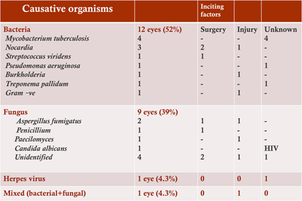
Table 3
Conclusion – Early diagnosis and antimicrobial treatment can result in complete resolution of infectious scleritis but visual prognosis depends on other complications.
References
- Jain V, Garg P, Sharma S. Microbial scleritis – experience from a developing country. Eye 2009;23(2):255-261.
- Jabs DA, Mudun A, Dunn JP, et al. Episcleritis and scleritis: clinical features and treatment results. Am J Ophthalmol. 2000;130:469-476
- Hodson KL, Galor A, Karp CL, Davis JL, Albini TA, Perez VL, Miller D, Forster RK.Epidemiology and visualoutcomes in patients with infectiousscleritis. 2013 Apr;32(4):466-72.
- Posterior scleritis associated with systemic tuberculosis Gupta A, Gupta V, Pandav SS, Gupta A.Indian J Ophthalmol. 2003 Dec;51(4):347-9.
- Latent tuberculosis related scleritis. Lhaj HA, Benjelloun A, Bouia Y, Bennouk Y, Mouzari Y, Kamouni YE, Kriet M. BMC Res Notes. 2016 Sep 20;9(1):446. doi: 10.1186/s13104-016-2251-8.
Figure 1
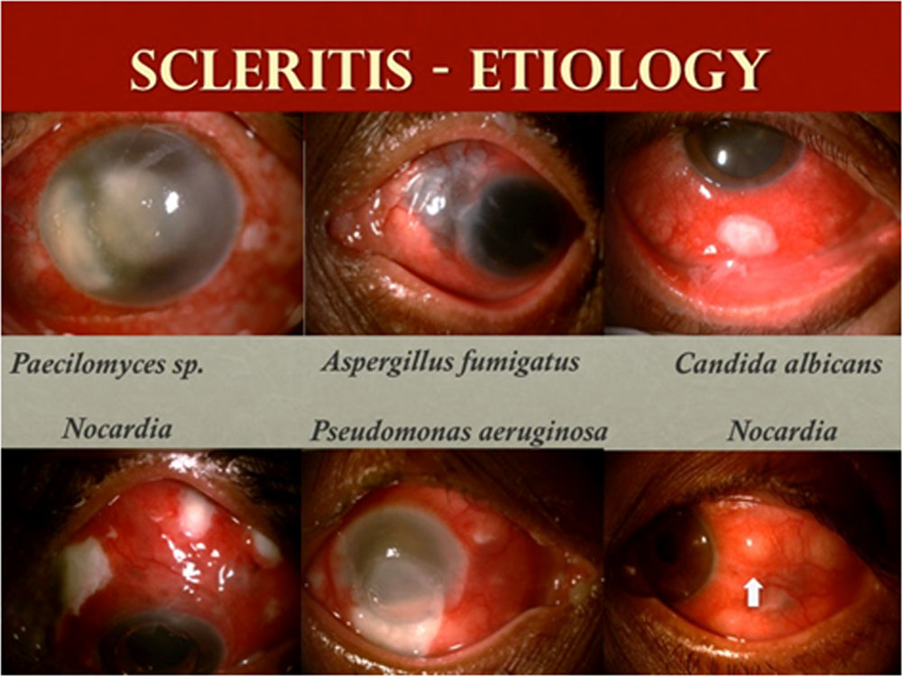
Figure 2 – Type of scleritis
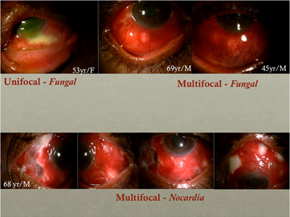
Figure 3 – Fungal scleritis
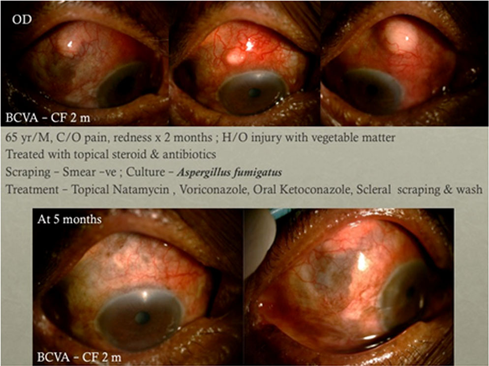
Figure 4 – Bacterial scleritis
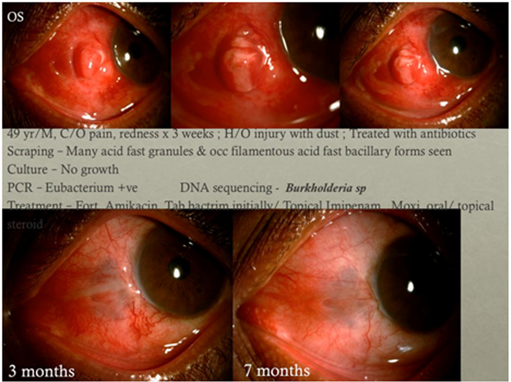
Figure 5 – Tubercular scleritis
Leave a Comment