

Dr.Sneha Kumari, S20179, Dr.Priti Sinha, Dr.Priyanka Bingi
ABSTRACT
AIM: To predict the window period of stroke (CVA) by analyzing ocular risk factors.
METHOD: A retrospective study was performed in all consenting patients with diagnosis of stroke made by the department of neurology between Dec 2017 to May 2018.
RESULTS: 24 cases with mean age of 60yrs were studied. 83.3% of the stroke cases showed ocular features of which 33% had Diplopia & 6thn palsy. Pre-stroke ocular findings were recorded from old records, it was found that 16.6% had HTN retinopathy for mean period of 18m & 16.5% had Diabetic retinopathy for mean period of 14m, retinal vasculopathy seen in 25% of patients which includes CRAO, CRVO for a period of 1-3m, and BRVO for 3-6m.
CONCLUSION: Ocular risk factors can predict imminent stroke. HTN retinopathy and Diabetic retinopathy are the long term risk factors (>1yr) whereas BRAO (<6m) and CRAO, CRVO (<3m) are short term risk factors. Hence early prediction can help in early management thus reducing the occurrence of stroke.
INTRODUCTION:
Stroke is a major health problem characterized by sudden onset of focal and neurological signs and symptoms caused by cerebrovascular origin by rapidly developing focal or global disturbances of cerebral function lasting >24hr.(1) Stroke was defined as a sudden focal neurologic deficit with a consistent cerebral ischemic or hemorrhagic lesion and it included transient ischemic attacks.
There is increasing evidence that small-vessel disease is a systemic vascular disorder that can be a major cause of stroke(2).Retinal vasculature is a circulatory system in the eye that can be observed without invasive procedures and provides useful information about the microcirculation system in the body(3).The retinal blood vessels, measuring 100 to 300um in size , offer a unique and easily accessible window to study correlation and consequences of cerebral microvascular diseases, because the retina and brain share similar anatomical and physiological properties. The retinal blood vessels are of the size and physiology similar to that of the cerebral small vessels(4). The quantified retinal microvascular abnormalities (HTN retinopathy, Diabetic Retinopathy, CRAO, CRVO, BRVO) might be useful as risk indicators for cerebrovascular disease(5). Some studies have shown that certain retinal microvascular abnormalities are associated with stroke and might act as surrogate markers for cerebral small-vessel diseases(6-7) .
In this study we aimed to predict the window period of stroke by above stated ocular risk factors.
- METHODOLOGY:
A Retrospective study was performed in all consenting patients with diagnosis of stroke made by the consultant of neurology unit of multispeciality hospital between December 2017 to May 2018.
INCLUSION CRITERIA:
- Conscious stable patients with diagnosis of stroke
- Patients with history of hypertension and/or diabetes
- Patients with recorded history of ocular co-morbidities like Hypertensive retinopathy, Diabetic retinopathy ,CRAO CRVO ,BRAO and BRVO.
EXCLUSION CRITERIA:
- Stroke patients with h/o trauma, unconscious patients, unstable patients.
- Patients with other systemic and comorbidities other than hypertension and diabetes.
- Patients without having previous complete ocular history records.
A total of 24 cases were included after taking informed consent. Patients data on age ,sex and history were taken regarding past significant medical and ocular pathology and detailed ocular examination was done.
Retinal vascular changes like HTN Retinopathy, Diabetic Retinopathy , CRAO, CRVO were recorded with duration of occurrence from old records. Also the time of occurrence of the stroke with other systemic findings were recorded. Window period of stroke is calculated by time of occurrence of first retinal vascular change to the time when diagnosis of stroke first made. Depending upon the calculated window period of stroke,the ocular risk factors into long term and short term risk factors. The ocular risk factor with window period more than 6m are classified as long term risk factors and those less than 6m are classified as short term risk factor.
After taking complete history patients were examined:-
Retinal vascular assesment:
Digital fundus photography was taken using a fundus camera(canon CR-Dgi with a 10D SLR digital camera)after pupil dilation using tropicamide .Retinal images were taken and confirmed the findings with the previous records of the patients.
RESULTS:
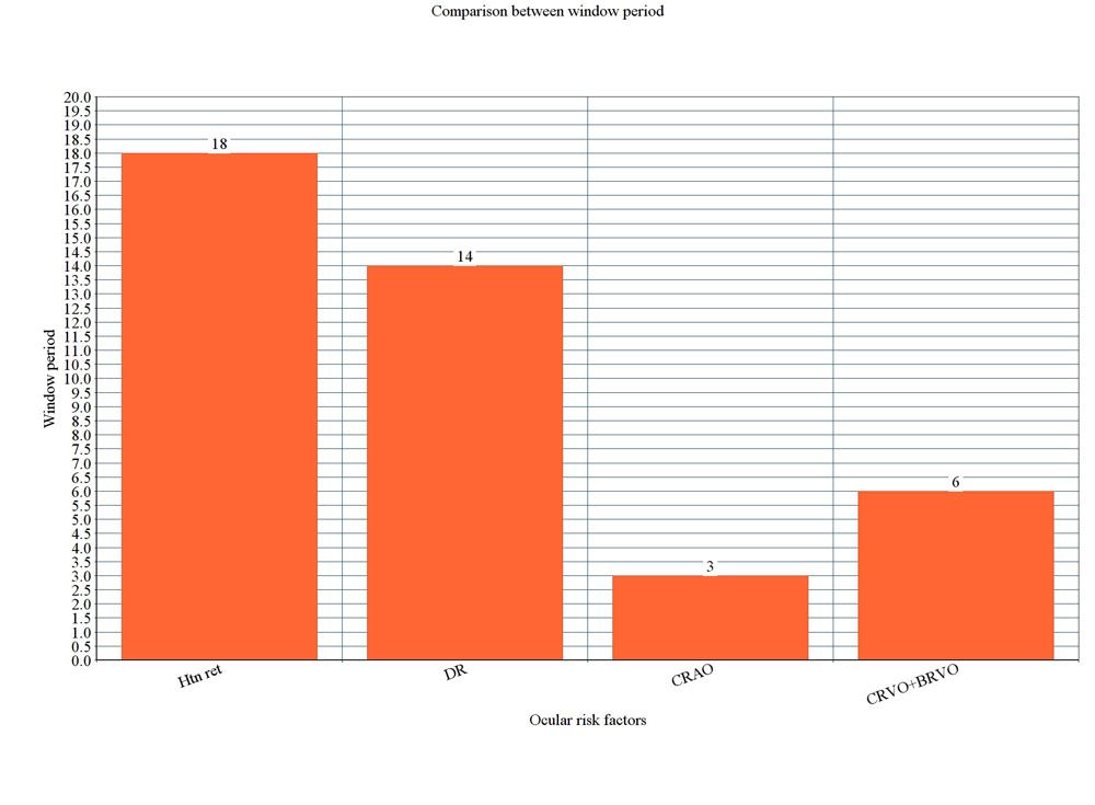
24 cases with mean age of 60 yr were included. Male: Female ratio- 3:1, Of these pre-stroke ocular findings from records shows 16.6% patients had HTN Retinopathy changes for mean period of 18 months and 16.5% had DR for period of 14 months, Retinal vasculopathy seen in 25% included CRAO,CRVO for period of 1-3 months and BRVO for 3-6 months.(table 1 and graph 1)
16.6% patients of total showed grade IV HTN Retinopathy changes for period of 18 months has visual aquity 6/36, Retinal images were taken with fundus camera and Retinopathy signs evaluated were retinal hemorrhages (blot and flame-shaped) microanerysms ,soft exudates, venous beading, optic disc swelling changes seen.
16.5% patients of total showed Diabetic retinopathy for period of 14 months has visual aquity 6/60,Retinal images were taken with fundus camera and findings evaluated for blot hemorrhages, hard exudates, neovascularisation of disc seen.
25% patients of total showed CRAO,CRVO for period of 3 months and BRVO for 3-6 months has Visual aquity counting figure 1 meter, Retinal images were taken with fundus camera and Retinopathy signs evaluated were occlusion of central retinal artery ,central retinal vein ,branch retinal vein, hemorrhages seen.
Table 1 and Graph 1
| ocular risk factors | % |
| HTN retino | 16.6% |
| Diab retin | 16.5% |
| CRAO + CRVO | 9% |
| BRVO | 16% |
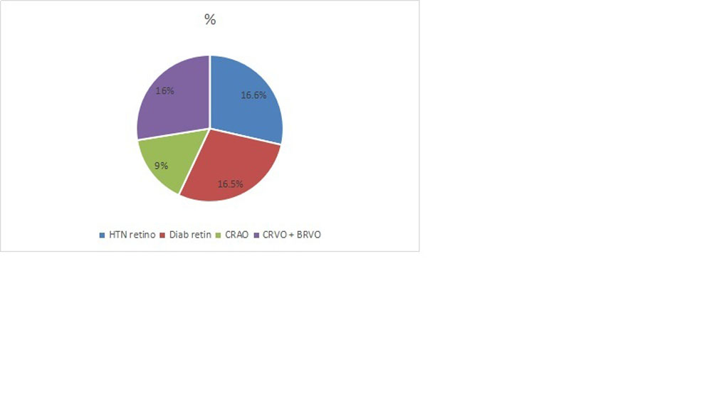 Table 2 and Graph 2:-
| Ocular Risk Factors | ||||
| Htn Retinopathy | DM Retinopathy | CRAO+ CRVO | BRVO | |
| Window period | 18 months | 14 months | 3 months | 6months |
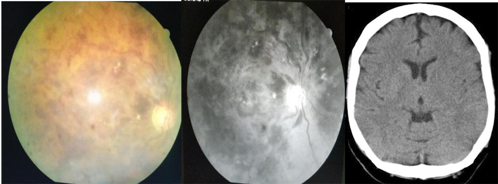
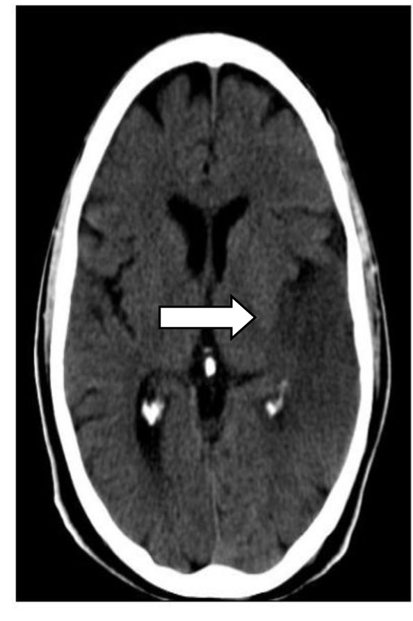
Coloured and redfree photograph of same patient with CRVO and CT image showing hypodensity of lenticular nucleus
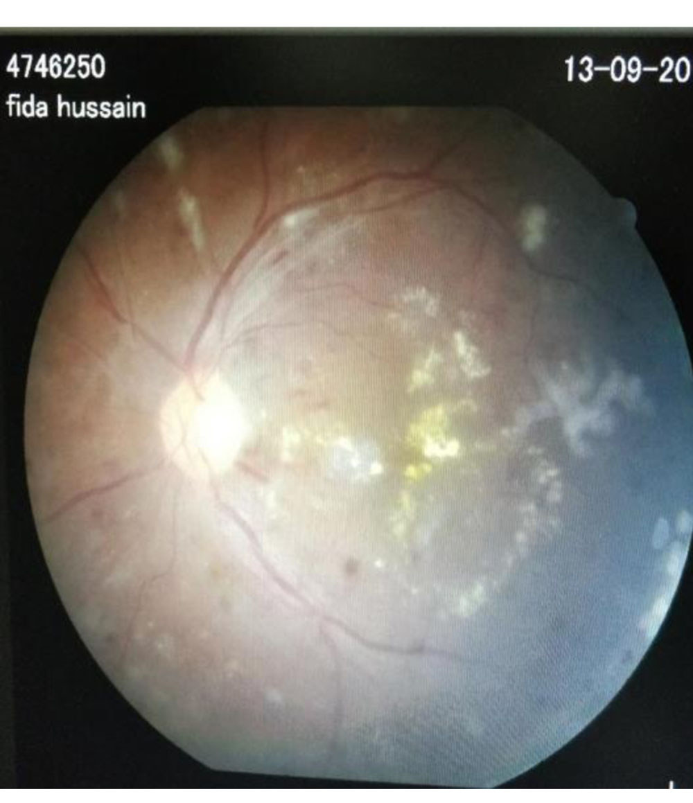
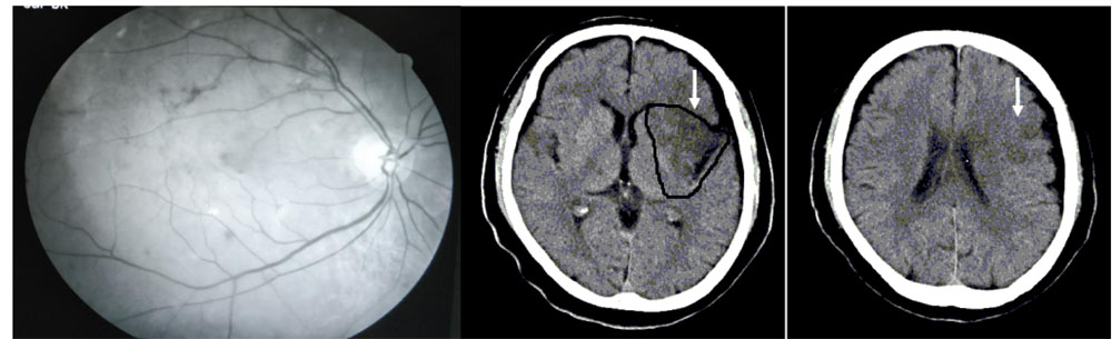
Fundus photograph of patient with diabetic retinopathy and CT image of the same patient showing ischemic changes in MCA region
Redfree Fundus photo of pt with BRVO (resolving) and CT finding of same patient showing hemorrhagic stroke which resolved over time.
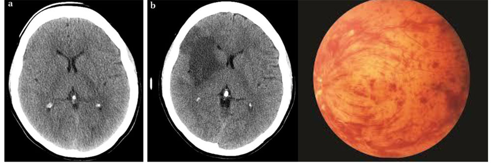
Axial CT of patient presenting with ischemic stroke and fundus finding of same patient showing CRVO.
DISCUSSION:
In this study we predicted the window period of stroke by ocular risk factors and classified them into long term and short term risk factors causing future risk of stroke.
In our study we observed that CRAO, CRVO, BRVO were the risk factors and also the short term risk factors ,as they cause future stroke within 1-3 months in CRAO,CRVO patients and within 3-6 months in BRVO patients.We found that these patients with ocular risk factors exhibited a higher risk of future stroke. In our study patients who had hypertensive retinopathy exhibited stroke after the window period of >1yr. So classified as low risk and long term predicting factor of stroke. Diabetic retinopathy exhibited the stroke after the window period >1yr so classified as low risk and also long term predicting factor of stroke.
Studies have also shown that patients with retinal occlusion are also at higher risk for stroke. Patients with RAO have higher prevalence of TIA as well as stroke(8).Recently a population based study in Taiwan found that 19.6% of patients with history of RVO went on to suffer an attack of stroke ,while only 10% of control group patients suffered from stroke(9).Stroke risk was particularly high in the first month following the artery occlusion and most strokes occurred within six months, suggesting the need to educate patients at the time of retinal finding. The study also found that patients with CRAO had higher rates of stroke than patients with BRAO(9).
Retinal emboli can be asymptomatic , but can also cause more severe visual sequelae in cases of retinal artery occlusion. With regard to stroke risk, the Beaver Dam Eye study found that retinal emboli were associated with threefold greater risk of fatal stroke over eight years(10).The Blue Mountain Eye study found that over 12yrs 30% of patients with retinal emboli died-with 4%of the deaths resulting from stroke. There was a moderate threefold increase in stroke related mortality rates with emboli(11).In general patients with emboli have a higher mortality rate compared to those without emboli.
A large population-based study in the United States recently found that the event rate for stroke in patients with RVO was increased approximately 2- fold relative to controls(12).
A well-designed population-based study recently reported that RVO was significantly associated with carotid artery plaque formation,a potential risk factor for ischemic stroke(13). Hypertensive retinopathy changes such as arteriolar nicking ,artery-vein nicking and artery opacification, have been shown to increase the risk of stroke in the Atherosclerosis Risk in communities (ARIC) study, artery vein nicking and artery narrowing were shown to increase the odds of MRI-defined subclinical infarction by nearly twofold(14).Severe retinopathy changes such as flame–shaped hemorrhages, cotton–wool spots exudates, optic nerve swelling were shown to increase relative risk of incident stroke two to three times compared to individuals without these retinal findings(14).
The ARIC found the risk incident stroke was two-three times higher in individuals with non-proliferative diabetic retinopathy vs individuals without diabetic retinopathy. In addition , the level of retinopathy also seemed to correlate with stroke risk. Of the 1,305 individuals in one study who had no diabetic retinopathy, only 3.9% went to on suffer an ischemic stroke .In contrast ,9.6% of individuals with mild to moderate diabetic retinopathy and 11.4%of individuals with severe diabetic retinopathy went on to suffer a stroke. Diabetics who have proliferative diabetic diabetic retinopathy (PDR) are not only at risk for devastating ocular sequale if not properly treated ,they are also at increased risk for stroke. The Wisconsin Epidemological Study in Diabetic Retinopathy found the risk of the stroke was six times higher in patients with PDR, and the risk of stroke mortality was doubled compared to patients without PDR(15).
It is important to note that in our study confounding factors like treatment and control of diabetes, hypertension, lipid profile were not take into consideration. Also due to small sample size application of its result to general population needs further evaluation.
CONCLUSION:
Ocular risk factors can be used to predict imminent stroke. HTN
Retinopathy and DR preceded stroke for a period >1yr of their occurrence. BRAO preceded stroke <6months of its occurrence and CRAO,CRVO within < 3months of its occurrence. So from above study it was found that HTN Retinopathy and DR are the long term risk factors whereas CRAO,CRVO,BRVO are short term risk factors. Hence early prediction of these ocular risk factors helps in early management and thus reducing the occurrence of stroke.
REFERENCES:
1.Alein CM ,Luek CJ,Dennis M,College NR,Walker BR,Ralston SH.Davidson’s principles and practice of medicine.21sted.Edinburg:Churchill-Livingstone;2010.P.1180-9. 2)Hagg S,Thom LM,Putaala J,Liebkind R,Harjutsalo V,Forsblom CM,Gordin D,Tatlisumak T,Groop PH:FinnDiane study Group.Incidence of stroke according topresence of dabetic nephropathy and severediabetic retinopathy in patients with type 1 diabetes.Diabetes Care 2013:36(12):4140-4146.
3)Gopinath B,Chiha J,Plant AJ,Thiagalingam A,Burlutsky G,Kovoor P, Liew G,Mitchell P.Associtions between retinal microvascular structure and the severity and extent of caronary artery disease.Atherosclerosis 2014:236(1):25-30.
4)Patton N,Aslam T,Macgillivray T,Pattie A,Deary IJ,Dhillon B,Retinal vascular image analysis as a potential screening tool for cerebrovascular disease:a rationale based on homology between cerebral and retinalmicrovasculature.J Anat 2005:206(4):319-348.
5)Ding J,Wai KL,McGeechan K,Ikram MK,Kawasaki R,Xie J,Klein R,Klein BB,Cotch MF,Wang JJ,Mitchell P,Shaw JE,Takamasa K, Sharrett AR,Wong TY;Meta-Eye study Group.Retinal vascular caliber and the development of hypertension:a meta analysis of individual participantdata.J Hypertens 2014:32(2):201-215. 6)Williams MA.McGowan AJ,Cardwell CR,Cheung CY,Craig D,Passmore P,Silvestri G,Maxwell AP,McKay GJ.Retinal microvascular network attenuation in Alzeimers disease.Alzheimers Dement(Anst)2015:1(2):229-235.
7).Jung HW,Kim SW,Yoon SJ,Choi JY,Kim KI,Kim CH.Associations betwwen frailty,retinal microvascular changes,and cerebral white matter abnormalities in Korean older adults.JamGeriatrSoc 2014;62(11):2209-2210.
8)Hayreh SS,Podhajsky P,Zimmerman MB.Retinal artery occlusion:associated systemic and ophthalmic abnormalities.Ophthalmology.2009 Oct:116(10):1928-36.
9)Chang YS,Jan RL,Weng SF,et alRetinal artery occlusion and the 3-year risk of stroke in Taiwan:a nationwide population based study.Am J Ophthalmol.2012 Oct:154(4):645-52.
10) Klein R,Klein BE,jensen SC,et al,Retinal emboli and stroke:the Beaver Dam Eye Study.Arch Ophthalmol1999 Aug:117(8):1063-8.
11)Wang JJ,Cugati S,Knudtson MD,et al.Retinal arteriolar emboli and longterm mortality:pooled data analysis from two older populations.Stroke.2006 jul:37(7):1833-6.
12)Werther W,Chu L, Holekamp N,et al.Myocardial infarction and cerebrovascular accident in patients with retinal vein occlusion.Arch Ophthalmol 2011:129:326-31.
13)Wong TY,Larsen EK,Klein R,et al.Cardiovascular risk factors for retinal vein occlusion and arterial emboli:the Atherosclerosis Risk in Communities & Cardiovascular Health Studies.Ophthalmology 2005:1125:540-7.
14)Cooper LS,Wong TY,Klein R,et al.Retinal microvascular abnormalities and MRI-defined subclinical infarction:the Atherosclerosis Risk in Communities study.Stroke 2006 jan 37(1):82-6.
15)Klein R,Klein BE,Moss SE,Epidemology of proliferative diabetic retinopathy,Diabetic care ,1992 Dec:15(12):1875-91.
Leave a Comment