

Dr. Prerna Agrawal, A17847, Dr. Ashutosh Kumar Singh, Dr. Ashok Kumar Grover
CYSTICERCOSIS-EPIDEMIOLGY
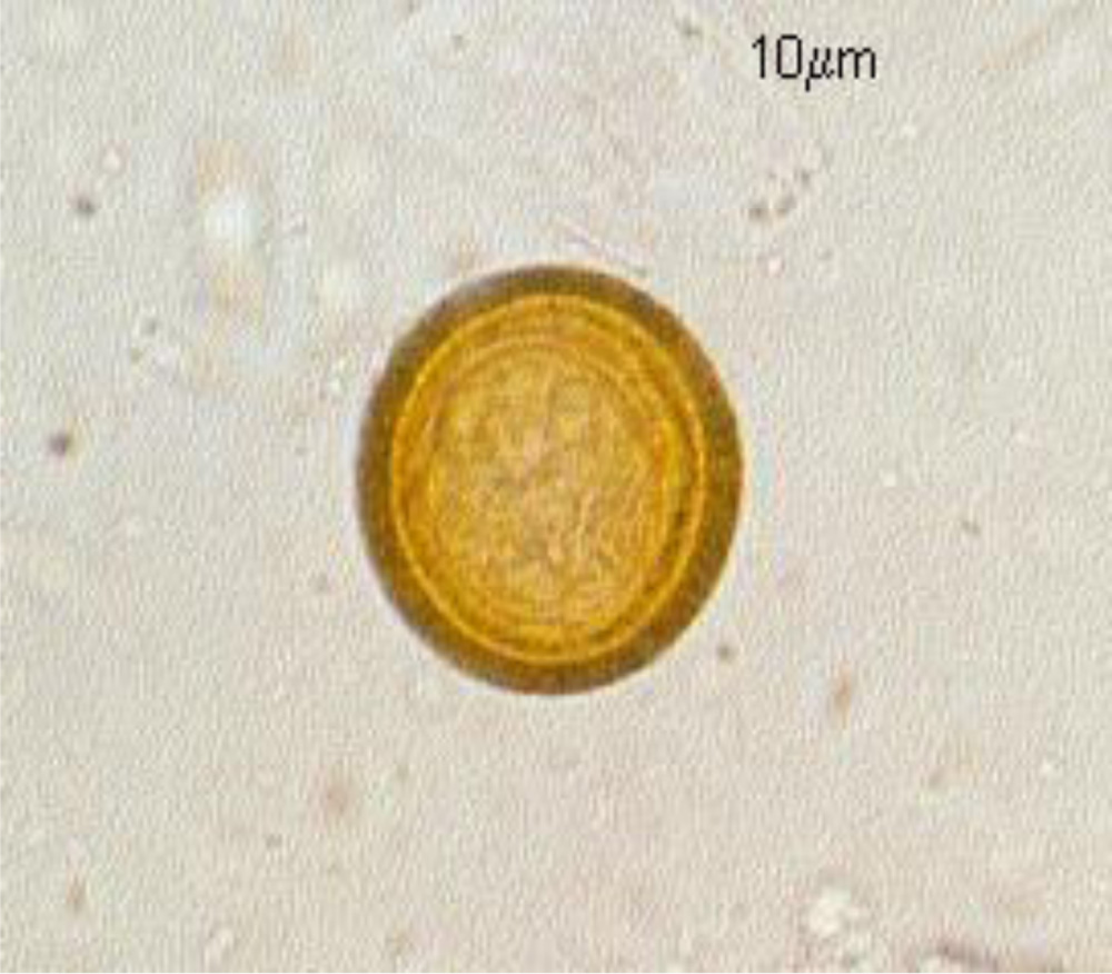
Caused by larval form of intestinal parasite Taenia solium
1. Prevalent in Eastern Europe, Central America, Mexico and Asia
2.Commoner in children and young adults.
LIFE CYCLE
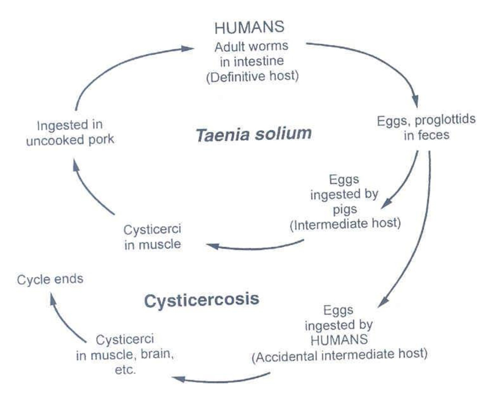
Source of human cysticercus infection
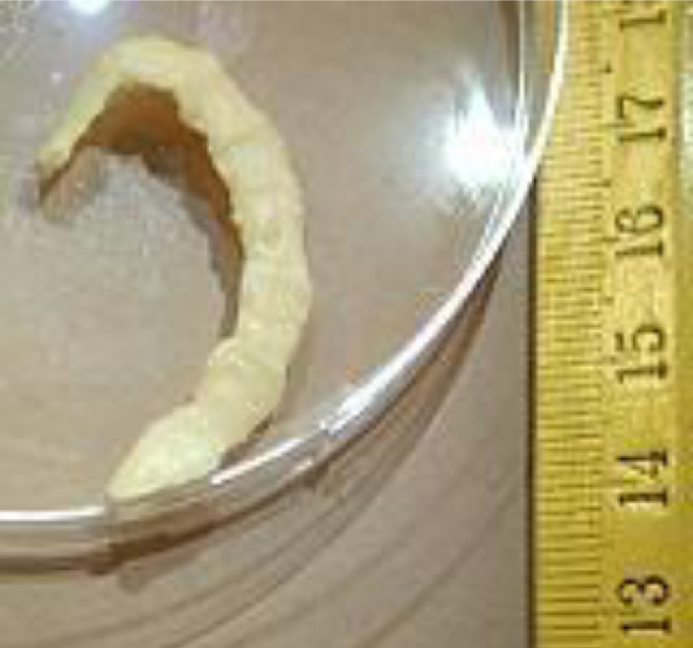
1. Ingestion of contaminated food-
* contaminated water
* vegetables,salads
2. Autoinfection-ingestion of eggs from intestinal parasite by reverse peristalsis
SYSTEMIC INVOLVEMENT
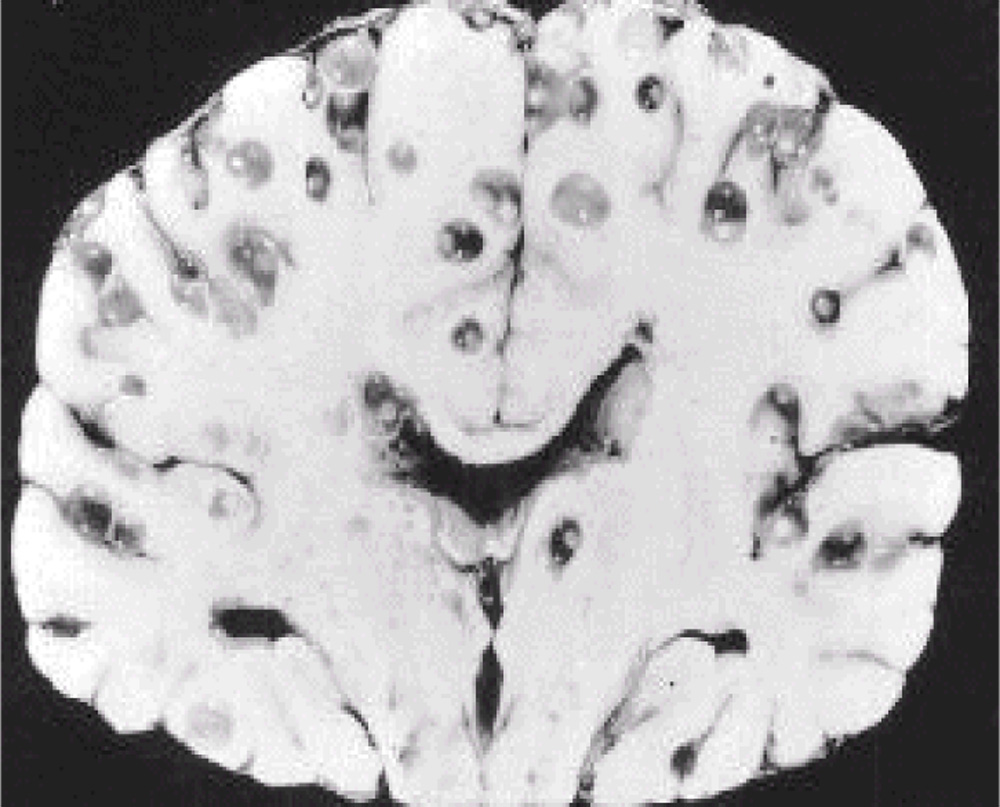
1. CNS
2. SUBCUTANEOUS TISSUE
3. SKELETAL MUSCLE
4. EYE
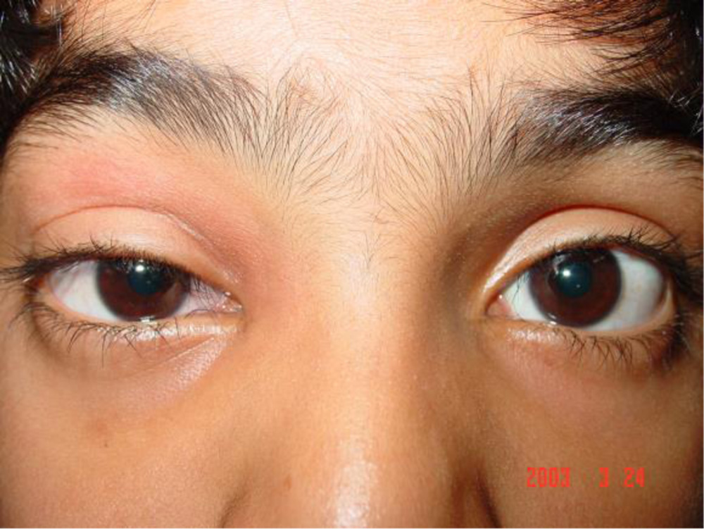
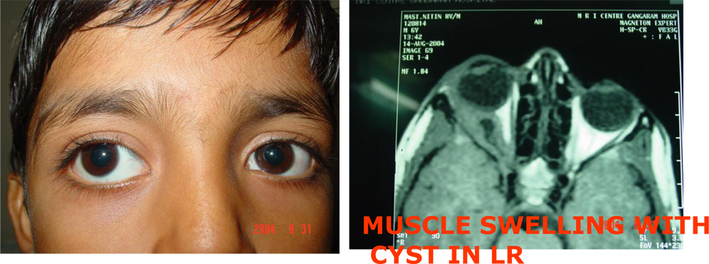
8 YR BOY WITH PROPTOSIS
MATERIALSAND METHODS
1. A retrospective study of 178 pa-tients of orbital myocysticercosis over a quarter century till March 2018 presented.
2. Slight male predilection(M-101,F-77)
3. Age 5 years to 48 years
RESULTS
ORBITAL MYOCYSTICERCOSIS- CLINICAL PRESENTATION (n-178)1. Ocular motility restriction with diplopia -138(77.52%)
2. Recurrent episodes of redness, swelling, pain -89(50%)
3. Proptosis -67(37.64%)
4. Recurrent Ptosis -26(14.60%)
5. Subconjunctival cyst -19(10.67%)
6. Presentation with picture like cellulitis-14(7.86%)
Extra ocular Myocysticercosis
(n-178)
1. EXTRAOCULAR MUSCLES
1. LATERAL RECTUS -74(41.57%)
2. MEDIAL RECTUS -51(28.65%)
3. SUPERIOR RECTUS -26(14.60%)
4. INFERIOR RECTUS -25(14.04%)
5. SUPERIOR OBLIQUE -1(0.56%)
2. LACRIMAL GLAND Nil
3. OPTIC NERVE -1(0.56%)
DIAGNOSIS
ANCILLARY INVESTIGATIONS
-
- Stool test-ova in rare cases
- Haematology- Eosinophilia
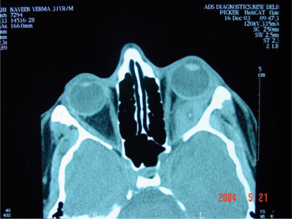
CT SCAN
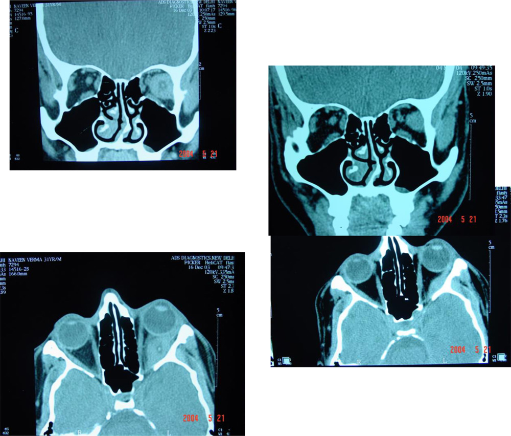
Shows hypodense cyst with hyperdense Scolex and muscle thickening with fuzzy margins
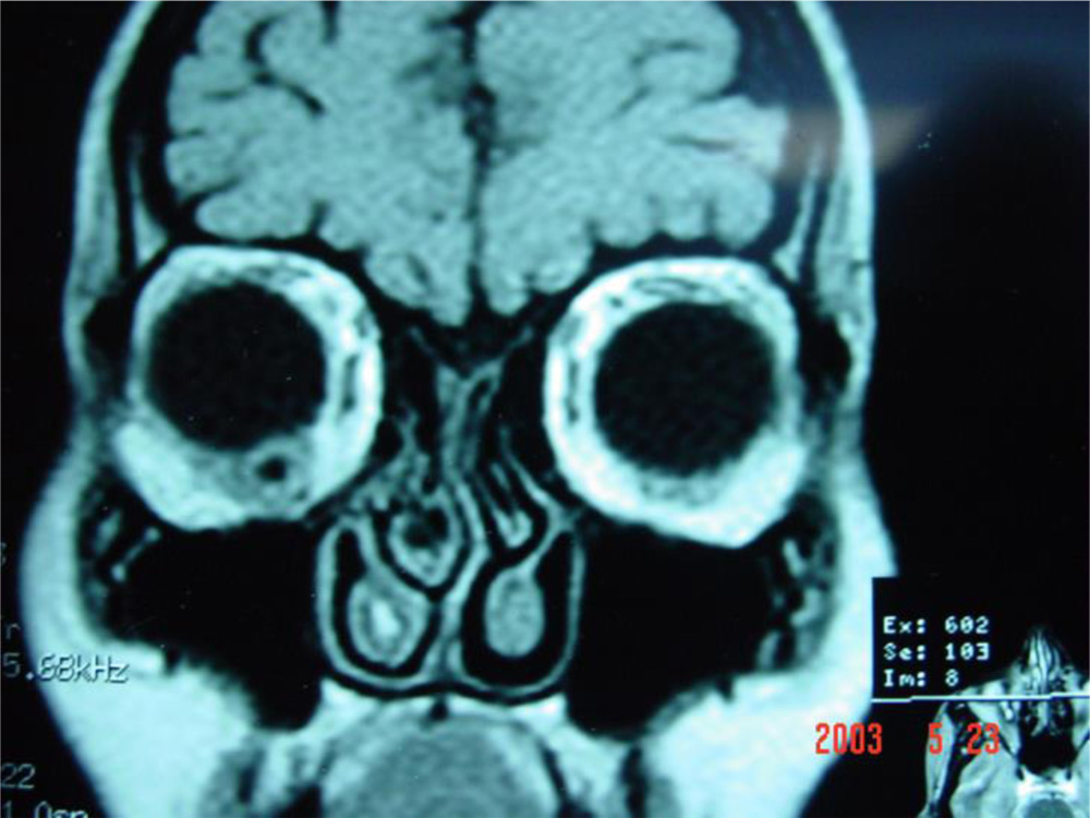
MRI
1. Good soft tissue delineation
2. -demonstates cyst with surrounding inflammation, causing muscle thickening.
ULTRASONOGRAPHY
1. A-scan ultrasonography shows high amplitude spikes corresponding to scolex and cyst wall.
2. B scan delineates the cyst and the scolex
3. Excellent for follow up
MANAGEMENT OF MYOCYSTICERCOS
1. Medical treatment is the mainstay of Management today
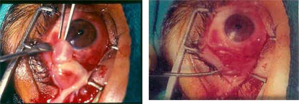
2. Surgical removal was the routine till 1993
MEDICALMANAGEMENT PROTOCOL
1. Albendazole- 30mg/kg/day for 15 days in divided doses
2. Oral steroids- Low dose ( 5-10 mg/day)
Medical management
1. Effective in 95% cases with a minimum follow up of 10 weeks with regression of clinical and imaging parameters
2. Clinical response begins to be noted in 1 to 2 weeks in most cases,full response may take 3 months or more
3. Alterations on imaging are noted in a few weeks,but regression of cyst may take upto 9 months
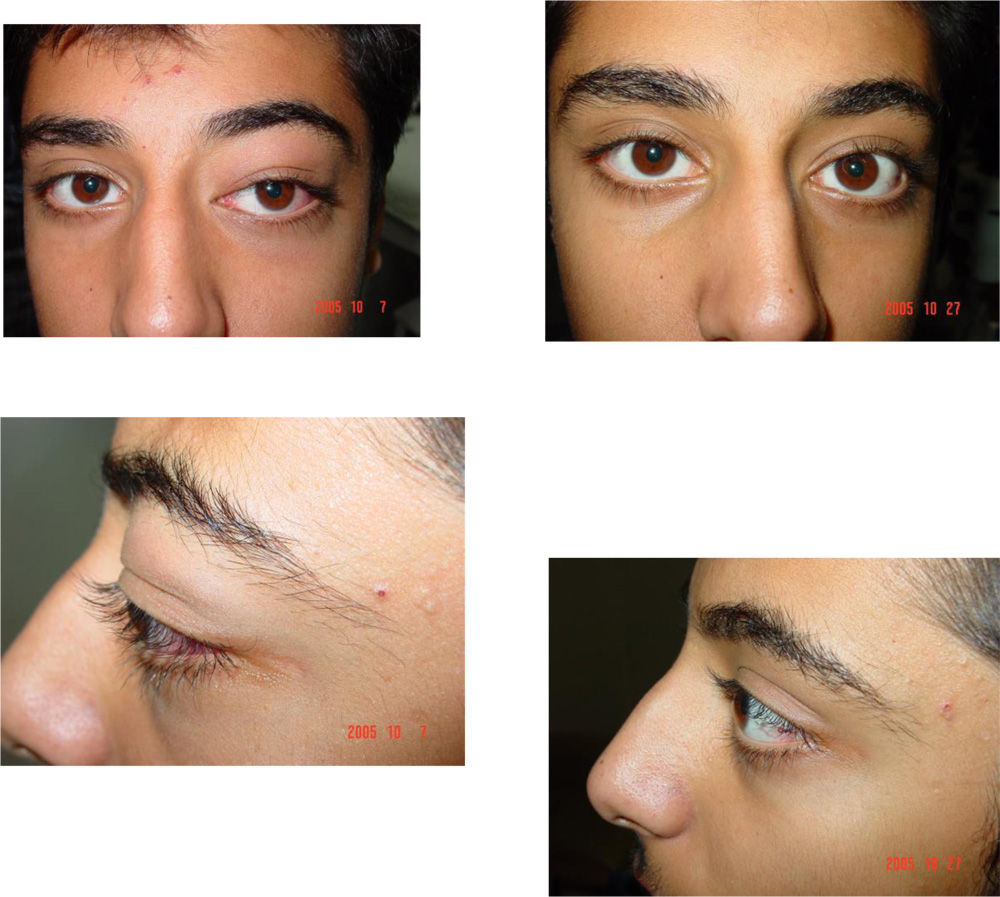
Medical Management Pre treatment Post treatment
Medical Management
Pre treatment Post treatment
CONCLUSION
1. The lessons for diagnosis and management based on one of the largest series in the world are presented.
2. A high index of suspicion for Myocysticercosis is needed in cases in or coming from endemic zones
3. Diagnosis can be easily established by imaging techniques
4. Medical treatment with Albendazole is an effective modality for treatment
THANK YOU
Leave a Comment