

Dr.Sriram Simakurthy, S18123, Dr.Rajesh Ramanjulu, Dr.Pradeep Sagar, Dr.Mahesh Shanmugam P
Abstract
Purpose: To assess the role of optical coherence angiography (OCTA) in monitoring the response to therapy in intraocular tumors
Methods: Retrospective series of intraocular tumors (vascular and non-vascular such as haemgioma, melanoma, astrocytoma) using OCTA both pre and post treatment
Results: Vascular tumors such as capillary haemangioblastoma and choroidal haemangioma showed reduction in intrinsic vascularity which was evident before morphological changes. In melanoma reduction in intrinsic vascularity was noted along with clinical reduction in size. In astrocytoma there was increase in vascular density which co related with increased exudates only in early post treatment period.
Conclusions: Changes in intrinsic vascularity of tumour visible on OCTA parallels treatment response and can be seen prior to change in size of the tumour or change in surface features. It can also identify inadequacy of treatment and guides in re-treatment.
Key words: OCTA, intraocular tumors
—————————————————————————————————-
Introduction
Vascular tumors of retina include cavernous hemangioma of retina, peripheral and juxtapapillary retinal capillary haemangioblastoma (RCH). Larger RCH needs multiple sessions of treatment and it is preferable to treat these tumors at an earlier stage when they are small.[1] Vascular tumors of choroid include solitary and diffuse choroidal hemangioma (DCH), and needs treatment if associated with exudative retinal detachment (RD). Pigmented tumours include melanoma and melanocytoma which if not correctly diagnosed and followed can have disastrous consequences.Optical coherence tomography angiography (OCTA) can identify the intrinsic vascularity of RCH[2] and choroidal hemangioma.[3]OCTA can also be used to differentiate between melanoma and melanocytoma based on vascular signal density as melanocytoma presents with signal void areas. Hence OCTA could help in identifying post-treatment changes in intrinsic vascularity of these tumors.
Methodology
This is a retrospective series of various intraocular tumors. Pretreatment and post treatment color photograph, OCT and OCTA of tumors were captured using swept source OCTA (Topcon DRI OCT Triton, Topcon Corporation, Japan). The available software in the swept source OCTA can measure the capillary density only in the superficial capillary plexus slab. Hence the segmentation lines in the superficial capillary plexus slab were modified manually to include the entire thickness of tumor that could be captured.
Results
In retinal capillary haemangioblastoma serial color photograph monitoring doesn’t show any change in tumor size but there is decrease in the macular edema at 2 months post laser photocoagulation, while OCTA shows the reduction in intrinsic vascularity of tumor (figure 1), similar response was noted even in cases of diffuse choroidal haemangioma post radiotherapy (figure 2). In a case of juxtapapillary melanoma, which was treated with Ru plaque brachytherapy, there was a decrease in the size of the tumor evident on color photographs and corresponding visits OCTA showed reduction in intrinsic vascularity as well as vessel caliber (figure 3). A case of retinal astrocytoma who underwent photodynamic therapy showed fluctuation in amount of exudation, while there was constant increase in amount of intrinsic vascularity which may indicate progression of disease and treatment failure (figure 4).
Discussion
In large RCH, decrease in capillary leak, as demonstrated by resolution of macular edema was evident after treatment as early as two months,but no significant changes in tumor morphology could be appreciated.OCTA could detect decrease in Mean tumor vascular density, but the reduction in vascular density was minimal, suggesting that laser photocoagulation (LP) would decrease capillary leak by mechanisms other than capillary closure. Exudation from RCH is due to the fenestrated nature of endothelial cells. [4] It would be possible that LP would alter the endothelial fenestrations or would induce changes in the basement membrane. In an experimental study, increase in number and length of choroidal endothelial cell processes was noted five days after retinal LP. This was associated with increase in thickness of endothelial basement membrane.[5] Hence it is possible that thickening of basement membrane can decrease leakage from the tumor and it appears inessential to destroy the intrinsic vasculature to achieve the same. Decrease in the tortuosity of larger vessels within the lesion was noted in our case, similar to the case reported in literature. [6]
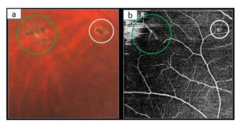
In our case, complete resolution of abnormal vessels was appreciated on OCTA in one small RCH (figure 5). Hence it is likely that LP can lead to capillary closure. Laser may not penetrate adequately in thicker tumor to achieve capillary closure while it is possible in smaller lesions. It is possible that milder burns lead to remodelling of capillary architecture and severe burns lead to capillary closure. Vascular loop was identified post-treatment in one small RCH, suggesting that OCTA can identify incomplete treatment.
In DCH, reduction in exudative retinal detachment and decrease in choroidal thickness were noted two months after treatment. Radiotherapy leads to apoptosis of endothelial cells and sloughing in early phase. Smooth muscle and endothelial proliferation occurs in late phase and leads to perivascular fibrosis. [7] The reduction in vascularity would parallel reduction in thickness of tumor. But the reduction in vascular density identified on OCTA was minimal. Hence there is a possibility that radiation induced vascular remodelling would decrease leak.
In non-vascular tumors such as melanoma and astrocytoma response to treatment is usually assessed by looking at co parameters such as tumor size, associated exudation, but in our analysis we noted that even though the exudate comes down there can be progressive increase in the vascular density of the tumor which in long term follow up may cause recurrence of the tumor as was evident in case of astrocytoma.
Changes in OCT characteristics, such as resolution of exudation, serve as early markers of response to therapy in intraocular tumors. But decrease in vascular density precedes morphological changes and also helps in assessment of response to treatment. Short comings of our study include retrospective nature and small sample size. Study of OCTA changes in early post-treatment period (prior to OCT changes) would help in better understanding. OCTA helps in identifying treatment inadequacy and understanding alternate mechanism involved in treatment response in vascular tumors.
References:
- Shanmugam PM, Ramanjulu R. Vascular tumors of the choroid and retina. Indian J Ophthalmol. 2015; 63:133-140.
- Sagar P, Rajesh R, Shanmugam M, Konana VK, Mishra D. Comparison of optical coherence tomography angiography and fundus fluorescein angiography features of retinal capillary hemangioblastoma. Indian J Ophthalmol. 2018; 66:872-6.
- Konana VK, Shanmugam PM, Ramanjulu R, Mishra KCD, Sagar P. Optical coherence tomography angiography features of choroidal hemangioma. Indian J Ophthalmol. 2018; 66:581-3.
- Green WR. Retina: capillary hemangioma. In: Spencer WH, ed. Ophthalmic pathology: An atlas and textbook, Philadelphia: WB Saunders, 1996; pp 709-718.
- Guymer RH, Hageman GS, Bird AC. Influence of laser photocoagulation on choroidal capillary cytoarchitecture. Br J Ophthalmol. 2001; 85:40-46.
- Chou BW, Nesper PL, Jampol LM, Mirza RG. Solitary retinal hemangioblastoma findings in OCTA pre-and post-laser therapy. Am J Ophthalmol Case Rep. 2018; 10:59-61.
- Girinsky T. Effects of ionizing radiation on the blood vessel wall. J Mal Vasc. 2000; 25:321-324.
Figure legends
Figure 1: case of capillary haemangioblastoma – colour photo of the lesion pre and post appear the same, while resolution of edema is noted in optical coherence tomography. OCTA pre and post via lesion shows reduction in intrinsic retinal vascularity
Figure 2: case of diffuse choroidal haemangioma treated with radiotherapy: shows resolution of exudation post radiotherapy with corresponding decrease in intrinsic vascularity of tumor on OCTA
Figure 3: case of juxta papillary melanoma treated with plaque brachytherapy: serial follow up shows decrease in size of tumor on colour photographs with corresponding decrease in intrinsic retinal vascularity and decrease in vessel calibre on OCTA
Figure 4: case of retinal astrocytoma treated with photodynamic therapy: At 2 months post treatment there is increase in exudation from tumor with corresponding increase in intrinsic vascularity of lesion, at 6 months post treatment there is decrease in amount of exudation but there is increase in intrinsic retinal vascularity indicating probable progression of tumor
Figure 5:A: Color photo showing chorioretinal scar at the site of laser photocoagulation. B: Absence of abnormal vasculature corresponding to the lesion (within the green circle) indicating complete treatment. Vascular lesion with feeder and draining vessel corresponding to the lesion (within white circle) indicating residual disease.
Figure1
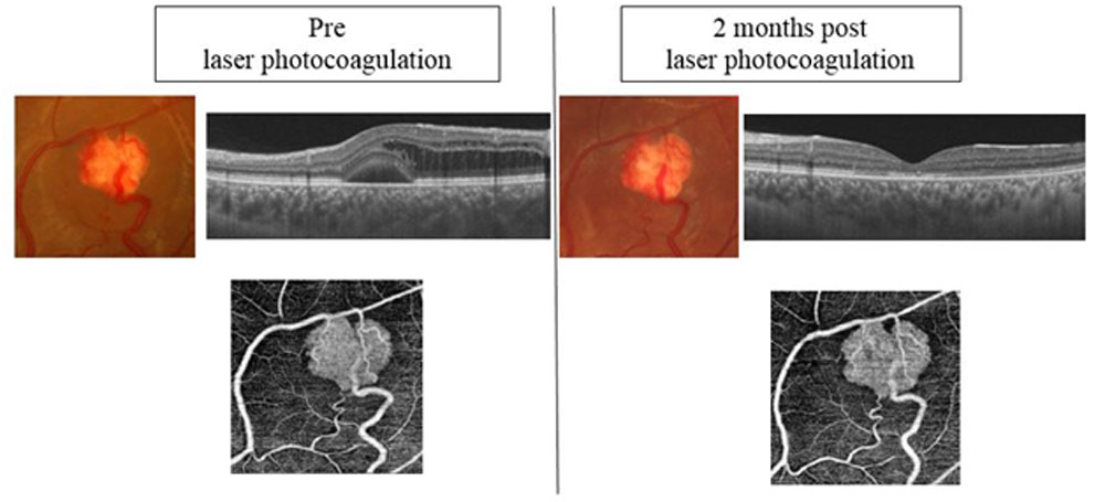
Figure2
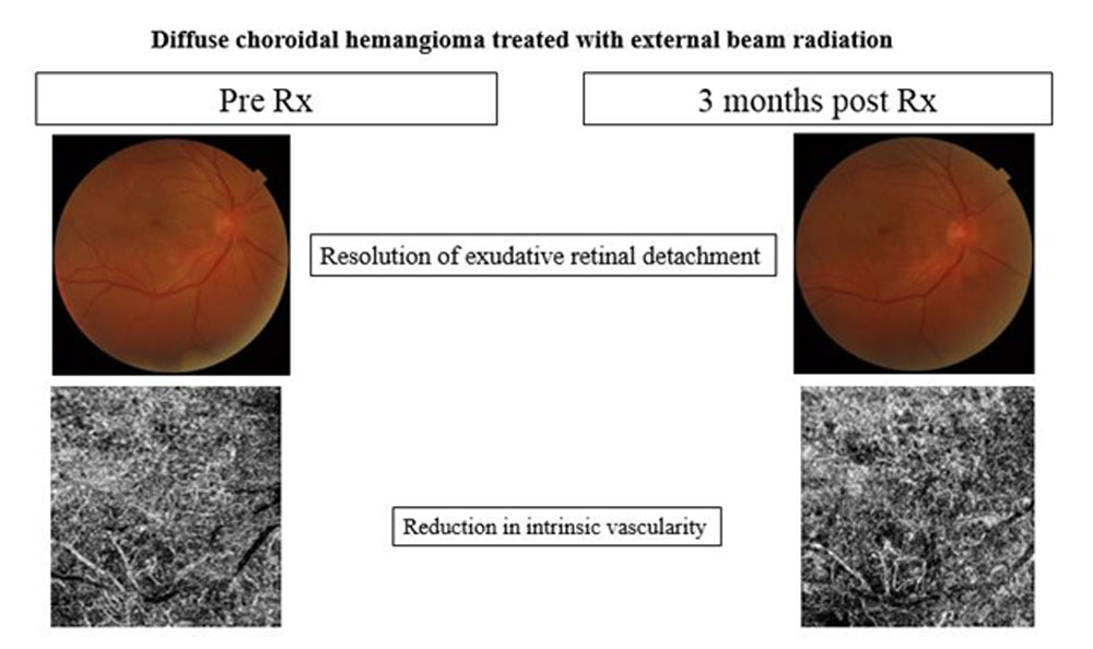
Figure3
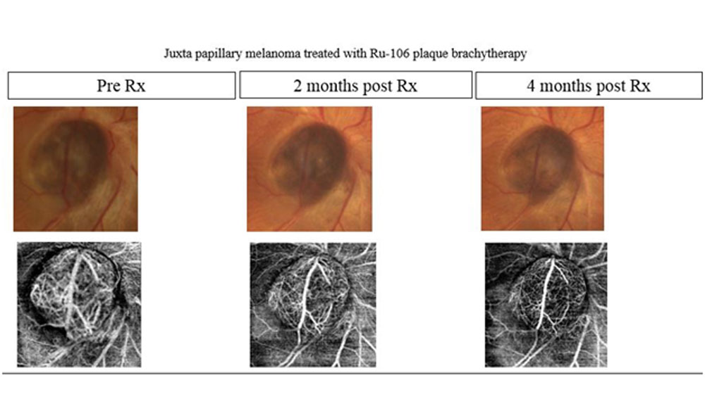
Figure4
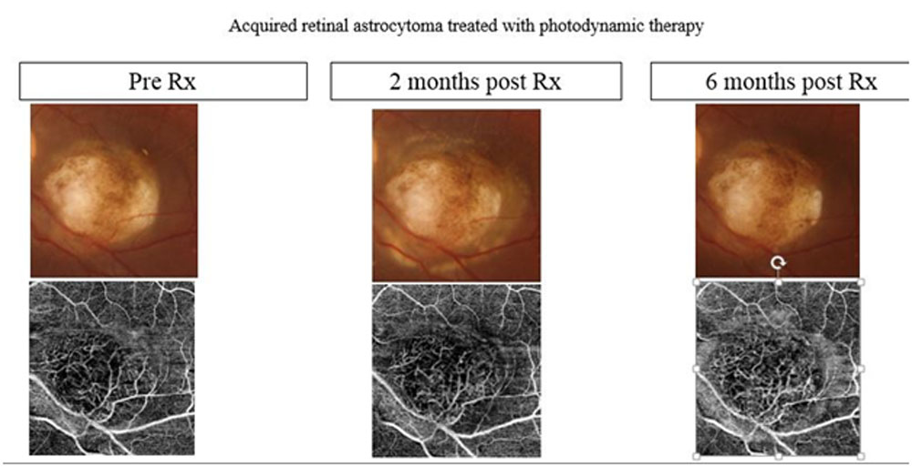
Figure 5
Leave a Comment