

Dr. Divya Alex, A15662, Dr. Mahesh G, Dr. Giridhar Anantharaman
ABSTRACT:
Purpose: Compare the effect of Combination therapy (PDT+antiVEGF) and antiVEGF monotherapy on choroidal vascularity parameters and morphological parameters in PCV
Method: Retrospective case analysis of 33 eyes diagnosed as PCV (Everest criteria) treated either with combination (PDT+antiVEGF; n=17) or antiVEGF monotherapy(n=16). Demographic details, visual acuity assessment and Double Layer Sign (DLS)width, PED height was considered from baseline to the 3rd and 6th month followup visits. Choroidal vascularity analysis including choroidal thickness, total surface area (TSA), total luminal area (TLA) and choroidal vascularity index (CVI) assessment was done for each visit using Enhanced Depth Imaging (EDI) using ImageJ software.
Results: Disease reactivation was significantly higher in the monotherapy arm compared to the PDT arm at 3,6months. There was statistically significant decrease in choroidal thickness in EDI, DLS, TSA&TLA (P<0.05) at both visits in the combination (PDT+AntiVEGF) therapy arm. Complete collapse of PED, reduction in DLS width which was achieved only in PDT arm showed negative correlation with the disease reactivation. Reduction in EDI, TSA, TLA did not correlate with disease activity.
Conclusion: This new choroidal imaging provides an insight to the vascular and morphological changes occurring after therapy. Our study proved PDT induce better choroidal vascular remodeling and thereby less disease recurrence.
INTRODUCTION:
Choroid, the vascular coating of the eye plays a predominant role in the ocular health. Choroid has the highest blood flow of any tissue in the body1 and has been implicated in the pathogenesis of many intraocular diseases such as age-related macular degeneration, myopic macular degeneration and especially pachychoroid disease spectrum including Polypoidal Choroidal Vasculopathy (PCV)2-5.
Unlike retina which has a consistent pattern, evaluating the morphometry of choroid is challenging because of its complex and variable architecture. Morphological and vascular analyses of the choroid have been revolutionised with the advent of enhanced depth imaging (EDI) Optical coherence tomography (OCT). EDI has shown increased choroidal thickness in eyes with Polypoidal Choroidal Vasculopathy and even in fellow eyes. Choroidal hyperpermeability is also an established pathological mechanism in PCV6. Sonodaet al firstdescribed a method for computing luminal and interstitial areas in the choroid as a means to quantify vascularstatus of the choroid by image binarization7. Recently, Choroidal Vascularity Index (CVI); (ratio of luminal area to total choroidal area) was introduced as an OCT marker to assess the vascularity of subfoveal choroid by Agarwal et al8.
Available treatment modalities for PCV include verteporfin photodynamic therapy (PDT), anti-VEGF therapy and thermal laser photocoagulation. PDT is thought to induce constriction of the choroidal vessels leading to hypoxia in both the choroid and retinal pigment epithelium, which causes upregulation of VEGF9-11. Combination therapy comprising PDT and anti VEGFwill inhibit VEGF expression, increase the efficacy of both PDT and pharmacologic agents. AntiVEGF also protect against choroidal ischemia and retinal pigment epithelium damage caused by PDT as well as result in resolution of exudative changes in the retina.The three landmark trials EVEREST12, LAPTOP13 and FUJISAN14 study established the role of combination therapy of full-fluence PDT (greater polyp closure rate) with anti-VEGF agents (better visual outcomes) in initiating therapy for this disease entity. In developing countries, cost remains a hindrance and many patients opt for anti VEGF monotherapy.
Sonoda etal7 by using the technique of binarization have proven that the choroidal remodelling after PDT is mainly due to a decrease in the luminal areas. But the vascular remodelling after the anti VEGF therapy is poorly understood. The purpose of the current study was to compare the effect of Combination therapy (PDT+antiVEGF) and antiVEGF monotherapy on choroidal vascular remodelling and retinal morphometrical analysis in PCV.
MATERIALS AND METHODS:
We retrospectively analysed the electronic medical records of patients diagnosed with PCV who presented to our institute between June 2014 and March 2018. Approval from the ethics committee review board was obtained. The research followed the tenets of the Declaration of Helenski.
Inclusion criteria:
Subjects diagnosed with PCV and with complete 6 month follow up visits and eyes with visible choroidal outer boundaries on spectral domain OCT (SD‑OCT) were included in the study.Diagnosis and case selection of PCV was made purely based on Everest criteria;Presenceof early subretinal nodular hyperfluorescence appearing within the first 5 min of ICG dye injection and at least one of the following diagnostic criteria: (1)Nodular appearance of the polyp on stereoscopic viewing (2)Hypofluorescent halo around the nodule (3)Abnormal vascular channel(s) supplying the polyps (4)Pulsatile filling of polyps (5)Orange subretinal nodules corresponding to the hyperfluorescent area on ICGA.
To avoid bias, we included cases with polyps within 2-disc dioptre area of the fovea in both the groups and in such a way that the greatest linear dimension circle in PDT involved fovea.
Exclusion criteria:
1)Coexisting macular/ vitreoretinal pathology
2) history of any vitreous surgery
3) history of previous treatment in the diseased eye in form of laser/ antivegf therapy.
4)recent ocular inflammation.
5) Pregnancy
6) Massive submacular haemorrhage //exudation which obscured the choroidal vasculature in SD OCT.
7) Any participants with poor visibility of choroidal boundaries.
8) patients who received focal laser to the polyps were also excluded from the study.
Participants were classified into two groups. Group A who underwent Full fluence Photodynamic therapy (FFPDT) followed by loading dose (3 doses) of anti VEGF therapy. Group B who received only antiVEGF injections 3 loading doses followed by pro re nata (PRN) regimen till 6 months. If group A had recurrence between 3rd and 6th month, PRN regimen was used.
All the participants underwent a comprehensive ophthalmic examination including best‑corrected VA (BCVA) using Snellen charts, intraocular pressure measurement using Goldmann applanation tonometer, and dilated fundoscopic examination using 78 D and 28 D.
Choroidal Imaging
As per defined protocol of the institution, Spectral domain optical coherence tomography (SD-OCT) was performed in each case using Heidelberg Spectralis HRA + OCT (Heidelberg Engineering, Germany) (1) macular dense scan (area 20° *20°, no of B scans: 49, distance between B scans:120 mm. (2) Enhanced depth imaging (EDI) at fovea. The EDI automatically sets the choroid closer to the zero-delay line and provides better visualization of the choroidal vasculature and sclero choroidal interface. Choroidal thickness was measured using the inbuilt caliper as the distance from the outer portion of hyperreflective RPE to the inner portion of hyperreflective zone corresponding to the sclerochoroid junction. Width of the Double layer sign (SDOCT finding defined as two hyperreflective lines corresponding to the separation of the Retinal pigment epithelium from the bruchs membrane) and PED height were analysed at each visit.
Image Binarization and Computation of Choroidal Vascularity Index
The EDI passing through the fovea was selected for image binarization. Central 3500microns was segmented using the protocol described by Sonoda et al7 with few modifications described by Agarwal etal8. The image binarization and computation of CVI was done using using ImageJ software (version1.49; National Institutes of Health, Bethesda, USA). The subfoveal choroidal area with a width of 3 mm, centred at the fovea, was selected and this constituted the region of interest. (Figure 1)
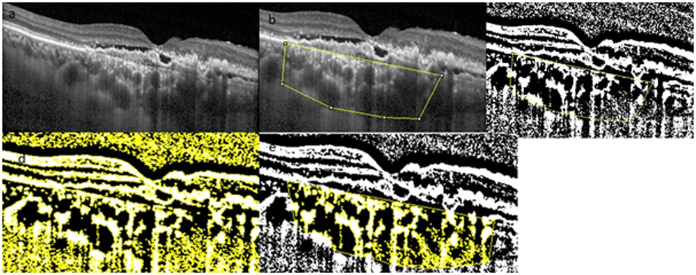
Figure1: Process of binarization of choroidal vasculature a. SDOCT EDI image through fovea. b. using polygon tool c.8bit converted autothreshold image. d)RGB color threshold image e)final binarization into total luminal area(black) and total stromal area(yellow).
INTER-RATER AND INTRA-RATER AGREEMENT.
Intraclass correlation coefficient for our study was 0.89 for intrarater agreement and 0.86 for
interrater agreement. Statistical analysis was done using SPSS 16.0 Version.
RESULTS:
Refining through the inclusion exclusion criteria, there were 33 participants. 17 in the combination arm and 16 in the Anti VEGF monotherapy arm.
Both the group received 3 Antivegf injections in the first 3 months and from 3rd to 6th month, combination arm received an average of 1.25 injections and monotherapy arm received an average of 2.5 injections. At the end of 3rd month, 77.7% eyes in the PDT arm was inactive and whereas only 35.4% remained inactive in the antiVEGF monotherapy arm. At 6 months, 66.7% remained inactive in the PDT group whereas only 41.2% remained inactive in the antivegf arm. On the contrary, more than 50% of the eyes had still activity at 3 and 6 months in the monotherapy arm.
Combination (PDT+AntiVEGF arm)
There was a statistically significant improvement in BCVA (P < 0.05) at the 3rd and 6th month visits. PDT arm showed statistically significant decrease in choroidal thickness in EDI, DLS, TSA & TLA (P<0.05) at both visits. However, there was no statistically significant difference in CVI (P > 0.05) at both the visits. Reduction in EDI, TSA, TLA did not correlate with disease activity.At 3rd month, complete resolution of the subretinal fluid and intraretinal fluid was significantly higher in the combination arm. (P< 0.05). Disease recurrence was significantly lower in the combination therapy arm at 6months. The mean subfoveal choroidal thickness decreased from 376 + 83u at baseline to 332 + 89 u (P=0.01) at 3 months and further 321+ 76u at 6 months. (P=0.008). Complete collapse of PED was achieved in 74% of the cases. The mean DLS width decreased from 2150 + 344u at baseline to 1860+ 278u (P=0.03) at 3 months and further 1664u+ 276u at 6 months. Complete collapse of PED, reduction in DLS width which was achieved only in PDT arm showed positive correlation with the disease reactivation (Figure2)
Monotherapy (AntiVEGF arm)
There was a statistically significant improvement in BCVA (P < 0.05) at the 3rd and 6th month visits. There were significantly more eyes with active disease in the monotherapy arm as compared to the PDT arm at 3 and 6months. Monotherapy arm showed significant increase in the DLS width in 3rd and 6th month visit. The mean subfoveal choroidal thickness increased from 358 + 102u at baseline to 361 + 97u (P=0.04) at 3 months and further increase to 368 + 89u(P=0.037) at 6 months. The mean DLS width increased from 2630 + 454u at baseline to 2786 + 478u at 3 months and further 2968u+276u at 6 months which showed a significant positive correlation with disease recurrence. (Table 1&2)
There was no statistically significant difference in CVI (P > 0.05) at both the visits in both the arms. Even though the reduction in total surface area, total luminal area and subfoveal choroidal thickness occured in both the arms, it was more statistically significant in the combination therapy arm.
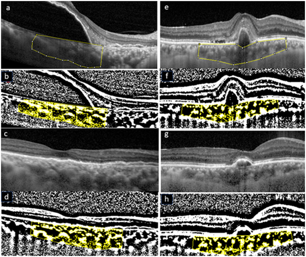
Figure2: (a) Extralarge serous PED and subretinalhyperreflectivity in PCV.(b) binarized image of the same (c,d) 6th month OCT showing complete collapse of the PED and resolution of subretinal hyper following Combination therapy ;binarized image.(e,f) thumb like polyp with subretinal hyper in PCV; binarized image (g,h) partial collapse of PED achieved with Antivegf monotherapy.
| Mean ±SD (P) | ||||||
| PDT | ANTIVEGF | |||||
| Baseline | 3 M | 6 M | Baseline | 3 M | 6 M | |
| BCVA | 0.45 ± 0.299 | 0.34 ± 0.358
(0.033**) |
0.31 ± 0.293
(0.010**) |
0.50 ± 0.281
|
0.43 ± 0.351
(0.389) |
0.39 ± 0.333
(0.004**) |
| CVI | 67.55 ± 4.755 | 68.77 ± 3.396
(0.327) |
67.97 ± 3.413
(0.560) |
68.90 ± 4.918 | 67.66 ± 2.809
(0.649) |
68.49 ± 3.026
(0.646) |
| TSA | 3654521.71 ± 1554771.579 | 3092534.25 ± 1273693.665
(0.025**) |
3123057.58 ± 1486757.960
(0.019**) |
3311239.61 ± 1088163.865 | 3240976.45 ± 1252898.299
(0.078) |
3142854.13 ± 1172042.467
(0.026**) |
| TLA | 2426521.13 ± 922220.126 | 2128087.70 ± 891624.348
(0.039**) |
2124248.68 ± 1016032.508
(0.047**) |
2578497.22 ± 700417.218 | 2501295.01 ± 787432.778
(0.078) |
2433945.28 ± 735574.848
(0.031**) |
| DLS Width | 2150.92(630.791) | 1860.00(666.071)
(<0.001**) |
2067.69(722.791)
(<0.001**) |
2630.38 (1162.635) | 2786.75 (722.635)
(<0.001**) |
2968.37 (693.932)
(<0.001**) |
Table 1: Baseline, 3rd and 6th month visit characteristics of PDT and Antivegf groups
| Disease activity at 6 months | PDT | ANTIVEGF | ||
| CorrelationCoefficient | P Value | CorrelationCoefficient | P Value | |
| Baseline EDI | 0.192 | 0.459 | -0.379 | 0.147 |
| Baseline DLS WIDTH | 0.577 | 0.039** | 0.516 | 0.028** |
| Baseline TSA | 0.481 | 0.051 | -0.379 | 0.147 |
| Baseline TLA | 0.457 | 0.065 | -0.348 | 0.187 |
| Baseline CVI | -0.241 | 0.352 | 0.095 | 0.727 |
Table2: Correlation of baseline parameters with disease activity of both the groups
DISCUSSION:
Segmentation of the choroid into different layers and its vascular components has been a significant challenge. The new automated segmentation software to binarize the choroidal structure has thrown light into the better understanding of the choroidal vasculature. It is a well-known fact that the recurrence rate of PCV with PDT therapy is much lower than that of PCV treated with anti VEGF alone13-15. Studying the choroidal remodelling after both these therapies will give a better clue in explaining the reason for the same. Our present study found the changes in the choroidal morphology and vascularity after the two different established treatment modalities in PCV.
Histologically, the choroid is composed of blood vessels (total luminal area) and interstitial tissues (total stromal area). For a clinical approach, we compared the effect of combination and AntiVEGF monotherapy on the luminal and interstitial areas of the choroid. Our results showed that both areas decreased in size, but the luminal area decreased more than the interstitial area after PDT. CVI did not show a statistical decrease; may be because of the proportional decrease in the luminal and interstitial area. Where as the anti VEGF arm showed a statistically significant decrease in the luminal area only at 6 months of follow-up. Anti VEGF arm did not show a statistically significant decrease in the total stromal area. Hence our study proves, combination therapy induces better choroidal remodelling than AntiVegf monotherapy.
The mechanism of action of PDT is postulated to be short-term choriocapillaris hypoperfusion and long-term choroidal remodelling leading to reductions in choroidal congestion, vascular hyperpermeability, and extravascular leakage.Schlötzer-Schrehardt et al detected choriocapillaris occlusion in the region of PDT application 1 week after standard PDT in human eyes by histopathological analysis16,17. These all facts are supported by our results in the combination arm in view of statistically significant reduction in TSA and TLA
As there is a decline in the luminal areas, it is logical to assume that either the number of vessels reduced or the diameter of vessels reduced. The other possibility is a decrease of the vascular diameter caused by an increase of vascular tone. VEGF can dilate vascular channels by upregulating endothelial nitricoxide synthase(eNOS)dependent pathways18, and thus downregulation of VEGF can explain our findings. Vascular endothelial growth factor plays an important role in maintaining homeostasis of the choroid, and it is secreted mainly on the basal side of the RPE.VEGF increases vascular permeability; this allows intravascular osmotically active molecules to move into the interstitial tissue, resulting in the interstitial tissue swelling. Thus, an inhibition of the release of VEGF might result in a decrease in the release of osmotically active molecules. So theoretically Antivegf monotherapy can also cause reduction in the total luminal area and total interstitial area. Our study proved the same even though it was not stasistically significant.
Even though the recurrence rates were significantly higher in the monotherapy arm, this group also achieved statistically significant improvement in the visual acuity in both 3 and 6 months followup. Combination therapy arm achieved a better visual acuity than the monotherapy arm, even though it was not stasistically significant. Recurrence rates in both the group were similar to study published by Maruko etal19.
Another important finding in the study was the coexistent retinal morphological changes in the combination therapy arm. Complete collapse of the PED was attained in 64.7% of the combination arm vs 23.5 % in the monotherapy arm. The same was associated with a strong positive correlation with the resolution of disease. Reduction in the DLS width which was attained only in the Combination arm showed a significant negative correlation with disease reactivation. Surprisingly, there was an increase in the DLS width in the monotherapy arm. Hence our study proves, combination therapy induces better outer retinal remodelling than AntiVegf monotherapy in PCV which may be the reason for the less recurrence rate. This is in consistent with Shimuzu etal20 study which proved PDT causes microstructural outer retinal changes.
CONCLUSION:
CVI could be a parameter demonstrating the choroidal vessel congestion and hyperpermeability, which is the primary pathogenic mechanism in PCV. Choroidal Vascular parameters could also be a useful to understand the effect of therapeutic intervention. Being primarily a type 1 choroidal neovascularization, administration of intravitreal anti-VEGF therapy should benefit in PCV. PDT addresses the choroid, the origin and root cause of the disease activity in PCV. Our study in view of better retinal and choroidal morphological changes and vascular remodelling, strongly support the superiority of combination therapy over Antivegf monotherapy.
REFERENCES
- Alm, A. & Bill, A. Ocular and optic nerve blood flow at normal and increased intraocular pressures in monkeys (Macacairus): a study with radioactively labelled microspheres including flow determinations in brain and some other tissues. Exp Eye Res 15,15–29 (1973).
- Spaide RF. Enhanced depth imaging optical coherence tomography of retinal pigment epithelial detachment in agerelated macular degeneration. Am J Ophthalmol. 2009;147: 644–652.
- Zarbin MA. Current concepts in the pathogenesis of agerelated macular degeneration. Arch Ophthalmol. 2004;122:598–614
- McLeod DS, Grebe R, Bhutto I, Merges C, Baba T, LuttyGA.Relationship between RPE and choriocapillaris in age-related macular degeneration. Invest Ophthalmol Vis Sci. 2009;50:4982–4991
- Uyama M, Wada M, Nagai Y, Matsubara T, Matsunaga H, Fukushima I, Takahashi K Polypoidal choroidal vasculopathy: natural history. Am J Ophthalmol,2002;133:639–648
- Sonoda, S. et al. Choroidal structure in normal eyes and after photodynamic therapy determined by binarization of optical coherence tomographic images. Invest Ophthalmol Vis Sci 55, 3893–3899.2014.
- Sonoda, S. et al. Luminal and stromal areas of choroid determined by binarization method of optical coherence tomographic images. Am J Ophthalmol159, 1123–1131,2015
- Agrawal R, Gupta P, Tan KA, Cheung CM, Wong TY, Cheng CY. Choroidal vascularity index as a measure of vascular status of the choroid: Measurements in healthy eyes from a population-based study. Scientific reports. 2016 Feb 12;6:21090.
- Kiss CG, Simader C, Michels S, Schmidt-Erfurth U. Combination of verteporfin photodynamic therapy and ranibizumab:effects on retinal anatomy, choroidal perfusion and visual function in the protect study. Br J Ophthalmol 2008;92(12):1620 –1627
- Kaiser PK; Registry of Visudyne AMD Therapy Writing Committee. Verteporfin photodynamic therapy combined with intravitreal bevacizumab for neovascular age-related macular degeneration. Ophthalmology 2009;116(4):747–755, 755.e1.
- Saito M, Shiragami C, Shiraga F, et al. Combined intravitreal bevacizumab and photodynamic therapy for retinal angiomatous proliferation. Am J Ophthalmol 2008;146(6):935–941.e1
- Koh A, Lee WK, Chen LJ, Chen SJ, Hashad Y, Kim H, Lai TY, Pilz S, Ruamviboonsuk P, Tokaji E, Weisberger A. EVEREST study: efficacy and safety of verteporfin photodynamic therapy in combination with ranibizumab or alone versus ranibizumab monotherapy in patients with symptomatic macular polypoidal choroidal vasculopathy. Retina. 2012 Sep 1;32(8):1453-64.
- Oishi A, Kojima H, Mandai M, Honda S, Matsuoka T, Oh H, Kita M, Nagai T, Fujihara M, Bessho N, Uenishi M. Comparison of the effect of ranibizumab and verteporfin for polypoidal choroidal vasculopathy: 12-month LAPTOP study results. American journal of ophthalmology. 2013 Oct 1;156(4):644-51.
- Gomi F, Oshima Y, Mori R, Kano M, Saito M, Yamashita A, Iwata E, Fujisan Study Group. Initial versus delayed photodynamic therapy in combination with ranibizumab for treatment of polypoidal choroidal vasculopathy: The Fujisan Study. Retina. 2015 Aug 1;35(8):1569-76.
- Anantharaman G, Sheth J, Bhende M, Narayanan R, Natarajan S, Rajendran A, Manayath G, Sen P, Biswas R, Banker A, Gupta C. Polypoidal choroidal vasculopathy: Pearls in diagnosis and management. Indian journal of ophthalmology. 2018 Jul;66(7):896.
- Blaauwgeers HG, Holtkamp GM, Rutten H, et al. Polarized vascular endothelial growth factor secretion by human retinal pigment epithelium and localization of vascular endothelial growth factor receptors on the inner choriocapillaris. Evidence for a trophic paracrine relation. Am J Pathol. 1999;155:421–428.
- Chan WM, Lam DS, Lai TY, Tam BS, Liu DT, Chan CK. Choroidal vascular remodelling in central serous chorioretinopathy after indocyanine green guided photodynamic therapy with verteporfin: a novel treatment at the primary disease level. British Journal of Ophthalmology. 2003 Dec 1;87(12):1453-8
- Izumi T, Koizumi H, Maruko I, Takahashi Y, Sonoda S, Sakamoto T, Iida T. Structural analyses of choroid after half-dose verteporfin photodynamic therapy for central serous chorioretinopathy. British Journal of Ophthalmology. 2017 Apr 1;101(4):433-7
- Maruko I, Iida T, Oyamada H, et al. Choroidal thickness changes after intravitreal ranibizumab and photodynamic therapy in recurrent polypoidal choroidal vasculopathy.Am J Ophthalmol 2013;156:548–556
- Shimizu K, Hashimoto Y, Azuma K, Nomura Y, Obata R, Takahashi H, Yanagi Y. Changes of outer retinal microstructures after photodynamic therapy for chronic central serous chorioretinopathy. Clinical ophthalmology (Auckland, NZ). 2017;11:1505-12
Leave a Comment